International Journal of Oral and Craniofacial Science
The erosion/infiltration technique made by a polymer based on TEGDMA to mask the white lesions of the tooth
Jean-Pierre Attal1,2*, Gil Tirlet3, Philippe François1,4, Elisa Caussin1,4 and Elisabeth Dursun1,5
2Charles Foix Hospital, 7 avenue de la République, 94200 Ivry-sur-Seine, France
3Private Practice, 280 Boulevard Raspail, Paris, France
4Bretonneau Hospital, 23 Rue Joseph de Maistre, 75018 Paris, France
05Henri Mondor Hospital, 1 Rue Gustave Eiffel, 94000 Créteil, France
Cite this as
Attal JP, Tirlet G, François P, Caussin E, Dursun E (2022) The erosion/infiltration technique made by a polymer based on TEGDMA to mask the white lesions of the tooth. Int J Oral Craniofac Sci 8(2):023-025. DOI: 10.17352/2455-4634.000055Copyright License
© 2022 Attal JP, et al. This is an open-access article distributed under the terms of the Creative Commons Attribution License, which permits unrestricted use, distribution, and reproduction in any medium, provided the original author and source are credited.It is common to observe anterior teeth that present white opacities of enamel in relation to hypomineralization. These lesions alter social life of children and adults. There is a recent non-invasive treatment that can remove these stains without loss of substance, keeping tooth structure intact. This treatment modifies the optical properties of the white spot by infiltrating a polymer based on trimethylene glycol dimethacrylate (TEGDMA). This short article presents a method called erosion/ infiltration, which our team first described. Dentists must be aware of it, in order to treat patients with either fluorosis, MIH or trauma, so that much more invasive treatments such as veneers and crown can be avoided.
Introduction
Enamel white spot lesions on anterior teeth are frequent and can impact patients’ quality of life whether among children [1] or adults when they alter the smile. Their prevalence on the teeth, although difficult to quantify, is high. Several dental anomalies can cause these defects: fluorosis; molar incisor hypomineralization; medicine intake during enamel mineralization; traumas; or initial caries.
Several techniques have been proposed to treat spots in the anterior region (remineralization, controlled microabrasion, laminate veneers, crown...). In this article, however, we will focus on the technique that we called ‘erosion/infiltration’ and that we described [2,3], a decade ago for treating fluorosis - and trauma-related lesions, which are most often superficial lesions in nature. Contrary to the treatments frequently proposed by dentists which are invasive including an important loss of substance (ceramic laminate veneers or even crown), the proposed strategy is not based on the elimination of enamel, but on masking the lesion by infiltrating the porous subsurface enamel with a polymer based on TEGDMA that has a refraction index close to sound enamel’s, after permeating the non- porous surface enamel through hydrochloric acid erosion. The aim of this short article was to present this very conservative treatment to treat these lesions.
Origin and principle of the technique
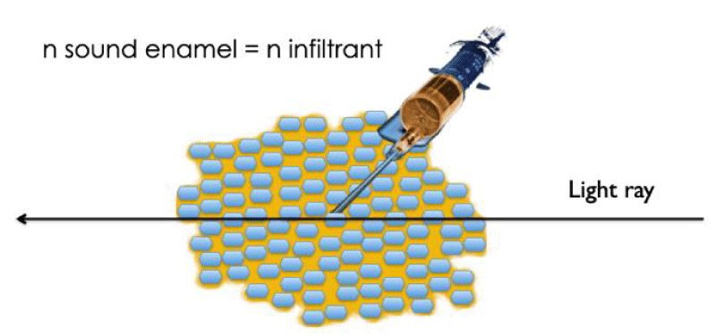
Erosion/infiltration is a technique that was proposed when a new product (Icon-DMG), designed to stop caries in the posterior segment, was launched on the market [4]. The first step, involving superficial demineralization by application of 15% hydrochloric acid solution, opens up the access to the hypomineralized lesion. Then, in a second stage, an extremely fluid TEGDMA-based polymer is infiltrated into the body of the lesion. As a result, this infiltration masks the white spot on the enamel, making this technique a new treatment for masking initial carious lesion [5] when took at an early stage [6]. Our team has extended the technique first described by Paris and Meyer Lueckel [5] to mask white carious lesions to others hypomineralization lesions (trauma, fluorosis) [3]. The explanation of the efficiency of this technique is a simple physical one: the white spot results of a complex optical phenomenon whereby an optical maze is formed within the lesion and contributes to the reflection of incident light (Figure 1).
Infiltration of the pores of the lesion with an infiltrant, a resin whose refractive index (n infiltrant = 1.52) is close to sound enamel’s (1.62) improves the transmission of photons through the hypomineralized enamel and restores its translucence (Figure 2).
Case report
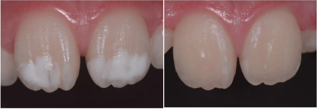
An 8-year-old female patient came to our dental office with a chief complaint of white lesions on the upper front teeth (Figure 3 left). After examination, symmetrical and large sized enamel white spots lesions were found in the right and left upper maxillary central incisors, characteristic of fluorosis. As this indication was not proposed by the manufacturer ICON (DMG, Germany), informed consent was obtained from the parents to use erosion infiltration technique. After obtaining a dry operating field, the surfaces of the lesion were, according to our protocol [7], conditioned twice with 15% hydrochloric acid gel (ICON ETCHANT, DMG, Germany) for 2 minutes, rinsed (30 s) using water spray and completely dried out with ethanol (ICON DRY, DMG, Germany). The TEGDMA resin (ICON INFILTRANT, DMG, Germany) was then applied in two steps and light cured for 40 seconds. To insure a good polymerization, we applied a layer of glycerin and photopolymerized through. A simple polishing protocol completes the sequence (Figure 3 right).
Discussion
Composition and properties of the polymer infiltrant
The infiltration resin mainly consists of TEGDMA (about 78%) and other monomers (trimethylolpropane triacrylate, or TMPTA for short: 20%), butylated hydroxytoluene (BHT), and photoinitiators. It is essentially a hydrophobic monomer, therefore it is imperative to remove as much residual water as possible from the lesion through an alcohol stage after the first step of erosion giving access the porosities. Another important point for the use of TEGDMA in dentistry concerns the toxic potential of the monomer since it can release formaldehyde [8]. However a perfect polymerization through glycerine should prevent any release of any kind. One must notice that in the case of incipient caries, some authors recently proposed a modified TEGDMA- based resin including fluorohydroxyapatite and fluoride- doped bioactive glass, to improve the mechanical and physical properties and induce a remineralizing potential [9]. Others had recently proposed the combination of TEGDMA with other monomers (HEMA) to obtain more suitable viscosity, handling characteristics, and general properties however none of these formulations were tried out by now in association with the erosion infiltration technique for the treatment of fluorosis or MIH [10].
Prognosis - Aging of the polymer infiltrant
Many authors raised the question of the aging of the resin used for infiltration and a recent in vivo study with a 6-year follow-up [11] demonstrated the excellent subjective and objective colorimetric stability of enamel treated with ICON. Our experience is similar, we can anticipate a good prognosis: in the case of superficial infiltration, we noticed an improvement of the result over time, and so had other authors [12]. This improvement could be explained by the resin absorption of water that had not been eliminated by the alcohol step. This absorption could lead to a smoothing in optical interfaces in the light path and thereby to an improvement in the translucency of enamel.
What to do if the lesion is deep and not superficial?
Molar Incisor Hypomineralization (MIH) is highly prevalent worldwide, with a rate around 14% [13]. In this case, the hypomineralized lesion is deep in the enamel, and treatment through superficial infiltration cannot be successful. To overcome these treatment-related difficulties, we devised a modified technique in 2012 and published it in 2013 [14]. We called it deep erosion/infiltration. This modified technique’s protocol can be the topic of the next article.
Conclusion
It is now possible, with a very simple technique called erosion infiltration, to mask white opacities of teeth without any loss of substance. The principle of the technique is based on a modification of the optical properties of enamel by infiltrating a TEGDMA-based polymer with a refractive index close to enamel hydroxyapatite. Dentists and patients should be aware of this possibility to avoid useless elimination of sound tissues of the tooth.
Conflict of interest disclosure
The two first Authors, as inventors of the technique to treat fluorosis, trauma and MIH, had financial help to support clinical and in vitro evaluation from DMG Companies but received no compensation for sales of the product Icon.
- Sujak SL, Abdul Kadir R, Dom TN. Esthetic perception and psychosocial impact of developmental enamel defects among Malaysian adolescents. J Oral Sci. 2004 Dec;46(4):221-6. doi: 10.2334/josnusd.46.221. PMID: 15901066.
- Tirlet G, Attal JP. A new therapy to hide white spots. Inf Dent. 2011;4‑7.
- Tirlet G, Chabouis HF, Attal JP. Infiltration, a new therapy for masking enamel white spots: a 19-month follow-up case series. Eur J Esthet Dent. 2013 Summer;8(2):180-90. PMID: 23712339.
- Paris S, Meyer-Lueckel H, Kielbassa AM. Resin infiltration of natural caries lesions. J Dent Res. 2007 Jul;86(7):662-6. doi: 10.1177/154405910708600715. PMID: 17586715.
- Paris S, Meyer-Lueckel H. Masking of labial enamel white spot lesions by resin infiltration--a clinical report. Quintessence Int. 2009 Oct;40(9):713-8. PMID: 19862396.
- Muthuvel P, Ganapathy A, Subramaniam MK, Revankar VD. Erosion Infiltration Technique': A Novel Alternative for Masking Enamel White Spot Lesion. J Pharm Bioallied Sci. 2017 Nov;9(Suppl 1):S289-S291. doi: 10.4103/jpbs.JPBS_150_17. PMID: 29284982; PMCID: PMC5731033.
- Attal JP, Denis M, Atlan A, Vennat E, Tirlet G. Deep infiltration: A new concept for masking white spots in enamel. Partie 1. Inf Dent. 2013;(19):74‑9.
- Emmler J, Seiss M, Kreppel H, Reichl FX, Hickel R, Kehe K. Cytotoxicity of the dental composite component TEGDMA and selected metabolic by-products in human pulmonary cells. Dent Mater. 2008 Dec;24(12):1670-5. doi: 10.1016/j.dental.2008.04.001. Epub 2008 May 16. PMID: 18486204.
- Hashemian A, Shahabi S, Behroozibakhsh M, Najafi F, Abdulrazzaq Jerri Al-Bakhakh B, Hajizamani H. A modified TEGDMA-based resin infiltrant using polyurethane acrylate oligomer and remineralising nano-fillers with improved physical properties and remineralisation potential. J Dent. 2021 Oct;113:103810. doi: 10.1016/j.jdent.2021.103810. Epub 2021 Sep 14. PMID: 34530057.
- Szczesio-Wlodarczyk A, Polikowski A, Krasowski M, Fronczek M, Sokolowski J, Bociong K. The Influence of Low-Molecular-Weight Monomers (TEGDMA, HDDMA, HEMA) on the Properties of Selected Matrices and Composites Based on Bis-GMA and UDMA. Materials (Basel). 2022 Apr 4;15(7):2649. doi: 10.3390/ma15072649. PMID: 35407980; PMCID: PMC9000443.
- Mazur M, Westland S, Ndokaj A, Nardi GM, Guerra F, Ottolenghi L. In-vivo colour stability of enamel after ICON® treatment at 6 years of follow-up: A prospective single center study. J Dent. 2022 Jul;122:103943. doi: 10.1016/j.jdent.2021.103943. Epub 2022 Jan 13. PMID: 35033596.
- Kim S, Kim EY, Jeong TS, Kim JW. The evaluation of resin infiltration for masking labial enamel white spot lesions. Int J Paediatr Dent. 2011 Jul;21(4):241-8. doi: 10.1111/j.1365-263X.2011.01126.x. Epub 2011 Mar 14. PMID: 21401750.
- Zhao D, Dong B, Yu D, Ren Q, Sun Y. The prevalence of molar incisor hypomineralization: evidence from 70 studies. Int J Paediatr Dent. 2018 Mar;28(2):170-179. doi: 10.1111/ipd.12323. Epub 2017 Jul 21. PMID: 28732120.
- Attal JP, Atlan A, Denis M, Vennat E, Tirlet G. White spots on enamel: treatment protocol by superficial or deep infiltration (part 2). Int Orthod. 2014 Mar;12(1):1-31. English, French. doi: 10.1016/j.ortho.2013.12.011. Epub 2014 Feb 3. PMID: 24503373.
Article Alerts
Subscribe to our articles alerts and stay tuned.
 This work is licensed under a Creative Commons Attribution 4.0 International License.


 Save to Mendeley
