Journal of Clinical Research and Ophthalmology
The association between primary open-angle glaucoma and helicobacter pylori infection
Mohammad Mehdi Raji, Alhaj Farhath Tasneem, Vittal I Nayak, Faiza Syed Jafar*, Zeba Ahmed and Yalavali Indraja
Cite this as
Raji MM, Tasneem AF, Nayak VI, Jafar FS, Ahmed Z, et al. (2021) The association between primary open-angle glaucoma and helicobacter pylori infection. J Clin Res Ophthalmol 8(2): 036-042. DOI: 10.17352/2455-1414.000091Background: Primary Open-Angle Glaucoma (POAG) the most common form of glaucoma is a neurodegenerative disease, which is the third most common cause of blindness worldwide. It is estimated that 60 million people in the world are affected by this disease and 8.4 million are bilaterally blind. Among the various factors that have been implicated in the pathophysiology of this disease is infection with Helicobacter pylori (HP), a Gram-negative bacterium that is commonly found in stomach and present in approximately one-half of the world’s population. Establishment of such a causal correlation will probably have important practical applications as the eradication of H. pylori might lead to developments in the treatment of glaucoma.
Objectives: To investigate the association between Primary Open-Angle Glaucoma (POAG) and Helicobacter Pylori infection and to observe fluctuations in intra ocular pressure after Helicobacter Pylori infection eradication.
Design: Duration based, prospective observational study.
Participants: 50 patients with documented Primary Open-Angle Glaucoma (POAG) as case group and 50 non-glaucoma participants as control group.
Methods: Upper gastrointestinal endoscopy was performed to evaluate macroscopic abnormalities, and gastric mucosal biopsy specimens were obtained for the presence of H. pylori infection tested by Rapid Urease Test (RUT). All subjects underwent detailed ocular examinations including visual acuity, slit-lamp examination, fundoscopy, intra-ocular pressure recording, gonioscopy, GHT to assess visual fields and OCT of optic nerve head.
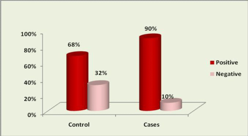
Results: In 90% of POAG patients of case group and in 68% of non-glaucoma participants of control group Helicobacter pylori infection was detected by RUT (P-Value=0.007).
Conclusion: H. pylori infection is more frequent in glaucoma patients, perhaps more so in those of Indian ethnicity. It may play a role as a secondary aggravating factor or even may be the primary cause. The establishment of such a causal relationship will probably have important practical applications as the eradication of H. pylori might lead to developments in the treatment of glaucoma.
Introduction
Glaucoma refers to a group of diseases that have in common a characteristic optic neuropathy with associated visual function loss. Although elevated Intraocular Pressure (IOP) is one of the primary risk factors, its presence and absence does not have a role in the definition of the disease.
The World Health Organization estimated the global population of POAG at 104.5 million with incidence of 2.4 million people per year. Blindness prevalence for all types of glaucoma as estimated at more than 8 million people, with 4 million cases caused by POAG. The different types of glaucoma were calculated to be responsible for 15% of blindness, placing glaucoma as the third leading cause of blindness worldwide [1].
Helicobacter pylori is a gram-negative spiral shape bacillus, it lives in gastric mucosa and colonizes the stomachs of about 50% of the world’s human population throughout their lifetimes and transmission takes place more often by the faecal-oral and oral-oral route.
Prevalence of H. pylori infection in India is about 69% and prevalence varies with age: about 50% of 60-year old persons, about 20% of 30- year old persons, and <10% of children are colonized.
Essentially all H. pylori colonized persons have gastric tissue responses (inflammations), but fewer than 15% develop associated illnesses such as peptic ulceration, gastric adenocarcinoma, or gastric lymphoma [2]. It should be noted that H pylori eradication regimen are not listed as medication changing IOP in Terminology and Guidelines of Glaucoma.
Recent research has focused on H. pylori colonization as a risk factor for some extragastric diseases. In a study in Greece by Kountouras, et al, shown that 87.5% of the Primary Openangle Glaucoma (POAG) patients had H. pylori infection which was histologically confirmed, hence Kontouras et al claimed there was a positive correlation between Helicobacter Pylori infection and glaucoma [3].
The goal of our study is to show any association between primary open-angle glaucoma and helicobacter pylori infection, and whether these two entities are linked by a causal relationship or whether they share common predisposing or precipitating factors. This association could have an important effect on pathophysiology and treatment of glaucoma.
Aim of the study
To investigate the association between Primary OpenAngle Glaucoma and Helicobacter Pylori infection as well as to observe fluctuations in intra ocular pressure after Helicobacter Pylori infection eradication.
Materials and methods
This study was conducted in Vydehi Institute of Medical Sciences and Research Center, Bangalore, on the subjects of age group 35 years old and above who visited the out-patient department of Ophthalmology from January 2014 to June 2017. All subjects were of Indian ethnicity and visited the center voluntarily.
This was a duration based, prospective observational study. The study population was composed of fifty primary openangle glaucoma (POAG) patients for the case group and a sample of fifty participants without history of primary openangle glaucoma as control group, all cases satisfied the inclusion and exclusion criteria.
All cases were evaluated for Helicobacter pylori infection, which is based on Upper Gastro-Intestinal Endoscopy and Rapid Urease Test (RUT) with gastric tissue biopsy specimen.
A pre-structured proforma was used to collect the baseline data and an informed written consent was obtained after explaining about the need of the study and the procedures that were to be performed for the collection of data. Detailed history was taken and examination (ocular and systemic) was performed as per the proforma for those who satisfied the inclusion and exclusion criteria.
Inclusion criteria
All patients aged 35 years and above, both male and female, with past history of primary open angle glaucoma as the case group from whom written informed consent was taken.
Exclusion criteria
(1) Age group less than 35 years old. (2) All eye diseases other than glaucoma. (3) Diabetes Mellitus (4) Hypertension (5) Myopic refractive error exceeding -8 diopters (6) Past history of any secondary glaucoma (post-traumatic, postuveitis, neovascular glaucoma and steroid-induced glaucoma) (7) Previous ocular surgery.
Investigations and interventions
The following investigations and interventions were done on glaucoma patients in case group and participants on control group:
1. Visual acuity test (with and without Pinhole) as assessed by Snellen’s chart
2. Best corrected visual acuity
3. Slit-lamp examination for anterior segment findings
4. Fundoscopy by direct ophthalmoscope and 78D lens
5. Fundus Photography
6. Goldmann applanation tonometry for measuring of Intra-Ocular Pressure
7. Gonioscopic examination by Ziess Goniolens to assess anterior chamber angles
8. Glaucoma Hemifield Test to assess visual fields
9. Spectral domain Optical Coherence Tomography of optic nerve head
10. Rapid Urease Test after Upper Gastro-Intestinal Endoscopy.
Study design
This was a duration based, prospective observational study. The data analysed 100 patients and 200 eyes (50 patients in case group and 50 participants in control group). The case group, consisted of 50 patients with documented primary open-angle glaucoma and the control group, consisted of 50 non-glaucoma participants who visited the out-patient department of Ophthalmology of Vydehi Institute of Medical Sciences and Research Center.
Proven glaucoma patients were included in the study using the following criteria: a history of intra-ocular pressure of 21 mm Hg or more; typical optic nerve head changes including cupping, neuro-retinal rim thinning, notching in the inferior or superior temporal or total glaucomatous cupping, and typical visual field loss including a paracentral, arcuate or Seidel’s scotoma or a nasal step.
The intra-ocular pressure was measured by a calibrated Goldmann applanation tonometer. None of the glaucoma patients received oral medications that could decrease intra- ocular pressure (e.g., carbonic anhydrase inhibitors).
All the glaucoma patients had prior experience with automated perimetry. Visual fields were evaluated using the 30-2 program of Glaucoma Hemifield Test by the same perimetrist. All 100 patients (50 glaucoma patients, 50 control participants) underwent elective diagnostic upper gastrointestinal endoscopy after informed consent. None of patients had received any Histamine2 receptor antagonists, bismuth compounds, proton pump inhibitors, non-steroidal anti-inflammatory drugs and antibiotics in the recent 4 weeks.
Participants reported at 9 AM after a 12-hour fast. Standard upper gastro-intestinal endoscopy was performed to identify evidence of macroscopic abnormalities. Two biopsy specimens were obtained, one from the antral region within 2 cm of the pyloric ring, and the other from the fundus. A biopsy specimen from each site was used for rapid urease slide testing of H. pylori infection.
To prevent contamination of specimens taken from different sites, biopsy specimens were taken from each site with a fresh pair of sterile forceps. The forceps were wiped with alcohol on withdrawal from the endoscope to remove any organism that might have been present in the biopsy channel. Endoscopes were sterilized between procedures according to the standard guidelines. Each biopsy specimen was placed in a tube containing 0.5 ml of 10% urea in deionized water to which had been added two drops of 1% phenol red as a pH indicator. The biopsy specimen test was read at 5 minutes, 1 hour, 3 hour, and 24 hours and was regarded as positive if the indicator changed from yellow to red at any time. H. pylori positive patients received a triple eradication regimen (Omeprazole, Clarithromycin and Metronidazole).
Statistical methods
Descriptive and inferential statistical analysis had been carried out in this study. It was assumed that dependent variables should be normally distributed throughout the data, samples drawn from the population should be random and cases of the samples should be independent.
Results on continuous measurements are presented on Mean ± SD (Min-Max) and results on categorical measurements are presented in Number (Percentage). The statistical analysis was performed by STAT11.2 (College station TX USA). Chi square test has been used to measure the association between the genders, age, place of residence, occupation, treatment of glaucoma, chief complaints, visual acuity, anterior segment findings, GI diagnosis and HP Rapid Urease Test (RUT) with both Case and control groups and is expressed as frequency and percentage.
Shapiro Wilk test has been used to check the normality. Student’s t-test was used to find the significance difference between the, intra- ocular pressure, Cup/Disc ratio and RNFL thickness of inferior, superior, nasal and temporal with groups (Cases and control) and it is expressed as mean and standard deviation.
P-value less than 0.05 is considered as statistically significant.
- Suggestive significance (P-Value: 0.05< P <0.10)
- Moderately significant (P-Value: 0.01< P ≤0.05)
- Strongly significant (P-Value: P ≤ 0.01)
Results
This study analysed 100 patients and 200 eyes (50 patients in case group and 50 participants in control group). Maximum number of patients (44%) in case group and (38%) in the control group belong to the age group of fifty-six years to sixty-five years.
In the case group, the range of age was 39-65 years with mean of 52.02 and standard deviation of ±7.26, while in the control group it was 40-60 years with mean of 50.04 and standard deviation of ±7.15 Table 1.
In the case group, thirty-one patients (62%) were males while in control group thirty-nine participants (78%) were males. In the case group, nineteen patients (38%) were females while in control group eleven participants (22%) were females. There is no statistically significant difference in age (p-value= 0.173) and gender (p-value= 0.081) between POAG patients in case group and control group in this study Table 2.
Thirty-two patients (64%) in case group and twenty-three participants (46%) in control group were from Bangalore. Rest of the patients (36% in case group and 54% in control group) were from West Bengal. Based on p-value (0.081), there is no statistically significant geographical difference in incidence of POAG between case and control group.
All patients in case group were known cases of documented POAG and have been taken anti glaucoma medications, while all participants in control group did not have POAG and have not received any anti glaucoma treatment. All glaucoma patients in case group and all participants in control group had complaints of dyspepsia.
The most common ocular complaints in case group were diminished distant vision (60%), while the maximum ocular complaints in control group were difficulty in near vision (52%). Only seven patients (14%) in case group and six participants (12%) in control group were symptom-free. Forty eyes (40%) in case group and sixty-seven eyes (67%) in control group had visual acuity of 6/6 or 6/9. Twenty-three eyes (23%) in case group and eighteen eyes (18%) in control group showed visual acuity of 6/60 or less. Rest of the eyes (37% in case group and 15% in control group) had visual acuity of 6/12 to 6/36.
After checking the visual acuity by using pinhole, the results showed that in the majority of eyes (80% in case group and 82% in control group), visual acuity improved to 6/6 or 6/9. The most common refractive error in both case and control group was hypermetropia (57% in case group and 49% in control group). Myopia was detected in 25% of eyes in case group and 21% of eyes in control group. Rest of eyes (18% in case group and 30% in control group) were emmetropic.
Forty patients (80%) in case group and forty-one participants (82%) in control group had anterior segment within normal limits, and only ten patients (20%) in case group and nine participants (18%) in control group had nuclear sclerosis grade II lens cataract based on LOCS III classification. p-value of the anterior segment findings in this study does not show any statistically significant difference between case and control groups. On gonioscopic examination, all the 200 eyes had grade 4 open angles in inferior, superior, nasal and temporal quadrants.
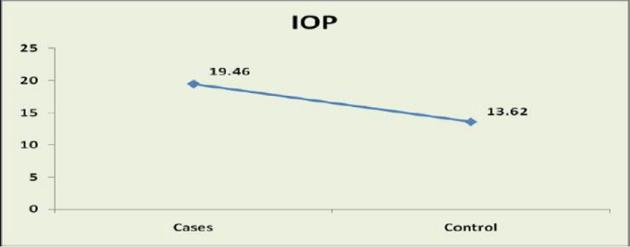
In this study, the mean intra-ocular pressure of case group was 19.46 mmHg with SD of ± 3.58 mmHg, while in control group the Mean intra-ocular pressure was 13.62 mmHg with SD of ± 1.57 mmHg. In comparison of Mean IOP between case group and control group, study shows statistically significant difference between glaucoma patients in case groups and participants of control group, because p-value is less than 0.001 Table 3, Figure 1.
On fundoscopy, all eyes in control group had normal optic nerve head, while in case group all eyes showed glaucomatous optic nerve head changes. The most common optic nerve head changes in case group was marked disc cupping in forty-one eyes (41%). In rest of eyes in case group the optic nerve head showed early disc cupping in twentyeight eyes (28%), moderate disc cupping in twenty-seven eyes (27%) and vertically oval cup in four eyes (4%) Table 4, Figure 2.
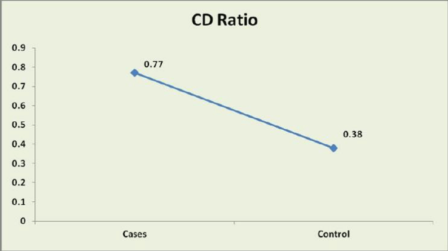
In this study, the Mean Cup/Disc Ratio of case group was 0.77 with SD of ± 0.09 while in control group, the mean Cup/Disc Ratio was 0.38 with SD of ± 0.08. In comparison of Mean C/D Ratio between case group and control group, study shows statistically significant difference between two groups of patients because p-value is less than 0.001 Table 5, Figure 3.
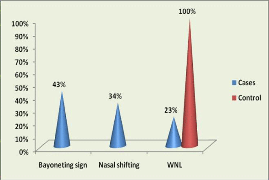
On fundoscopy, no eyes of control group showed any abnormal changes in optic disc vessels, while in case group only twenty-three eyes (23%) had normal optic disc vessels. The most common optic disc vessels change in case group was bayoneting sign in forty-three eyes (43%). In rest of the eyes in case group (34%), nasal shifting of optic disc vessels presented.
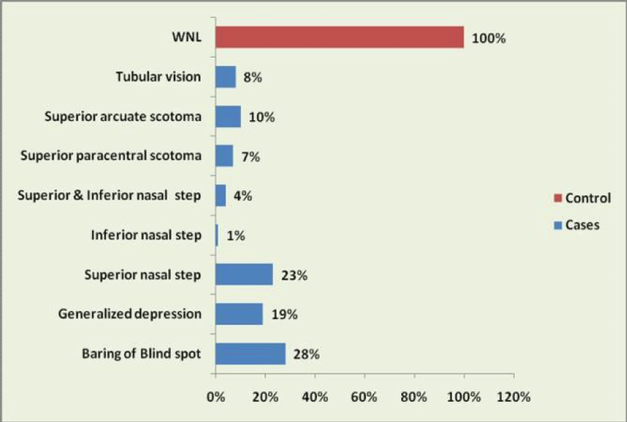
The visual fields as assessed with HFA by the same perimetrist showed, all eyes in control group had normal visual fields, while all the eyes in case group showed visual fields defects. The most common visual fields defects among eyes in case group was baring of blind spot, which was detected in twenty-eight eyes (28%). Only one eye, presented with inferior nasal step in the case group Table 6, Figures 4,5.
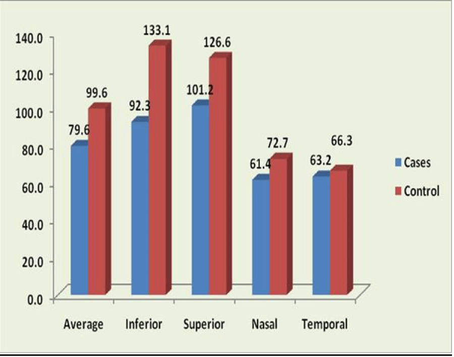
Spectral domain OCT was used to measure the average RNFL thickness of optic nerve head in both case and control groups (Table 5). Because P-value is < 0.001, study shows the statistically significant thinning of average RNFL thickness in inferior, superior and nasal quadrants of optic nerve head of eyes in case group compared to average RNFL thickness of same quadrants in eyes of control group.
However, this study does not show any significant thinning of average RNFL thickness in temporal quadrants of optic nerve head between case and control groups. (P-value=0.132) The “ISNT Rule” can explain these results. Based on this rule, in the course of glaucoma disease thinning of RNFL first will occurs in inferior quadrant of optic nerve head, followed by superior, nasal and temporal quadrants, respectively Table 7, Figure 6.
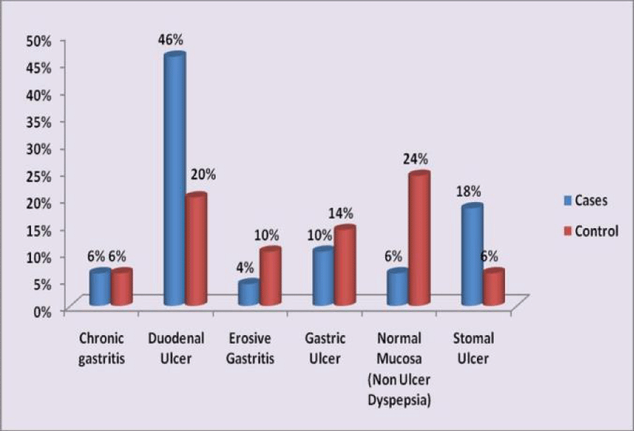
Study shows endoscopic evidence of chronic gastritis, duodenal ulcer, erosive gastritis, stomal ulcer and normal gastric mucosa appearance (called as non- ulcer dyspepsia), in glaucoma patients of the case group and participants of the control group. When compared with the participants in control group, glaucoma patients exhibited more often stomal ulcer and less often endoscopic normal appearance of gastric mucosa. Comparison for endoscopic evidence of chronic gastritis, duodenal ulcer, erosive gastritis and gastric ulcer did not reveal statistically significant differences between glaucoma patients in case group and participants of the control group.
Forty-five glaucoma patients (90%) in case group exhibited histologically proven H. Pylori infection, while infection detected in thirty-four participants (68%) of control group. Study showed a statistically significant difference in prevalence of H. Pylori infection between glaucoma patients in case group and control group Table 8, Figures 7,8.
Discussion
Helicobacter pylori is a curved spiral gram-negative bacterium which colonizes in the gastric mucosa and is associated with various upper gastrointestinal diseases as well as extra-digestive disorders [4-7].
In recent decades, H. Pylori infection has been considered as an important risk factor involved in primary open angle glaucoma, suggesting the existence of a possible link between the two.
The present study reinforces this link as 90% of the glaucoma patients exhibited histologically proven H. pylori infection whereas the rate of infections was significantly lower in the control group.
Our study has relied on Rapid Urease Test (RUT) for documentation of H. pylori infection. Mucosal biopsy and histological examination as well as RUT of the gastric biopsy specimen is the actual gold standard for the diagnosis of H. pylori infection [8]. This requires an upper GI endoscopy which is invasive, costly and time-consuming and should be done by a professional. Therefore, non-invasive tests such as 13C-urea breath test or serological detection of specific IgG antibodies against H. pylori may be preferable for screening purposes which is relatively inexpensive and requires only sampling of blood. However, it does not discriminate between a current and an old infection [9,10].
The 13C-urea breath test, though it is non-invasive, it requires the individual to be fasting as well as an expensive radioisotope-ratio mass spectrometer or a non-dispersible infrared spectrometer and produces a long half-life waste [11]. This test has false negative and false positive results if the patient has been using antibiotics for the past 4 weeks or from the presence of urease in the mouth [10]. However, it is a useful test for follow-up after eradication of H. pylori infection, hence, endoscopic histological analysis of gastric mucosa and RUT appears to be the most reliable diagnostic procedures, though it is invasive and costly.
H. pylori infection may increase the incidence or severity of glaucoma through the following mechanisms:
i. Stimulate the platelet and platelet–leukocyte aggregation, which leads to the decrease in ocular blood flow and results in ocular ischemia which have been proposed to play pathophysiologic roles in the development of glaucoma [12].
ii. Releasing large amounts of proinflammatory and vasoactive substances, such as cytokines (interleukin [IL]-1, IL-6, IL-8, IL-10, IL-12, tumor necrosis factor-α, interferon-γ), eicosanoids (leukotrienes, prostaglandins), and acute phase proteins (fibrinogen, C-reactive protein). In particular, increased endothelin-1 (a potent constrictor of arterioles and venules), nitric oxide, and nitric oxide synthase (iNOS) levels are associated with H. pylori infection. Endothelin-1-induced vasoconstriction of the anterior optic nerve vessels and nitric oxide modulation of vascular tone in the ophthalmic artery may produce glaucomatous damage [13].
iii. Stimulating mononuclear cells to produce a tissue factorlike procoagulant that converts fibrinogen into fibrin. iv. Producing reactive oxygen metabolites such as lipid peroxides which may be involved in the pathophysiology of glaucoma [14].
iv. H. pylori induce a systemic autoimmune response and release of a large number of inflammatory substances, which can induce apoptotic process of the trabecular meshwork cells.
v. H. pylori antibodies are considered to cross-react with ciliary body epithelial antigens.
vi. Studies proved that glaucoma patients have a common genetic factor that makes them more susceptible to H. pylori infection which has been reported in monozygotic twins.
vii. The toxic materials secreted by H. pylori may influence the glaucoma and cause antibody-induced apoptosis, which is attributed to inflammation in the retrobulbar area [15].
Our results show a high prevalence of H. pylori infection in glaucoma, however it did not establish causality, because this requires us to show that eradication of H. pylori alters the course of glaucoma. Also, the glaucoma patients in this study received only topical anti-glaucoma medications such as timolol (0.5%), brinzolamide (1%), brimonidine (0.2%) and latanoprost (0.005%). These drugs have no known effect on infectious manifestations or outcome. Kountouras et al. were the first investigators to report an association between H. pylori and glaucoma, and they subsequently reported that H. pylori IgG antibody levels were elevated in the aqueous humours of POAG patients. In addition, they showed that H. pylori eradication therapy had a positive effect on visual field parameters and IOP in POAG patients.
In our study, we found that H. pylori infection was associated with risk for POAG and more POAG patients were positive for H. pylori than participants of control group.
Conclusion
In conclusion, it is extremely difficult to compare the results of studies that currently are present in the literature but we need a based study population, including thousands of patients, to really know the prevalence of glaucoma in patients infected with H. pylori. It is possible to say that H. pylori infection may be associated with an increased risk for POAG, particularly in those of Indian ethnicity as discussed in this study, by releasing various proinflammatory and vasoactive substances, as well as by influencing the apoptotic process, parameters that may also exert their own effects in the induction and/or progression of glaucomatous neuropathy. In glaucoma, H. pylori may play a role as a secondary aggravating factor because of coexistence of other main causes or it may be the primary cause. The “link” might be the oxidative damage that recurs in circulation disorders, inflammation and glaucoma. It is clear that an increase in oxidative damage occurs in H. pylori-infected subjects. This phenomenon does not occur only locally in the infected gastric mucosa, but may be exported to the systemic level, thereby affecting a variety of organs and tissues, including the eye, with damage to the TM and optical nerve head that result in glaucoma, however the exact pathophysiology is still unclear.
Clinicians who care for patients with H. pylori infection should also consider that H. pylori could cause not only digestive illness but also eye disease.
The establishment of such a causal relationship will probably have important practical applications as the eradication of H. pylori might lead to developments in the treatment of glaucoma. To elucidate the role of H. pylori in the development of POAG and to observe fluctuations in intra-ocular pressure after H. pylori eradication, further studies with larger sample size and longer period of followup are required.
- Cioffi GA, Durcan FJ, Girkin CA, Gross RL, Netland PA, et al. (2010) Glaucoma. In: Gregory LS, Cantor LB, Weiss JS. Basic and Clinical Science Courses. 2010 ed. San Francisco: American Academy of Ophthalmology 2011- 2012: 3-83.
- Atherton JC, Blaser MJ (2011) Helicobacter Pylori Infections. In: Fauci AS, Hauser SL, Jameson JL, Kasper DL, Longo DL, Loscalzo. Harrison’s Principles of Internal Medicine. 18 ed. New York: McGraw-Hill 12611265.
- Kountouras J, Mylopoulos N, Boura P, Bessas C, Chatzopoulos D, et al. (2001) Relationship between helicobacter pylori infection and glaucoma. Ophthalmology 108: 599-604. https://bit.ly/3BJwsha
- Mendall MA, Goggin PM, Molineaux N, Levy J, Toosy T, et al. (1994) Relation of Helicobacter pylori infection and coronary heart disease. Br Heart J 71: 437-439. Link: https://bit.ly/3x6GKEK
- Kountouras J, Halkides F, Hatzopoulos D (1997) Decrease in plasma fibrinogen after eradication of Helicobacter pylori infection in patients with coronary heart disease. Hell J Gastroenterol 10: 113-117.
- McColl KE (1997) What remaining questions regarding Helicobacter pylori and associated disease should be addressed by future research? View from Europe. Gastroenterology 113: S158- S162. Link: https://bit.ly/3kWI0rv
- Gasbarrini A, Franceschi F, Arnuzzi A, Ojettib V, Candellib M, et al. (1999) Extradigestive manifestations Of Helicobacter pylori gastric infection. Gut 45: I9-12. Link: https://bit.ly/3y9b42O
- Peterson WL, Graham DY (1998) Helicobacter pylori. In: Sleisenger and Fordtran’s Gastrointestinal and Liver Disease. Philadelphia: Saunders 604-619.
- Hopkins RJ, Morris JG (1994) Helicobacter pylori: the missing link in perspective. Am J Med 97: 265-277. Link: https://bit.ly/3x2Gzdj
- Fennerty MB (1994) Helicobacter pylori. Arch Intern med 154: 721-727. Link: https://bit.ly/3iKyPre
- Koletzko s, Haisch M, Seeboth I, Braden B, Hengels K, et al. (1995) Isotope-selective non-dispersive infrared spectrometry for detection of Helicobacter pylori infection with 13C-urea breath test. Lancet 345: 961-962. Link: https://bit.ly/3x7YUpF
- Samarai V, Sharifi N, Nateghi S (2014) Association between helicobacter pylori infection and primary open angle glaucoma. Glob J Health Sci 6: 13-17. Link: https://bit.ly/2UWix6N
- Sugiyama T, Moriya S, Oku H, Azuma I (1995) Association of endothelin-1 with normal tension glaucoma: clinical and fundamental studies. Surv Ophthalmol 39: S49- S56. Link: https://bit.ly/3BDtoTI
- McNamara J, Fridovich I (1993) Human genetics. Did radicals strike Lou Gehring? Nature 362: 20-21. Link: https://bit.ly/2Vgx2C9
- Kim JM, Kim SH, Park KH, Han SY, Shim HS (2011) Investigation of the association between Helicobacter pylori infection and normal tension glaucoma. Invest Ophthalmol Vis Sci 52: 665-668. Link: https://bit.ly/3xhQvQB

Article Alerts
Subscribe to our articles alerts and stay tuned.
 This work is licensed under a Creative Commons Attribution 4.0 International License.

 Save to Mendeley
