Archives of Hematology Case Reports and Reviews
Synchronous seminoma and Langerhans cell histiocytosis with a multilocular thymic cyst: A rare case report
Ji Hye Kim1 and Hee Jeong Cha1,2*
2University of Ulsan College of Medicine, Ulsan, South Korea
Cite this as
Kim JH, Cha HJ (2022) Synchronous seminoma and Langerhans cell histiocytosis with a multilocular thymic cyst: A rare case report. Arch Hematol Case Rep Rev 7(1): 015-018. DOI: 10.17352/ahcrr.000039Copyright License
© 2022 Kim JH, et al. This is an open-access article distributed under the terms of the Creative Commons Attribution License, which permits unrestricted use, distribution, and reproduction in any medium, provided the original author and source are credited.A 28-year-old man with an anterior mediastinal mass underwent total thymectomy. The surgical specimen showed multilocular cysts and nodules. On microscopic examination, the cysts were lined by squamoid epithelium, consistent with thymic cysts and tumors showing two different histologic features were identified. One tumor component was composed of sheets of monotonous, oval-to-round nuclei with pale cytoplasm and indistinct cell borders. The other tumor component comprised sheets of grooved cells admixed with eosinophils. Immunohistochemical analysis revealed different staining patterns in each component: the former was positive for D2-40, c-kit and OCT3/4; and the latter was positive for S100 protein and CD1a.
Based on these histopathological and immunohistochemical findings, we diagnosed the tumor as a synchronous seminoma and Langerhans cell histiocytosis along with a thymic cyst. To the best of our knowledge, this is the first case of the coexistence of these lesions in the thymus. Coronavirus Disease 2019 (COVID-19) is caused by the Severe Acute Respiratory Syndrome Coronavirus 2(SARS-CoV-2) that is associated with several inflammatory and vascular endothelial complications. Ocular vascular occlusive events were reported in COVID-19 patients including both Central Retinal Vein Occlusion (CRVO) and Central Retinal Artery Occlusion (CRAO). We report a case of a 34-years-old patient with right CRVO after 14 days of COVID-19 infection presenting with decreased vision. In addition, we provide a summary of both CRVO and CRAO reported in the literature.
Abbreviations
LCH: Langerhans Cell Histiocytosis; MS-LCH: Multi-Systemic Langerhans Cell Histiocytosis
Introduction
Cases of mediastinal seminoma or Langerhans cell histiocytosis (LCH) with coexistent thymic cysts are rare, although there is no report of simultaneous presentation of these types. Moreover, cases of mediastinal seminoma associated with unilocular or rarely multilocular cysts have been reported [1]. A limited number of case reports of LCH with thymic cysts have also been published [2,3], although most cases were in children or young adults; moreover, the thymus was typically unaffected by the tumorous cellular infiltrate of Langerhans cells. Owing to the rarity of these two disease conditions, herein, we report an extremely rare case of a primary mediastinal tumor showing both seminoma and LCH with the coexistence of a thymic cyst in a 28-year-old man.
Case presentation
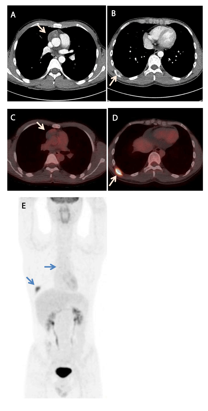
A 28-year-old man presented with heterogeneous mass in the anterior mediastinum on axial chest computed tomography (CT) (Figure 1A), with mild hypermetabolism on 18F-fluorodeoxyglucose (FDG)-positron emission tomography (PET)/CT (the maximum standardized uptake value [SUVmax] was 2.0; Figure 1C,E). Although the patient had no specific medical or surgical history, he had clinical symptoms such as cough and chest wall pain. Moreover, there was a bone-destroying soft tissue lesion in the posterior arc of the right ninth rib (Figure 1B), which was hypermetabolic on PET/CT (the SUVmax was 8.3; Figure 1D,E). The patient was suspected to have a malignant mediastinal tumor with bone metastasis, for which he underwent extended total thymectomy along with resection of the right ninth rib.
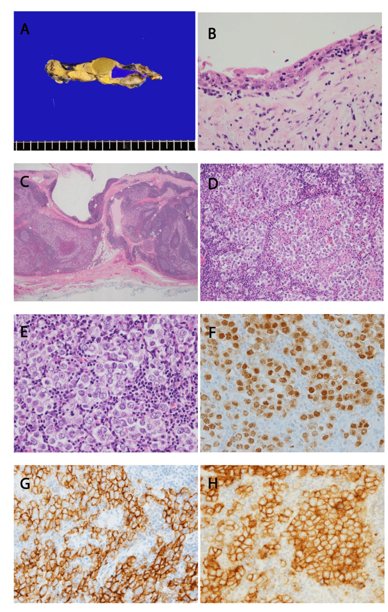
The thymus specimen revealed a 5.0- × 4.0- × 3.1-cm mass with a multilocular cyst having viscous contents as well as several small adjacent nodules (Figure 2A). The inner layer of the cystic wall was lined by flat-to-cuboidal squamoid epithelium with inflammatory changes, consistent with the histology of a thymic cyst (Figure 2B). On low-power magnification, the cyst wall showed tumor components composed of sheets of monotonous tumor cells with lymphocyte infiltrations in the background (Figure 2C). On high-power magnification, we observed large polygonal tumor cells with clear cytoplasm, round or oval nuclei and prominent nucleoli surrounding the thin fibrous septa (Figure 2D and E).
The several nodules around the thymic cyst walls revealed a variety of distinct histologic features on microscopic examination. One of the nodules had the same histologic features as those of the tumor cells in the multilocular cystic wall. The differential diagnosis for thymic tumors comprising monotonous, round-to-ovoid cells includes thymic seminoma, thymoma, thymic Hodgkin lymphoma, and mediastinal large B cell lymphoma. We performed immunohistochemistry to differentiate between these neoplasms. In both the tumor cells from the cystic wall and the nodule, immunohistochemical staining was positive for OCT3/4, c-kit and D2-40 (Figure 2F, G and H), while staining was negative for TdT, CK, CD3, CD20 and CD30. Therefore, this lesion was diagnosed to be a primary thymic seminoma associated with a thymic cyst.
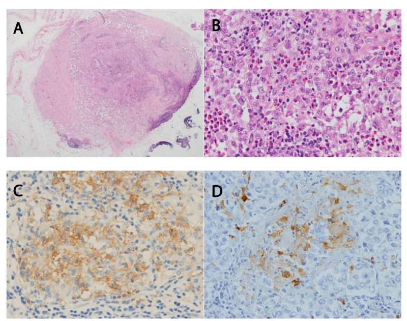
Another thymic nodule and intramedullary mass in the rib specimen were composed of a diffuse proliferation of histiocytic cells with moderate infiltration of eosinophils (Figure 3A). The histiocytic cells displayed nuclei of uniform size with a striking irregularity of convolutions, linear grooves, indentations and fine chromatin (Figure 3B) and showed positivity for S-100 protein and CD1a (Figure 3C and D). These findings were consistent with the cytomorphology of the proliferation of Langerhans cells. We diagnosed the tumors as multi-systemic LCH (MS-LCH).
The final diagnosis was a synchronous seminoma and multi-systemic LCH along with a thymic cyst. The patient was discharged on the fifth postoperative day without any complications. Moreover, no abnormality was identified in a chromosomal study conducted to exclude Klinefelter syndrome, which is well known to be associated with seminoma. Follow-up chest CT after 5 months showed no evidence of recurrence. The patient was transferred to a tertiary hospital to receive adjuvant chemotherapy.
Discussion
Mediastinal germ cell tumors account for only 1% to 5% of all germ cell tumors [4]. Seminomas account for only 0.4% of all mediastinal neoplasms in adults, with most being typically solid and lobulated in appearance [5,6]. Thymic cystic changes have also been observed frequently as a secondary process of underlying neoplasms, including thymoma, Hodgkin lymphoma, and germ cell tumor (e.g., mature cystic teratoma, seminoma and non-seminomatous malignant germ cell tumor) [7]. So, when an anterior mediastinal tumor with cysts is observed, primary seminoma should be considered in the differential diagnosis.
Seminomas arising in the mediastinal compartment show similar immunohistochemical characteristics to those of testicular seminomas considering many of the novel markers [8]. Mediastinal seminomas are consistently positive for OCT3/4, SALL4, SOX17 and MAGEC2, while being negative for SOX2, glypican 3, GATA-3 and CK5/6. These results support the diagnosis of the current case. The initial management of primary mediastinal seminoma is cisplatin-based chemotherapy [9]. According to the International Germ-Cell Cancer Collaborative Group risk classification, patients without evidence of nonpulmonary organ metastases are classified as having a good prognosis.
LCH is a clinically heterogeneous disease with the potential to affect a wide spectrum of body sites, including ‘risk organs’ (hematopoietic system, spleen and/or liver) or other organs (skin and bones, or the skin and pituitary gland) [10]. But, the thymus is rarely involved by LCH. According to the standard pediatric protocol, vinblastine/prednisone had toxicity in almost all patients and suboptimal efficacy in adults [11].
LCH can sometimes occur along with another hematologic or non-hematologic malignancy [12,13]. Rarely, the synchronous occurrence of germ cell tumors and LCH and LCH following chemotherapy or radiation therapy are reported in the literature. Few cases of mediastinal germ cell tumor and LCH have been reported [14-17] (Table 1).
In the present case, both seminoma and LCH were present simultaneously along with a thymic multilocular cyst. The patient received chemotherapy that mainly focused on the LCH over the seminoma for 2 years outside the hospital. The chemotherapy regimen is unknown. The patient is currently free of disease for 48 months.
Conclusion
In the present case, both seminoma and LCH were present simultaneously along with a thymic multilocular cyst. To the best of our knowledge, this is the first case of a synchronous presentation of seminoma and LCH within the thymus. As LCH can recur or is associated with other malignancies, regular follow-up is necessary for patients. Because this case showed a rare coincidence of seminoma and LCH, further studies are needed to establish the optimal treatment regimen, prognosis, and possible association.
- Inui M, Nitadori JI, Tajima S, Yoshioka T, Hiyama N, Watadani T, Shinozaki-Ushiku A, Nagayama K, Anraku M, Sato M, Fukayama M, Nakajima J. Mediastinal seminoma associated with multilocular thymic cyst. Surg Case Rep. 2017 Dec;3(1):7. doi: 10.1186/s40792-016-0278-7. Epub 2017 Jan 5. PMID: 28054283; PMCID: PMC5215007.
- Wakely P Jr, Suster S. Langerhans' cell histiocytosis of the thymus associated with multilocular thymic cyst. Hum Pathol. 2000 Dec;31(12):1532-5. doi: 10.1053/hupa.2000.20410. PMID: 11150381.
- Kawano R, Hata E, Ikeda S, Yokota T, Tagawa K, Sato F. Langerhans cell histiocytosis: coexistence of bronchogenic and thymic cysts in the thymus. Gen Thorac Cardiovasc Surg. 2008 Feb;56(2):74-6. doi: 10.1007/s11748-007-0191-x. Epub 2008 Feb 24. PMID: 18297462.
- Hainsworth JD. Diagnosis, staging, and clinical characteristics of the patient with mediastinal germ cell carcinoma. Chest Surg Clin N Am. 2002 Nov;12(4):665-72. doi: 10.1016/s1052-3359(02)00031-5. PMID: 12471870.
- Dulmet EM, Macchiarini P, Suc B, Verley JM. Germ cell tumors of the mediastinum. A 30-year experience. Cancer. 1993 Sep 15;72(6):1894-901. doi: 10.1002/1097-0142(19930915)72:6<1894::aid-cncr2820720617>3.0.co;2-6. PMID: 7689921.
- Strollo DC, Rosado-de-Christenson ML. Primary mediastinal malignant germ cell neoplasms: imaging features. Chest Surg Clin N Am. 2002 Nov;12(4):645-58. doi: 10.1016/s1052-3359(02)00026-1. PMID: 12471868.
- Kim JH, Goo JM, Lee HJ, Chung MJ, Jung SI, Lim KY, Lee MW, Im JG. Cystic tumors in the anterior mediastinum. Radiologic-pathological correlation. J Comput Assist Tomogr. 2003 Sep-Oct;27(5):714-23. doi: 10.1097/00004728-200309000-00008. PMID: 14501362.
- Weissferdt A, Rodriguez-Canales J, Liu H, Fujimoto J, Wistuba II, Moran CA. Primary mediastinal seminomas: a comprehensive immunohistochemical study with a focus on novel markers. Hum Pathol. 2015 Mar;46(3):376-83. doi: 10.1016/j.humpath.2014.11.009. Epub 2014 Nov 26. PMID: 25576290.
- Fizazi K, Culine S, Droz JP, Terrier-Lacombe MJ, Théodore C, Wibault P, Rixe O, Ruffié P, Le Chevalier T. Initial management of primary mediastinal seminoma: radiotherapy or cisplatin-based chemotherapy? Eur J Cancer. 1998 Feb;34(3):347-52. doi: 10.1016/s0959-8049(97)10021-1. PMID: 9640220.
- Chu T, Jaffe R. The normal Langerhans cell and the LCH cell. Br J Cancer Suppl. 1994 Sep;23:S4-10. PMID: 7521202; PMCID: PMC2149705.
- Allen CE, Ladisch S, McClain KL. How I treat Langerhans cell histiocytosis. Blood. 2015 Jul 2;126(1):26-35. doi: 10.1182/blood-2014-12-569301. Epub 2015 Mar 31. PMID: 25827831; PMCID: PMC4492195.
- Howarth DM, Gilchrist GS, Mullan BP, Wiseman GA, Edmonson JH, Schomberg PJ. Langerhans cell histiocytosis: diagnosis, natural history, management, and outcome. Cancer. 1999 May 15;85(10):2278-90. doi: 10.1002/(sici)1097-0142(19990515)85:10<2278::aid-cncr25>3.0.co;2-u. PMID: 10326709.
- Ma J, Laird JH, Chau KW, Chelius MR, Lok BH, Yahalom J. Langerhans cell histiocytosis in adults is associated with a high prevalence of hematologic and solid malignancies. Cancer Med. 2019 Jan;8(1):58-66. doi: 10.1002/cam4.1844. Epub 2018 Dec 30. PMID: 30597769; PMCID: PMC6346231.
- Ladanyi M, Roy I. Mediastinal germ cell tumors and histiocytosis. Hum Pathol. 1988 May;19(5):586-90. doi: 10.1016/s0046-8177(88)80209-0. Erratum in: Hum Pathol 1988 Sep;19(9):1121. Landanyi M [corrected to Ladanyi M]. PMID: 2453444.
- Takahashi S, Asamoto M, Nakazawa T, Kosaki T, Katsumi K, Shirai T. Robb-Smith type malignant histiocytosis associated with a mediastinal germ cell tumor. Jpn J Clin Oncol. 1994 Dec;24(6):327-30. PMID: 7830338.
- Sood R, Mehta A, Bansal D, Singh N, Purohit SR. Mediastinal mixed germ cell tumor with splenic histiocytosis: A rare coincidence. Indian J Pathol Microbiol. 2019 Apr-Jun;62(2):341-342. doi: 10.4103/IJPM.IJPM_691_18. PMID: 30971575.

Article Alerts
Subscribe to our articles alerts and stay tuned.
 This work is licensed under a Creative Commons Attribution 4.0 International License.


 Save to Mendeley
