Open Journal of Orthopedics and Rheumatology
Scaphoid Non-Union Treated by Zaidemberg’s Vascularized Bone Graft: About 30 Cases
Mohamed Ali Sbai1*, Mayssa El M’chirgui1, Riadh Maalla2 and Adel Khorbi2
2Plastic Surgery Department, La Rabta Hospital, Tunis, Tunisia
Cite this as
Ali Sbai M, El M’chirgui M, Maalla R, Khorbi A (2016) Scaphoid Non-Union Treated by Zaidemberg’s Vascularized Bone Graft: About 30 Cases. Open J Orthop Rheumatol 1(1): 018-023. DOI: 10.17352/ojor.000005Introduction: Scaphoid fractures evolves in 10% of cases to nonunion. Untreated, it progresses to arthrosis of the wrist that may compromise the function of the hand. The recently described vascularized bone grafts have helped to expand the armamentarium of management of scaphoid nonunion. We wanted to verify those data by studying the results of Zaidemberg’s graft made in our orthopedic department.
Materials and methods: 30 scaphoid nonunion cases treated by a vascularized bone graft using Zaidemberg’s procedure were studied retrospectively. The clinical criteria studied were: range of motion, the Mayo Wrist Score, the Quick Dash, and PWRE. The radiographs have controlled the consolidation and performed a full radiometry.
Results: Our series is made up of young adults (average age 28 years), a male-dominated manual workers. The dominant side is attained in 60% of cas.57% of patients are smokers. The seniority of the nonunion was 4 years on average. Nonunion sat, according to Schernberg’s classification in zone 3 in 30% of cases. We had 50% in stage 2a and 30% in stage 2b according to Alnot’s classification. Fixation was realized by pins followed by immobilization during 6 weeks on average. We had a consolidation in all cases. The tobacco intoxication had a deleterious effect, a delayed union was observed in smokers’ patients. Our patients had a Mayo Wrist Mean score 72%, a PRWE to 11% and a Quick DASH 10%. Analysis of radiometry showed an improvement of the analyzed parameters.
Conclusion: Zaidemberg’s graft is a reliable vascularized bone graft; it requires a learning curve, these results are better than inert transplants and Kuhlmann’s graft, it is indicated in the old nonunion, stage 2 Alnot the “proximal pole necrosis and changing of the scaphoid shape”.
Introduction
Despite the effective diagnostic and treatment options of scaphoid fracture available today, its evolution towards pseudarthrosis remains important.
Untreated, the scaphoid pseudarthrosis progress to carpal instability and at long term, to a degenerative wrist arthrosis wrist type “Scaphoid Non-union Advanced Collapse” (SNAC) [1-3].
The appearance of vascularized bone grafts allowed reopening discussion of the treatment modalities of this nonunion. A meta-analysis of the literature [4], has indeed demonstrated the superiority of vascularized bone grafts to conventional grafts [5,6].
Several types of vascular grafts were described through a better knowledge of the vascular anatomy of the wrist and the evolution of microsurgery; however Kuhlmann’s and Zaidemberg’s grafts remain the most used.
Whatever type of graft, surgery has to keep three goals: restoring the height and morphology of the scaphoid, obtaining bone healing and correction of the lunate dorsal tilt, corresponding to carpal instability.
We carried out a retrospective study of 30 cases of scaphoid nonunion treated by a vascularized bone grafts using Zaidemberg’s procedure. The objective of this study is to evaluate our results and to define the place of this process of reconstruction of the scaphoid compared to the other surgical treatments.
Material and Methods
This is a retrospective study of 30 cases conducted in the orthopedic department of Nabeul durant past decade. The patient age, the proximal pole necrosis, nonunion reoperated, smoking and the age of the nonunion have been noted. We have realized a nonunion carpal scaphoid cure by the described Zaidemberg’s technique; a styloidectomy was done routinely and internal fixation by kierchner pins No. 14. The reviewed patients had received a detailed assessment. It includes mobility and clinical strength measurements, subjective functional assessment using questionnaires and a recognized radiological review comparing preoperative images to those in the revision. We included all patients operated in our orthopedic department by a Zaidemberg’s vascular graft for nonunion of the scaphoid with at least 1 year after surgery. We excluded patients with Zaidemberg’s graft in another center and transcapholunate dislocations of the carpal.
Results
Our series is made up of young adults; the mean age was 28 years, ranging from 19 to 52, male-dominated manual workers. The dominant side is attained in 60% of cases .57% of patients are smokers. The etiology is dominated by sports injuries and work accidents which represent 60% of cases. The seniority of the nonunion was 4 years average. Three patients are operated after percutaneous pinning type Galluccio failure. Nonunion sat Schernberg’s [7], classification in zone 3 in 30% of cases. We had 50% stage 2a and 30% stage 2b according to Alnot’s classification [8]. Fixation was achieved by pins followed by 6 weeks of immobilization on average. Rehabilitation was also applied for all patients during at least 1 month. We noted a decrease in flexion mobility extension arc of 25 ° and 15 ° of arc mobility inclinations postoperatively all zones. We had a consolidation in all patients with 12 weeks on average. The tobacco intoxication had a deleterious effect, a delayed union was observed in smokers’ patients. Our patients had 92% Mayo Wrist Mean score, a PRWE to 11% and a Quick DASH 10%. Analysis of radiometry showed improvement in the analyzed parameters: Improved index and Youm Mac Murtry increased from 0.517 to 0.524, decreasing the radiolunate angle from 11.56 ° to 9.9 ° postoperatively and improved scapholunate angle from 60.2 ° to 55 °. We deplored two infections pin, malunion of the radial styloid and sensory disturbances in the sensory branch of the radial nerve in a patient (Table 1)
Discussion
Superiority of vascularized bone grafts to conventional grafts is well established, but the contribution of the Zaidemberg’s graft compared to other vascular grafts including the Kuhlmann’s graft taken by Mathoulin will be analyzed. Since the description of Zaidemberg [9] and Sheetz [10], the graft based on supra retinacular artery is increasingly used in scaphoid nonunions, with varying rates depending on the series, usually exceeding 90 % of consolidations. Zaidemberg et al. [9], Series, of 11 patients shows a consolidation ratio to 100% by 6.2 weeks. The concept of proximal pole necrosis is a fundamental concept to know. It is a pejorative factor to be evaluated systematically. MRI confirms the diagnosis of nonunion and can assess the vascular status of the proximal pole preoperatively [10].
The results of scaphoid nonunion with proximal pole necrosis treated by vascularized grafts are better in comparison with conventional bone grafts.Thus Munk Larsen [6], in a meta-analysis has found similar rates for conventional grafts either with or without osteosynthesis (respectively 84 and 80% of consolidation, no significant difference). However, they found a significant increase in consolidation ratios when the graft was vascularized (91%).
It is found that Steinmann studies [12] and Uerpairojkit [13] concerning nonunion never operated with a 100% success, respectively on 14 and 10 patients. Tsai et al. [14], reported 5 cases of the proximal pole necrosis consolidating in 100% of cases in 4 months. Waters et al. [15], report a series of three proximal pole necrosis.
Merrell et al. [4], found a deleterious effect of age on consolidation (95% consolidation before 20 years and 78% after 50 years). Chang et al. [16], also found a correlation between the twice .The average age of patients who consolidated was 28 years. Chang et al. [16], show consolidation ratios significantly above 80% in men against 30% in women.
The difference in proportions of smokers among the general population and patients of our study confirm the derogatory role of tobacco for the consolidation of scaphoid fractures. The relative risk of nonunion in smoker is 3.7 [37]. Then, in our series, we found consolidation delay in our smokers, despite the use of a vascular graft. The series of Chang et al. [16], found a relative failure risk of 2.69 in case of smoking.
The analysis comparing nonunion under 5 years in those over 5 years showed no significant difference in terms of consolidation. However, it is difficult to establish the exact date of the initial trauma, it is sometimes disregarded time surgical management is therefore imprecise.
Shah and Jones [18], Boyer et al. [19], Straw et al. [20] showed that prior history of surgery gave less good results on their scaphoid nonunion. The meta-analysis of Merrell et al. [4] and the series of Chang et al. [16], showed no significant difference in terms of consolidation.
The consolidation period is a given difficult to evaluate. Indeed, it is entirely dependent on the frequency of the radio-clinical tests and radiographs appreciation explaining the difference of results in the literature. Thus Zaidemberg et al. [9] found a consolidation period of 6.2 weeks whereas it was 22.1 weeks in Waitayawinyu et al. [21] in never operated patients, 6.5 weeks with Uerpairojkit et al. [13], 11.1 weeks with Steinmann et al. (12), 14.7 weeks with Waters et al. [15], 15.6 weeks with Chang et al. [16], 18.4 weeks with Boyer et al. [19], and 21.7 weeks with Ong et al. [22].
Our series include, in turn, a good result of 12 weeks on average. Further, we left the osteosynthesis material indefinitely in place, ensuring a longer internal capital. The consolidation was considered acquired but the equipment was not necessarily removed. Consolidation must be certain and sustainable. Most authors report their consolidation ratios without addressing the success of the angular correction and thus the restoration of the scaphoid anatomy. It is established that the success of the treatment of nonunion also depends on the correction of the “humpback deformity” and DISI. The work of Fisk [23] has shown the consequences of the deformation of the scaphoid on the alteration of biomechanics on the carp. So even if consolidation gained a malunion may have functional consequences and impact on pain.
Some authors have noted an improvement in mobility compared to preoperative status [9,24] or stability of the latter [12,21]. Others have reported a decrease in mobility [15,19]. In our series, there is a persistent deficit of 25 ° on the arc of mobility of flexion-extension and 15 ° on the arc of mobility of radial and ulnar inclination significantly. Hankins et al. [25], demonstrated on a cadaver study that a good positioning of the vascularized graft did not result in significant reduction of range of motion. Postoperative stiffness has other causes: technical problems of proper positioning of the graft while maintaining anatomical reduction, biological factors involving tissue healing and prolonged postoperative immobilization. Waitayawinyu et al. [21], found a restored power to 99% compared to the healthy side on a series of operated patients but never with necrosis of the proximal pole. Zaidemberg et al. [9] found a 95% power compared to the healthy side, Boyer et al. 95% compared to the healthy side [19], Uerpairojkit et al. to 77% compared to the healthy side [13] and Malizos et al., to 86% compared to the healthy side [24]. The strength found in our series is comparable to literature with a rate of 80% compared to the healthy side. However, despite a high rate of recovery in surgical patients (28%), there was no difference in results might ultimately based on the number of previous interventions.
Waitayawinyu et al. [21], found a significant improvement in the satisfaction score of patients compared with the preoperative situation. There was no difference in preoperative and postoperative DASH. Ong et al. [22], found a DASH 10.3% comparable to our results. Chen et al. [26] found 91% excellent and good results, according to the Mayo Wrist Score, Tsai et al. 100% good results. These data are to be qualified because of the small size of these series. Our average score was 92%, corresponding to a satisfactory overall result. We had 50% excellent and good results. Uerpairojkit et al. [13] and Malizos [24] et al., have 100% of their patients who were able to return to their prior work. We have a slightly lower rate of 91% return to the former job, similar to the results of Waitayawinyu et al. [21]/ 93%.
We find that low postoperative loss of grip strength (-20% compared to the contralateral side) will not cause a major professional harm, even in manual workers, given the frequent return to regular work. The duration of total work stoppage is 3 months in our series. This period is only indicative. In fact, this data is difficult to obtain accurately.
Nonunion and classification Zone: Chang et al. [16] showed no difference in consolidation based on the location of the fracture as in our series. Ong et al. [22], found a consolidation rate of 80% when the proximal pole nonunion interested and 66% when it is located at the waist of the scaphoid, but on a staff of only 10 patients.
The Waitayawinyu et al. [21], found an improvement in 6 of scapholunate angle from 56 ° to 50 ° postoperatively and report on height scaphoid length of 0.75 to 0.62 significantly. Chen et al. [26], reported an improvement of 10 ° scapholunate angle from 55.9 ° to 45.9 °, the lateral intrascaphoïdien angle ranging from 32.3 ° to 20, 5 °. Tsai et al. [14] found a significant improvement in 7 of scapholunate angle and a correction of 11 intrascaphoidien side angle. Our series shows no significant improvement in carpal height of + 1.75% according to index and Youm Mc Murtry a significant correction radiolunaires angles of 1.66 ° and 5.2 ° scapholunate. These different series found concordant results, even if the main defect is the variability of these measures intra- and interobserver.
Radiometry improvement (significant improvement in scapholunate angle, and radiolunate intrascaphoïdien side, non-significant increase in the index of Youm and Mac Murthry) allow approaching an anatomical restoration. This limits further deterioration and arthrosis in particular to a SNAC wrist.
Chang et al. [16], found 9% of graft migration, 6% of sepsis (2% deep, superficial 4%) 2% default mounting. The low caliber of the 1,2 Intercompartmental Supraretinacular Artery (ICSRA) prohibits dissection with isolation of the vascular pedicleof the graft. Therefore we must take the artery in a fatty cellular rail downstream of the radial styloid to avoid any risk of dissection. The average number of perforating branches from the pedicle was 5.5 [3-9]. A 1 cm graft located between 8 -18 mm proximal to the articular surface of the distal radius has as many branches piercing or 4 may correspond to the ideal point for a vascular graft [27].
The cadaveric study of Waitayawinyu et al. [28] demonstrated that the pedicle length of 1.2 ICSRA was sufficient to allow placement of bone graft in both dorsal and palmar in all cases studied. Yet another study found that the graft cannot always be located on the palmar surface of the scaphoid [27]. His path is tortuous in neutral and wrist extension. This reserve length disappears with wrist flexion. There is a significant decrease of 2mm the length of the pedicle between the point of rotation and the proximal scaphoid, during the passage of the radial inclination of 20 ° to the ulnar inclination of 30 ° [27]. The tension on the pedicle is substantially reduced by carrying out a radial styloidectomy [27]. These findings suggest that the radial tilt position and the flexion of the wrist should be avoided during surgery and postoperative immobilization.
For the realization of osteosynthesis, the pins are more manageable but also more prone to migration. The screws are theoretically more stable but at the cost of considerable bulk, with a risk of graft expulsion during tightening. The use of pins is the only option when the proximal pole is too small. The meta-analysis of Merrell et al. [4], showed that the bone screw, with 94% of consolidation is greater than that achieved by pins (77%). In the literature, the rate of consolidation of non-vascularized grafts, with the use of screws, vary between 94% and 100% [29-31]. The results with the pins are more variable. Indeed, Barton et al. [32], found 56% of consolidation with pins and screws with 78% of Herbert in conventional grafts. For vascularized grafts Zaidemberg, the study by Chang et al. [16], found consolidation in 88% of cases with the use of screws and pins with 53%; this difference was significant (odds ratio low 0.087 and p <0.004). However, when comparing the consolidation according to the necrosis of the proximal pole and the osteosynthesis technique, there is more statistical difference. Series Waitayawinyu [21] found 100% consolidation with two different models of cannulated screws. Steinmann series [12] and Uerpairojkit [13] using screws and pins have 100% consolidation ratio, regardless of the techniques do not allow comparing these osteosynthesis methods. Straw et al, [20], found no difference between consolidation Herbert screws and pins. Our series only included osteosynthesis pin by making impossible any comparison. The quality of fixation is essential, but it remains difficult. Indeed, osteosynthesis can fragment the graft and detach cancellous bone fragments which then become avascular. The osteosynthesis material rests in the spongy part of the graft whose holding is limited. The goal remains to have a stable mounting.
In our series, there was no significant difference of consolidation or failure depending on the number of pins. However in view of the large number of hardware migration, it is appropriate to use at least 2 pins. These must be removed once the consolidation is acquired formally. Series Straw et al. [20], with a consolidation ratio of 27%, the lowest found in the literature, was marked by the use of only one pin in 63% of cases and removed systematically eight weeks after surgery. Graft and inadequate fixation are pejorative factors difficult to bring out. Furthermore it is not possible to prove that the blood supply to the bone graft can continue. All these factors are “invisible” to all studies and contribute to the differences in results. However the results of this study should be confirmed more specifically described by the Zaidemberg’s graft: Incorporation is better, revascularization of avascular bone is facilitated by a vascular graft, a faster consolidation and only one route of all, allowing in the same operation, taking the graft and the treatment of nonunion. The regional anesthesia allows an outpatient surgery [33]. The anatomy of the Zaidemberg’s artery is constant, the pedicle is long enough to reach the scaphoid without tension providing a reliable and reproducible technique
Conclusion
The Zaidemberg’s bone graft is a reliable technique, it requires a learning curve, these results are better than inert transplants and of Kuhlmann’s graft, it is indicated in the old nonunion, stage 2 of Alnot’s classification (36)“proximal pole necrosis and changing of the scaphoid shape”.
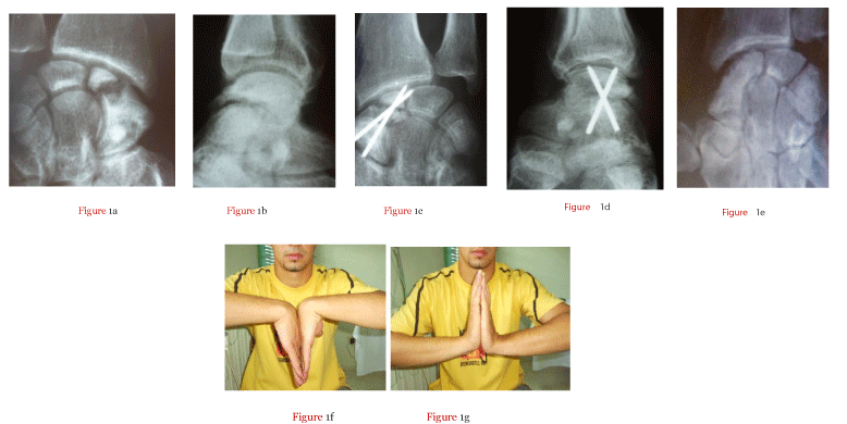
Case 1: proximal pole nonunion IIb stage treated by graft Zaidemberg with excellent clinical and radiological result correction DISI (Figure 1a-g).
Case 2: Nonunion old stage IIIa treated graft Zaidemberg with excellent clinical and radiological outcome (Figure 2a-e).
Case 3: Nonunion old stage IIIa treated graft Zaidenberg with an average clinical outcome (Figure 3a-e).
- Ruby LK, Stinson J, Belsky MR (1985) The natural history of scaphoid non-union. A review of fifty-five cases. J Bone Joint Surg Am 67: 428–432. Link: https://goo.gl/0U3TBF
- Vender M (1987) Degenerative change in symptomatic scaphoid nonunion. J Hand Surg Am 12: 514–519. Link: https://goo.gl/F3XnIc
- Berdia S, Wolfe S (2001) Effects of scaphoid fractures on the biomechanics of the wrist. Hand Clin 17: 533–540. Link: https://goo.gl/8WGcA9
- Merrell GA, Wolfe SW, Slade JF (2002) Treatment of scaphoid nonunions: Quantitative metaanalysis of the literature. The Journal of Hand Surgery 27: 685–691. Link: https://goo.gl/4JNcyJ
- Rizzo M, Moran S (2008) Vascularized Bone Grafts and Their Applications in the Treatment of Carpal Pathology. Seminars in Plastic Surgery 22: 213–227. Link: https://goo.gl/47lqDR
- Munk B, Larsen CF (2004) Bone grafting the scaphoid nonunion: a systematic review of 147 publications including 5,246 cases of scaphoid nonunion. Acta Orthop Scand 75: 618–629. Link: https://goo.gl/PQNtXj
- Schernberg F (1988) Classification of fractures of the carpal scaphoid. An anatomo-radiologic study of characteristics. Rev Chir Ortho Reparatrice appar mot 74: 693-695. https://goo.gl/Xl2mVr
- Alnot JY (1988) Sympossium sur les fractures et pseudarthrose du scaphoide carpien. Rev Chir Orthop: 714-717.
- Zaidemberg C, Siebert JW, Angrigiani C (1991) A new vascularized bone graft for scaphoid nonunion. J Hand Surg Am 16: 474–478. Link: https://goo.gl/dva9w6
- Sheetz KK, Bishop AT, Berger RA (1995) The arterial blood supply of the distal radius and ulna and its potential use in vascularized pedicled bone grafts. J Hand Surg Am 20: 902–914. Link: https://goo.gl/89xKdw
- Ciprian S, Iochum S, Kohlmann R, Dautel G, Dap F, et al. (2004) Valeur de l’IRM dans l’évaluation du potentiel de guérison des pseudarthroses du scaphoïde traitées par greffe osseuse. Journal de Radiologie 85: 1699–1706. Link: https://goo.gl/1Uw2CZ
- Steinmann SP, Bishop AT, Berger RA (2002) Use of the 1,2 intercompartmental supraretinacular artery as a vascularized pedicle bone graft for difficult scaphoid nonunion. The Journal of Hand Surgery 27: 391–401. Link: https://goo.gl/jmqfQm
- Uerpairojkit C, Leechavengvongs S, Witoonchart K (2000) Primary vascularized distal radius bone graft for nonunion of the scaphoid. J Hand Surg Br 25: 266–270. Link: https://goo.gl/QXHorT
- Tsai TT, Chao EK, Tu YK, Chen ACY, Lee MSS, et al. (2002) Management of scaphoid nonunion with avascular necrosis using 1, 2 intercompartmental supraretinacular arterial bone grafts. Chang Gung Med J 25: 321–328. Link: https://goo.gl/ht6ZPn
- Waters PM, Stewart SL (2002) Surgical treatment of nonunion and avascular necrosis of the proximal part of the scaphoid in adolescents. J Bone Joint Surg Am 84: 915–920. Link: https://goo.gl/4gQJ0y
- Chang MA, Bishop AT, Moran SL, Shin AY (2006) The Outcomes and Complications of 1,2- Intercompartmental Supraretinacular Artery Pedicled Vascularized Bone Grafting of Scaphoid Nonunions. J Hand Surg Am 31: 387–396. Link: https://goo.gl/bv1k6t
- Little CP, Burston BJ, Hopkinson-Woolley J, Burge P (2006) Failure of surgery for scaphoid non-union is associated with smoking. J Hand Surg Br 31: 252–255. Link: https://goo.gl/1PB9ic
- Shah J, Jones WA (1998) Factors affecting the outcome in 50 cases of scaphoid nonunion treated with Herbert screw fixation. J Hand Surg Br 23: 680–685. Link: https://goo.gl/9swjxp
- Boyer MI, von Schroeder HP, Axelrod TS (1998) Scaphoid nonunion with avascular necrosis of the proximal pole. Treatment with a vascularized bone graft from the dorsum of the distal radius. J Hand Surg Br 23: 686–690. Link: https://goo.gl/5rCI8x
- Straw RG, Davis TRC, Dias JJ (2002) Scaphoid nonunion: treatment with a pedicled vascularized bone graft based on the 1,2 intercompartmental supraretinacular branch of the radial artery. J Hand Surg Br 27: 413. Link: https://goo.gl/PXvlU6
- Waitayawinyu T, McCallister WV, Katolik LI, Schlenker JD, Trumble TE (2009) Outcome after vascularized bone grafting of scaphoid nonunions with avascular necrosis. J Hand Surg Am 34: 387–394. Link: https://goo.gl/3nppqE
- Ong HS, Tan G, Chew WYC (2011) Treatment of scaphoid non-union with 1,2 intercompartmental supraretinacular artery (1,2 ICSRA) vascularised graft. Singapore Med J 52: 658–661. Link: https://goo.gl/aXS1ct
- Fisk GR (1970) Carpal instability and the fractured scaphoid. Ann R Coll Surg Engl 63–76. Link: https://goo.gl/E0Pgcu
- Malizos KN, Dailiana ZH, Kirou M, Vragalas V, Xenakis TA, et al. (2001) Longstanding nonunions of scaphoid fractures with bone loss: successful reconstruction with vascularized bone grafts. J Hand Surg Br 26: 330–334. Link: https://goo.gl/y7L0cs
- Hankins CL, Budoff JE (2011) Analysis of Wrist Motion Following Vascularized Bone Graft to the Proximal Scaphoid. J Hand Surg Am 36: 583–586. Link: https://goo.gl/tzauJ0
- Chen ACY, Chao EK, Tu YK, Ueng SWN (2006) Scaphoid nonunion treated with vascular bone grafts pedicled on the dorsal supra-retinacular artery of the distal radius. J Trauma 61: 1192–1197. Link: https://goo.gl/9Kjw6k
- Saint Cast Y, Césari B, Dagregorio G, Le Bourg M, Gazarian A, et al. (2012) Technique simplifiée de reconstruction du scaphoïde par le greffon vascularisé radial de Zaidemberg. Revue de Chirurgie Orthopédique et Traumatologique 98: S167–S173. Link: https://goo.gl/tnQK5q
- Waitayawinyu T, Robertson C, Chin SH, Schlenker JD, Pettrone S, et al. (2008) The Detailed Anatomy of the 1,2 Intercompartmental Supraretinacular Artery for Vascularized Bone Grafting of Scaphoid Nonunions. The Journal of Hand Surgery 33: 168–174. https://goo.gl/FfsD6q
- Tsuyuguchi Y, Murase T, Hidaka N, Ohno H, Kawai H (1995) Anterior wedge-shaped bone graft for old scaphoid fractures or non-unions. An analysis of relevant carpal alignment. J Hand Surg Br 20: 194–200. Link: https://goo.gl/VhaKFw
- Trumble TE, Clarke T, Kreder HJ (1996) Non-union of the scaphoid. Treatment with cannulated screws compared with treatment with Herbert screws. J Bone Joint Surg Am 78: 1829–1837. Link: https://goo.gl/6TLWPJ
- Daly K, Gill P, Magnussen PA, Simonis RB (1996) Established nonunion of the scaphoid treated by volar wedge grafting and Herbert screw fixation. J Bone Joint Surg Br 78: 530–534. Link: https://goo.gl/kOejgA
- Barton NJ (1997) Experience with scaphoid grafting. J Hand Surg Br 22: 153–160. Link: https://goo.gl/XFYIiD
- Obert L, Lemaire B, Lepage D, Clappaz P, Garbuio P, et al. (2007) 257 Analyse des échecs du greffon vascularisé de Zaidenberg en cas de pseudarthrose du scaphoïde : difficultés technique ou erreur chirurgicale ? Rev Chir Ortho et Rép de l’Appareil Moteur 93: 140–141. Link: https://goo.gl/wWS7Nw
Article Alerts
Subscribe to our articles alerts and stay tuned.
 This work is licensed under a Creative Commons Attribution 4.0 International License.


 Save to Mendeley
