Journal of Dental Problems and Solutions
Miths and solutions in digital dental impression
Guilherme Cabral1*, Luciana M Soares2, Hilton Riquieri3, Adriano Baldotto Barbosa4, Renato Sartori5, Eric Padovani6, Fabio Costa7, Jonas U.R. Schindler8, Juliano Arantes Vasconcelos9 and Diego Henrique de Lima10
2Private Practice Los Angeles, USA
3Director of Crow & Bridge Department of Midwest Dental Art, Palm bay, USA
4Master Ceramist of Midwest Dental Art, Palm Bay, USA
5Brazilian Society of Digital Dentistry, Brazil
6Private Practice Araraquara, Brazil
7Brazilian Society of Digital Dentistry, Brazil
8Schindler Studio Dental, Brazil
9Dental Atelie, Brazil
10Diego Lima Studio Dental, Brazil
Cite this as
Cabral G, Soares LM, Riquieri H, Barbosa AB, Sartori R, et al. (2022) Miths and solutions in digital dental impression. J Dent Probl Solut 9(2): 025-027. DOI: 10.17352/2394-8418.000113Copyright License
© 2022 Cabral G, et al. This is an open-access article distributed under the terms of the Creative Commons Attribution License, which permits unrestricted use, distribution, and reproduction in any medium, provided the original author and source are credited.Nowadays, the process of transferring intra-oral information from the dental office to dental laboratories has been a current reality in dentistry. The number of advantages presented in this process include work accuracy, patient acceptance, a transfer, and storage information facility, three-dimensional visualization, and an increase in time efficiency [1]. Among the disadvantages observed are the learning curve [2] and the cost of acquisition and maintenance of the IOS (Intra-oral Scanner). However, the main challenge is not actually related to the methodology for acquiring the information, either using the digital or conventional methods, but the control of factors performed previously in the impression-taking procedure or the intra-oral scanning. This article explains these factors present in both methods that are fundamental to increase the potential to solve some frequent problems in the dental laboratory (Figure 1).
Introduction
It is indisputable the number of benefits when using digital impression in dentistry, which has gained more popularity and acceptance in the daily routine as to be considered the method of choice for future generations in every dental specialty [3]. When capturing the intra-oral information, the tools used for impressions with elastomers materials compared to light-optical acquisition represents a totally different domain in terms of the method to acquire this information. However, the intra-oral matter remains the same and yet the biological components (hard tissues - tooth and bone and soft tissues - gingiva, tongue, cheek, lips) play a role under equal principles.
Managing these important principles in dentistry haven’t changed throughout the years and should always be managed properly in order to guarantee the effectiveness of the information acquired [4-6].
The most common error is mainly due to human inaccuracy in the process of environment preparation [1] which leads to an inadequate impression or a deficient intra-oral scanning. Ninety percent of the problems that dental laboratories have faced are related either to poor teeth preparation or difficulties to visualize and identify the margins [2,7]. These problems are often present in both techniques, the conventional, using impression materials, and the digital impression.
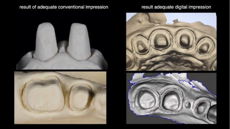
“We are going from: not having problems with conventional impression to having problems with intra-oral scanning” (Figure 2).
Digital impressions eliminate some of the drawbacks of conventional elastomer impressions; however, proper soft-tissue management, the isolation of teeth preparation, and defined margins are still necessary [8].
The aim of this study review is to present the main factors that can prevent common errors of intra-oral optical scanning in the production of dental restoration when working with digital impression CAD-CAM technology.
We should understand that information acquisition is a process, not a solo step. It will depend on the quality of intra-oral hard and soft tissue. Also, it is necessary to determine that all the parameters are under control as well as the key steps be well managed by dentists to provide adequacy of the oral environment before the acquisition act:
- Tooth preparation (design, polishing, and cleaning)
- Proper provisionalization (polishing and ideal emergence profile)
- Healthy soft tissue contour (biological width)
- Proper gingival retraction (bleeding control and marginal clearance)
There is nothing new in these fundamentals. By managing these period-restorative pillars, we simply prioritize the health of biological tissues protecting them and creating a proper foundation before moving forward to the final restoration.
1. Adequate tooth preparation design helps to establish a clear and evident margin, an ideal restorative material thickness, crown and bridge path of insertion as well as improving marginal adaptation [9,10,11] (Figure 3).
3. When the dentist controls and respect the relationship between the emergence profile and the new restoration using a correct soft tissue management protocol, the specific gingival biotype presents a favorable response by perfectly adapting to the material without any inflammation or invasion of biological width [10,15-17] (Figure 5).
4. When acquiring adequate intra-oral Information, a clear design for the tooth preparation combined with a healthy and remodeled marginal gingiva facilitates the gingival retraction procedure providing clearance for the dental impression material or the light of the IOS device.
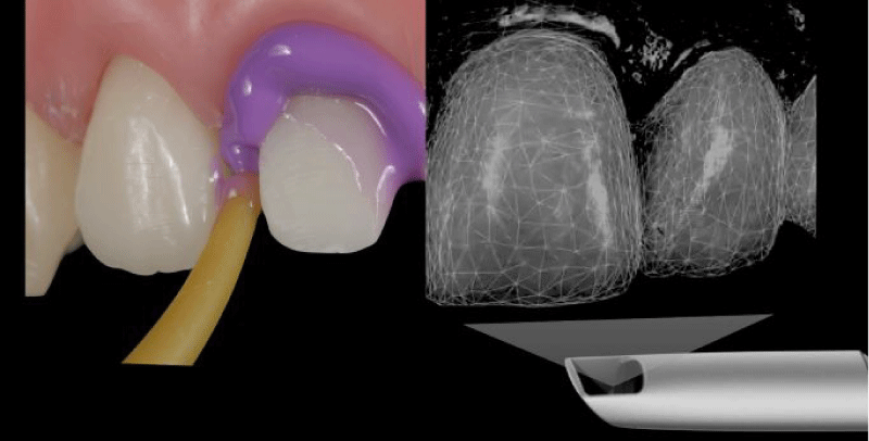
Once we manage all these previous steps, the two-cord technique seems to be a superior option because promotes an effective horizontal displacement. The dentist should be able to see the first cord in place while the light from the IOS device reads the preparation margins 360-degree (Figures 6,7).
Conclusion
During the last decade, we have been distracted mostly by doing comparisons between the advantages and the disadvantages of new technologies in indirect restorative dentistry, and, the points that have a major impact on the oral health care of our patients, have been missed.
The approach for data acquisition is a process and not a single act and it is paramount to change the clinicians’ mindset toward taking particular care and performing all crucial steps that are related to the biology behind dentistry helping to ensure the most predictable and satisfactory outcomes. As this article has demonstrated, these combined efforts diminish most part of the problems that dental laboratories have faced particularly regarding to inaccurate conventional and digital impressions.
- Samet N, Shohat M, Livny A, Weiss EI. A clinical evaluation of fixed partial denture impressions. J Prosthet Dent. 2005 Aug;94(2):112-7. doi: 10.1016/j.prosdent.2005.05.002. PMID: 16046964.
- Christensen GJ. Improving the quality of fixed prosthodontic services. J Am Dent Assoc. 2000 Nov;131(11):1631-2. doi: 10.14219/jada.archive.2000.0094. PMID: 11103584.
- Patel N. Contemporary dental CAD/CAM: modern chairside/lab applications and the future of computerized dentistry. Compend Contin Educ Dent. 2014 Nov-Dec;35(10):739-46; quiz 747, 756. PMID: 25454527.
- Rudolph H, Salmen H, Moldan M, Kuhn K, Sichwardt V, Wöstmann B, Luthardt RG. Accuracy of intraoral and extraoral digital data acquisition for dental restorations. J Appl Oral Sci. 2016 Jan-Feb;24(1):85-94. doi: 10.1590/1678-775720150266. PMID: 27008261; PMCID: PMC4775014.
- Güth JF, Keul C, Stimmelmayr M, Beuer F, Edelhoff D. Accuracy of digital models obtained by direct and indirect data capturing. Clin Oral Investig. 2013 May;17(4):1201-8. doi: 10.1007/s00784-012-0795-0. Epub 2012 Jul 31. PMID: 22847854.
- Lee SJ, Gallucci GO. Digital vs. conventional implant impressions: efficiency outcomes. Clin Oral Implants Res. 2013 Jan;24(1):111-5. doi: 10.1111/j.1600-0501.2012.02430.x. Epub 2012 Feb 22. PMID: 22353208.
- TodorovićA, Lisjak D, Lazić V, Gostović A. Possible Errors During the Optical Impression Procedure. Serbian Dental Journal. 2010; 57:1; 30-37.
- Christensen GJ. Will digital impressions eliminate the current problems with conventional impressions? J Am Dent Assoc. 2008 Jun;139(6):761-3. doi: 10.14219/jada.archive.2008.0258. PMID: 18520000.
- Tarnow DP, Magner AW, Fletcher P. The effect of the distance from the contact point to the crest of bone on the presence or absence of the interproximal dental papilla. J Periodontol. 1992 Dec;63(12):995-6. doi: 10.1902/jop.1992.63.12.995. PMID: 1474471.
- Silness J. Periodontal conditions in patients treated with dental bridges. 3. The relationship between the location of the crown margin and the periodontal condition. J Periodontal Res. 1970;5(3):225-9. doi: 10.1111/j.1600-0765.1970.tb00721.x. PMID: 4254186.
- Lima FF, Neto CF, Rubo JH, Santos GC Jr, Moraes Coelho Santos MJ. Marginal adaptation of CAD-CAM onlays: Influence of preparation design and impression technique. J Prosthet Dent. 2018 Sep;120(3):396-402. doi: 10.1016/j.prosdent.2017.10.010. Epub 2018 Mar 15. PMID: 29551386.
- Baba NZ, Goodacre CJ, Jekki R, Won J. Gingival displacement for impression making in fixed prosthodontics: contemporary principles, materials, and techniques. Dent Clin North Am. 2014 Jan;58(1):45-68. doi: 10.1016/j.cden.2013.09.002. PMID: 24286645.
- Chaudhari J, Prajapati P, Patel J, Sethuraman R, Naveen YG. Comparative evaluation of the amount of gingival displacement produced by three different gingival retraction systems: An in vivo study. Contemp Clin Dent. 2015 Apr-Jun;6(2):189-95. doi: 10.4103/0976-237X.156043. PMID: 26097353; PMCID: PMC4456740.
- Franco EB, da Cunha LF, Herrera FS, Benetti AR. Accuracy of Single-Step versus 2-Step Double-Mix Impression Technique. ISRN Dent. 2011;2011:341546. doi: 10.5402/2011/341546. Epub 2011 Jul 25. PMID: 21991468; PMCID: PMC3169190.
- Gargiulo AW, Wentz FM, Orban B. Dimensions and rela- tions of the dento-gingival junction in humans. J Periodontol 1961; 32:261-267.
- Kois JC. Altering gingival levels.The restorative connection. Part I: Biologic variables. J Esthet Dent. 1994; 6(1):3-9.
- Anusavice KJ, Kenneth J. Phillips ’ science of dental materials. 11th edition. Elsevier 2003; 12.
Article Alerts
Subscribe to our articles alerts and stay tuned.
 This work is licensed under a Creative Commons Attribution 4.0 International License.


 Save to Mendeley
