Archives of Community Medicine and Public Health
Histopathologically atypical astroblastoma with MN1-CXXC5 fusion transcript diagnosed by methylation classifier
Gerald C Wallace1*, Robert JB Macaulay2, Arnold B Etame2, Kenneth Aldape3 and Yolanda Pina2
2H. Lee Moffitt Cancer Center, Tampa, FL, USA
3National Institute of Health, Bethesda, USA
Cite this as
Wallace GC, Macaulay RJB, Etame AB, Aldape K, Pina Y (2022) Histopathologically atypical astroblastoma with MN1-CXXC5 fusion transcript diagnosed by methylation classifier. Arch Community Med Public Health 8(3): 113-117. DOI: 10.17352/2455-5479.000185Copyright License
© 2022 Wallace GC, et al. This is an open-access article distributed under the terms of the Creative Commons Attribution License, which permits unrestricted use, distribution, and reproduction in any medium, provided the original author and source are credited.Adult astroblastoma is an exceedingly rare primary brain tumor. Previous reports have suggested various radiographic and histological features typical for these tumors, but the diagnosis can be challenging. We present a unique case of astroblastoma diagnosed after 13 years of treatment as a CNS embryonal neoplasm. Histologically, this tumor lacked previously identified astroblastic features such as pseudorosettes, trabeculated patterns, and hyalinized vessels. The tumor was synaptophysin positive which further confounded the diagnosis in this case. Methylation classification was performed with a high confidence match to a high-grade neuroepithelial neoplasm with a CXXC5-MN1 fusion. Molecular characterization confirmed a CXX5-MN1 fusion transcript which has been seen in at least one other instance. Though known to be involved in tumorigenesis, the roles of CXXC5 and MN1, in this case, remain unclear. We discuss the unusual histopathological features of this tumor and the value of recent updates to the WHO molecular diagnosis scheme for central nervous system tumors. We also briefly review the literature related to astroblastoma. The current case highlights our evolving recognition of atypical histological patterns for astroblastoma and the importance of new molecular profiles which can aid in the diagnosis.
Case
The patient is a 36-year-old woman who presents in 2007 with intractable headache plus nausea and vomiting. A left mesial parietal mass was identified by MRI and subsequently resected. The preliminary pathology was unclear, but the tumor was classified as a Primitive Neuroectodermal Tumor (PNET), WHO grade 4. Her postoperative course was complicated by wound site infection that delayed subsequent radiation and chemotherapy and which ultimately required another surgery for washout. After the resolution of her infection, she received craniospinal radiation followed by temozolomide, etoposide, carboplatin and cyclophosphamide. The first radiographic recurrence occurred 5 years after the initial presentation. She underwent a second resection to debulk and clarify the diagnosis, but the histological classification remained unclear. A provisional diagnosis of CNS embryonal type neoplasm was made, and a gross total resection was achieved. Post-operatively, she was treated with bevacizumab and carboplatin. Subsequently, she underwent induction chemotherapy with carboplatin, etoposide, and thiotepa followed by an autologous stem cell transplant. The course was again complicated by chronic craniotomy site wound infection which required definitive craniectomy and cranioplasty with a titanium plate. At this time, her primary deficits were cognitive and attributable to craniospinal radiation.
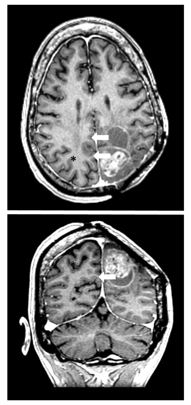
She developed a second radiographic recurrence in 2021 (Figure 1). Given the refractory nature of the tumor, and given the unclear histological diagnosis, she underwent a third surgery with the goal of making a definitive diagnosis and debulking the tumor. Histological features were similar to previous resections (Figure 2). Her tumor was sent for methylome profiling followed by next-generation sequencing which disclosed a CXXC5-MN1 fusion transcript. Based on accumulated methylome data and recent updates to WHO classifications of primary tumors, the MN1 fusion, and methylome profile confirmed diagnosis of Astroblastoma, MN-1 altered, WHO Grade 4 (Louis 2021). No other actionable mutations were discovered by sequencing using a 170 gene in-house screening assay or with Foundation Medicine CDx testing. Surgery was followed by concurrent radiation/temozolomide followed by 6 cycles of adjuvant temozolomide. At last encounter, she had symptoms of cognitive slowing and a 6-month history of subacute progressive ataxia and length-dependent peripheral neuropathy. While it is tempting to blame her symptoms on previous therapy, she was found to have thiamine deficiency due to self-imposed dietary restrictions to which ataxia and neuropathy may be attributable.
Evidence of 2 prior resections includes cystic cavities (arrows) into which the tumor is growing from the posterior medial aspect of the largest cavity. As with other cases of astroblastoma, there are bubbly features (asterisk) of recurrent enhancing tumor and minimal associated edema.
Discussion
Only a handful of astroblastomas have been reported that share the CXXC5-MN1 fusion, and none with the atypical histological features of the current case [1-3]. The current case highlights the importance of methylation profiling in diagnosing primary brain tumors. The new WHO diagnostic criteria based on methylation profiling [4] have led to other astroblastoma diagnoses with CXXC5-MN1 fusions [3,5]. Radiographic imaging was non-specific (Figure 1) and histopathology from both earlier and later resections failed to generate a clear diagnosis (Figure 2). Purported histological hallmarks of astroblastoma are radially arrayed neoplastic cells with papillary or pseudopapillary formations in ribbon-like/trabecular alignment called astroblastic pseudorosettes [1,3,6-9]. The tumor in this case had no rosettes, papillary or pseudopapillary features, nor trabeculae. Instead, there were vague streams and nests of cells with round mildly pleomorphic nuclei. Features of high-grade embryonal neoplasm were also present, including high mitotic rate, anaplastic morphology, high cellularity with cellular atypia, and endothelial hyperplasia without vessel wall sclerosis [10]. Moreover, extensive synaptophysin staining is unusual in astroblastoma and further obscured the final diagnosis. The most recent pathologic sample from the 2021 resection showed up to 5 mitoses in a single high-powered field, Ki-67 of 35%, high cellularity with atypia, and anaplastic features. Endothelial hyperplasia was absent. Only methylome-based profiling and sequencing which found meningioma 1 (MN1)-CXXC5 fusion transcript allowed the diagnosis of astroblastoma [4,8].
The function of MN1 in astroblastoma and other solid tumors is unknown. However, the oncogenic role of MN1 overexpression has been confirmed in AML [11]. Elevated MN1 protein levels strongly correlate with chemotherapy resistance and confer a poor prognosis among these patients [11-19]. Oncogenic MN1 depends upon poly-glutamine stretches, particularly at the N-terminal region, and SMARCA4 to stabilize BAF-chromatin binding [11]. The BAF complex mediates ATP-dependent nucleosome remodeling and maintains the active state of enhancer regions [20-22]. MN1-knockout mouse models exhibit neonatal lethal palatal abnormalities [23]. Similar features are seen in infants with MN1 C-terminal truncation syndrome which is phenotypically characterized by craniofacial malformations and impaired rhombencephalon formation [24].
MN1 amplification is associated with meningioma, astroblastoma, and particularly with acute myeloid leukemia (AML) where it influences the prognosis for patients with MN1 alterations [7,13-15,25,26]. Increased expression of MN1 in patients with AML and the otherwise normal karyotype is associated with shorter survival and reduced response to treatments [13]. However, the role of MN1 alterations in astroblastoma remains unclear. The CXXC5 gene codes for a transcriptional regulator which is ubiquitously expressed and connected to a number of human neoplasms [27-31]. It is a member of the CXXC-type zinc-finger protein family. CXXC5 activation appears to negatively feedback against the WNT/β-catenin pathway, regulating glucose metabolism [32]. Though seen here and in other cases of astroblastoma, it is unclear how the CXXC5-MN1 fusion affects astroblastoma survival. Equally unclear is whether the fusion product has prognostic significance in addition to the known diagnostic significance.
Radiographically, astroblastomas appear well demarcated with a “bubbly” appearance due to signal voids caused by atypical vascular architecture on MRI [33,34]. There may be minimal perilesional edema relative to their large size which may be related to the non-infiltrating nature of these tumors and the (generally) slow growth. They are hyperintense on T2-weighted imaging and hypo- to isointense on T1. Imaging was not available from the time of diagnosis in 2007. However, subsequent recurrences were radiographically consistent with previous publications. Radiographic features are highly non-specific, and in this case, they were confounded by previous resections.
The lack of clarity regarding the diagnosis and prognosis of astroblastoma, particularly among adults, directly relates to the rarity of the diagnosis. It is among the rarest adult primary neoplasms accounting for fewer than 3% of all diagnosed neurologic tumors. The actual incidence of astroblastoma based on a broad literature review has been estimated at 1.6 new patients/per year [35]. These tumors have a predilection for women over men with a reported male-to-female ratio ranging from 1:1.7 to as high as 1:11 [35,36]. Symptoms suggesting tumor growth depend upon anatomic location but may also be non-specific for patients who present first with headaches. The current case fits neatly into this epidemiological pattern, though at 36, our patient was in the upper age range for adults diagnosed with astroblastoma. Age at diagnosis with astroblastoma is generally bimodal with peaks in the first and third decades of life; however, MN1 alterations are mostly seen among patients diagnosed under 10 years of age [2]. While it is tempting to hypothesize various mechanisms for tumorigenesis on the basis of MN1 genetic and epigenetic regulation, it may simply be that we have an incomplete understanding of MN1 alterations in astroblastoma. Indeed, the sub-classification of astroblastoma continues to evolve. It is unclear whether MN1, CXXC5, or other genes (such as BEND1 or EWSR1) may be most influential in these tumors.
Regardless of age at diagnosis, surgery remains the mainstay of treatment for astroblastoma. A large national cancer database was unable to show significant increases in overall survival relating to post-operative radiation and/or chemotherapy, but there was a trend suggesting benefits for patients who had tumors with high-grade features [37]. Our patient has survived > 14 years since initial treatment in 2007 following aggressive maximal safe resection and craniospinal radiation (at that time for presumed high-grade PNET). Our patient also received multiple rounds of chemotherapy even up to ablative chemotherapy requiring a stem cell transplant. While the current case is insufficient to support the use of any particular therapy, treatment with platinum-based therapy and etoposide is common when dealing with embryonal-type tumors. Given the intensity of prior chemotherapies used in this case and based on the lack of efficacy data for particular regimens for recurrent astroblastomas, we chose temozolomide for its well-known efficacy against various types of intracranial neoplasms and low toxicity profile. Prognosis is generally favorable for astroblastoma. Overall survival for patients with astroblastoma is approximately 80% after 5 years and may be better than 60% at 10 years [37]. Data are lacking for long-term survival at recurrence.
Conclusion
The current case is unique histologically for the presence of synaptophysin in astroblastoma without other typical features. Moreover, this case highlights the value of recent advances in methylome based tumor profiling which accurately identifies MN1-altered astroblastoma.
- Lehman NL, Usubalieva A, Lin T, Allen SJ, Tran QT, Mobley BC, McLendon RE, Schniederjan MJ, Georgescu MM, Couce M, Dulai MS, Raisanen JM, Al Abbadi M, Palmer CA, Hattab EM, Orr BA. Genomic analysis demonstrates that histologically-defined astroblastomas are molecularly heterogeneous and that tumors with MN1 rearrangement exhibit the most favorable prognosis. Acta Neuropathol Commun. 2019 Mar 15;7(1):42. doi: 10.1186/s40478-019-0689-3. PMID: 30876455; PMCID: PMC6419470.
- Lehman NL, Spassky N, Sak M, Webb A, Zumbar CT, Usubalieva A, Alkhateeb KJ, McElroy JP, Maclean KH, Fadda P, Liu T, Gangalapudi V, Carver J, Abdullaev Z, Timmers C, Parker JR, Pierson CR, Mobley BC, Gokden M, Hattab EM, Parrett T, Cooke RX, Lehman TD, Costinean S, Parwani A, Williams BJ, Jensen RL, Aldape K, Mistry AM. Astroblastomas exhibit radial glia stem cell lineages and differential expression of imprinted and X-inactivation escape genes. Nat Commun. 2022 Apr 19;13(1):2083. doi: 10.1038/s41467-022-29302-8. PMID: 35440587; PMCID: PMC9018799.
- Sturm D, Orr BA, Toprak UH, Hovestadt V, Jones DTW, Capper D, Sill M, Buchhalter I, Northcott PA, Leis I, Ryzhova M, Koelsche C, Pfaff E, Allen SJ, Balasubramanian G, Worst BC, Pajtler KW, Brabetz S, Johann PD, Sahm F, Reimand J, Mackay A, Carvalho DM, Remke M, Phillips JJ, Perry A, Cowdrey C, Drissi R, Fouladi M, Giangaspero F, Łastowska M, Grajkowska W, Scheurlen W, Pietsch T, Hagel C, Gojo J, Lötsch D, Berger W, Slavc I, Haberler C, Jouvet A, Holm S, Hofer S, Prinz M, Keohane C, Fried I, Mawrin C, Scheie D, Mobley BC, Schniederjan MJ, Santi M, Buccoliero AM, Dahiya S, Kramm CM, von Bueren AO, von Hoff K, Rutkowski S, Herold-Mende C, Frühwald MC, Milde T, Hasselblatt M, Wesseling P, Rößler J, Schüller U, Ebinger M, Schittenhelm J, Frank S, Grobholz R, Vajtai I, Hans V, Schneppenheim R, Zitterbart K, Collins VP, Aronica E, Varlet P, Puget S, Dufour C, Grill J, Figarella-Branger D, Wolter M, Schuhmann MU, Shalaby T, Grotzer M, van Meter T, Monoranu CM, Felsberg J, Reifenberger G, Snuderl M, Forrester LA, Koster J, Versteeg R, Volckmann R, van Sluis P, Wolf S, Mikkelsen T, Gajjar A, Aldape K, Moore AS, Taylor MD, Jones C, Jabado N, Karajannis MA, Eils R, Schlesner M, Lichter P, von Deimling A, Pfister SM, Ellison DW, Korshunov A, Kool M. New Brain Tumor Entities Emerge from Molecular Classification of CNS-PNETs. Cell. 2016 Feb 25;164(5):1060-1072. doi: 10.1016/j.cell.2016.01.015. PMID: 26919435; PMCID: PMC5139621.
- Louis DN, Perry A, Wesseling P, Brat DJ, Cree IA, Figarella-Branger D, Hawkins C, Ng HK, Pfister SM, Reifenberger G, Soffietti R, von Deimling A, Ellison DW. The 2021 WHO Classification of Tumors of the Central Nervous System: a summary. Neuro Oncol. 2021 Aug 2;23(8):1231-1251. doi: 10.1093/neuonc/noab106. PMID: 34185076; PMCID: PMC8328013.
- Lake JA, Donson AM, Prince E, Davies KD, Nellan A, Green AL, Mulcahy Levy J, Dorris K, Vibhakar R, Hankinson TC, Foreman NK, Ewalt MD, Kleinschmidt-DeMasters BK, Hoffman LM, Gilani A. Targeted fusion analysis can aid in the classification and treatment of pediatric glioma, ependymoma, and glioneuronal tumors. Pediatr Blood Cancer. 2020 Jan;67(1):e28028. doi: 10.1002/pbc.28028. Epub 2019 Oct 8. PMID: 31595628; PMCID: PMC7560962.
- Brat DJ, Hirose Y, Cohen KJ, Feuerstein BG, Burger PC. Astroblastoma: clinicopathologic features and chromosomal abnormalities defined by comparative genomic hybridization. Brain Pathol. 2000 Jul;10(3):342-52. doi: 10.1111/j.1750-3639.2000.tb00266.x. PMID: 10885653; PMCID: PMC8098511.
- Wood MD, Tihan T, Perry A, Chacko G, Turner C, Pu C, Payne C, Yu A, Bannykh SI, Solomon DA. Multimodal molecular analysis of astroblastoma enables reclassification of most cases into more specific molecular entities. Brain Pathol. 2018 Mar;28(2):192-202. doi: 10.1111/bpa.12561. Epub 2017 Oct 27. PMID: 28960623; PMCID: PMC5843525.
- Hirose T, Nobusawa S, Sugiyama K, Amatya VJ, Fujimoto N, Sasaki A, Mikami Y, Kakita A, Tanaka S, Yokoo H. Astroblastoma: a distinct tumor entity characterized by alterations of the X chromosome and MN1 rearrangement. Brain Pathol. 2018 Sep;28(5):684-694. doi: 10.1111/bpa.12565. Epub 2017 Nov 9. PMID: 28990708; PMCID: PMC8028274.
- Mhatre R, Sugur HS, Nandeesh BN, Chickabasaviah Y, Saini J, Santosh V. MN1 rearrangement in astroblastoma: study of eight cases and review of literature. Brain Tumor Pathol. 2019 Jul;36(3):112-120. doi: 10.1007/s10014-019-00346-x. Epub 2019 May 20. PMID: 31111274.
- Bonnin JM, Rubinstein LJ. Astroblastomas: a pathological study of 23 tumors, with a postoperative follow-up in 13 patients. Neurosurgery. 1989 Jul;25(1):6-13. PMID: 2755581.
- Riedel SS, Lu C, Xie HM, Nestler K, Vermunt MW, Lenard A, Bennett L, Speck NA, Hanamura I, Lessard JA, Blobel GA, Garcia BA, Bernt KM. Intrinsically disordered Meningioma-1 stabilizes the BAF complex to cause AML. Mol Cell. 2021 Jun 3;81(11):2332-2348.e9. doi: 10.1016/j.molcel.2021.04.014. Epub 2021 May 10. PMID: 33974912; PMCID: PMC8380056.
- Haferlach C, Kern W, Schindela S, Kohlmann A, Alpermann T, Schnittger S, Haferlach T. Gene expression of BAALC, CDKN1B, ERG, and MN1 adds independent prognostic information to cytogenetics and molecular mutations in adult acute myeloid leukemia. Genes Chromosomes Cancer. 2012 Mar;51(3):257-65. doi: 10.1002/gcc.20950. Epub 2011 Nov 10. PMID: 22072540.
- Heuser M, Beutel G, Krauter J, Döhner K, von Neuhoff N, Schlegelberger B, Ganser A. High meningioma 1 (MN1) expression as a predictor for poor outcome in acute myeloid leukemia with normal cytogenetics. Blood. 2006 Dec 1;108(12):3898-905. doi: 10.1182/blood-2006-04-014845. Epub 2006 Aug 15. PMID: 16912223.
- Langer C, Marcucci G, Holland KB, Radmacher MD, Maharry K, Paschka P, Whitman SP, Mrózek K, Baldus CD, Vij R, Powell BL, Carroll AJ, Kolitz JE, Caligiuri MA, Larson RA, Bloomfield CD. Prognostic importance of MN1 transcript levels, and biologic insights from MN1-associated gene and microRNA expression signatures in cytogenetically normal acute myeloid leukemia: a cancer and leukemia group B study. J Clin Oncol. 2009 Jul 1;27(19):3198-204. doi: 10.1200/JCO.2008.20.6110. Epub 2009 May 18. PMID: 19451432; PMCID: PMC2716941.
- Metzeler KH, Dufour A, Benthaus T, Hummel M, Sauerland MC, Heinecke A, Berdel WE, Büchner T, Wörmann B, Mansmann U, Braess J, Spiekermann K, Hiddemann W, Buske C, Bohlander SK. ERG expression is an independent prognostic factor and allows refined risk stratification in cytogenetically normal acute myeloid leukemia: a comprehensive analysis of ERG, MN1, and BAALC transcript levels using oligonucleotide microarrays. J Clin Oncol. 2009 Oct 20;27(30):5031-8. doi: 10.1200/JCO.2008.20.5328. Epub 2009 Sep 14. PMID: 19752345.
- Pogosova-Agadjanyan EL, Moseley A, Othus M, Appelbaum FR, Chauncey TR, Chen IL, Erba HP, Godwin JE, Jenkins IC, Fang M, Huynh M, Kopecky KJ, List AF, Naru J, Radich JP, Stevens E, Willborg BE, Willman CL, Wood BL, Zhang Q, Meshinchi S, Stirewalt DL. AML risk stratification models utilizing ELN-2017 guidelines and additional prognostic factors: a SWOG report. Biomark Res. 2020 Aug 12;8:29. doi: 10.1186/s40364-020-00208-1. PMID: 32817791; PMCID: PMC7425159.
- Thol F, Yun H, Sonntag AK, Damm F, Weissinger EM, Krauter J, Wagner K, Morgan M, Wichmann M, Göhring G, Bug G, Ottmann O, Hofmann WK, Schambach A, Schlegelberger B, Haferlach T, Bowen D, Mills K, Ganser A, Heuser M. Prognostic significance of combined MN1, ERG, BAALC, and EVI1 (MEBE) expression in patients with myelodysplastic syndromes. Ann Hematol. 2012 Aug;91(8):1221-33. doi: 10.1007/s00277-012-1457-7. Epub 2012 Apr 10. PMID: 22488406.
- Wang T, Chen X, Hui S, Ni J, Yin Y, Cao W, Zhang Y, Wang X, Ma X, Cao P, Liu M, Chen KN, Wang F, Zhang Y, Nie D, Yuan L, Liu H. Ectopia associated MN1 fusions and aberrant activation in myeloid neoplasms with t(12;22)(p13;q12). Cancer Gene Ther. 2020 Nov;27(10-11):810-818. doi: 10.1038/s41417-019-0159-x. Epub 2020 Jan 6. PMID: 31902945; PMCID: PMC7661342.
- Xiang L, Li M, Liu Y, Cen J, Chen Z, Zhen X, Xie X, Cao X, Gu W. The clinical characteristics and prognostic significance of MN1 gene and MN1-associated microRNA expression in adult patients with de novo acute myeloid leukemia. Ann Hematol. 2013 Aug;92(8):1063-9. doi: 10.1007/s00277-013-1729-x. Epub 2013 Mar 21. PMID: 23515710.
- Alver BH, Kim KH, Lu P, Wang X, Manchester HE, Wang W, Haswell JR, Park PJ, Roberts CW. The SWI/SNF chromatin remodelling complex is required for maintenance of lineage specific enhancers. Nat Commun. 2017 Mar 6;8:14648. doi: 10.1038/ncomms14648. PMID: 28262751; PMCID: PMC5343482.
- Mittal P, Roberts CWM. The SWI/SNF complex in cancer - biology, biomarkers and therapy. Nat Rev Clin Oncol. 2020 Jul;17(7):435-448. doi: 10.1038/s41571-020-0357-3. Epub 2020 Apr 17. PMID: 32303701; PMCID: PMC8723792.
- Wang X, Lee RS, Alver BH, Haswell JR, Wang S, Mieczkowski J, Drier Y, Gillespie SM, Archer TC, Wu JN, Tzvetkov EP, Troisi EC, Pomeroy SL, Biegel JA, Tolstorukov MY, Bernstein BE, Park PJ, Roberts CW. SMARCB1-mediated SWI/SNF complex function is essential for enhancer regulation. Nat Genet. 2017 Feb;49(2):289-295. doi: 10.1038/ng.3746. Epub 2016 Dec 12. PMID: 27941797; PMCID: PMC5285474.
- Meester-Smoor MA, Vermeij M, van Helmond MJ, Molijn AC, van Wely KH, Hekman AC, Vermey-Keers C, Riegman PH, Zwarthoff EC. Targeted disruption of the Mn1 oncogene results in severe defects in development of membranous bones of the cranial skeleton. Mol Cell Biol. 2005 May;25(10):4229-36. doi: 10.1128/MCB.25.10.4229-4236.2005. PMID: 15870292; PMCID: PMC1087735.
- Mak CCY, Doherty D, Lin AE, Vegas N, Cho MT, Viot G, Dimartino C, Weisfeld-Adams JD, Lessel D, Joss S, Li C, Gonzaga-Jauregui C, Zarate YA, Ehmke N, Horn D, Troyer C, Kant SG, Lee Y, Ishak GE, Leung G, Barone Pritchard A, Yang S, Bend EG, Filippini F, Roadhouse C, Lebrun N, Mehaffey MG, Martin PM, Apple B, Millan F, Puk O, Hoffer MJV, Henderson LB, McGowan R, Wentzensen IM, Pei S, Zahir FR, Yu M, Gibson WT, Seman A, Steeves M, Murrell JR, Luettgen S, Francisco E, Strom TM, Amlie-Wolf L, Kaindl AM, Wilson WG, Halbach S, Basel-Salmon L, Lev-El N, Denecke J, Vissers LELM, Radtke K, Chelly J, Zackai E, Friedman JM, Bamshad MJ, Nickerson DA; University of Washington Center for Mendelian Genomics, Reid RR, Devriendt K, Chae JH, Stolerman E, McDougall C, Powis Z, Bienvenu T, Tan TY, Orenstein N, Dobyns WB, Shieh JT, Choi M, Waggoner D, Gripp KW, Parker MJ, Stoler J, Lyonnet S, Cormier-Daire V, Viskochil D, Hoffman TL, Amiel J, Chung BHY, Gordon CT. MN1 C-terminal truncation syndrome is a novel neurodevelopmental and craniofacial disorder with partial rhombencephalosynapsis. Brain. 2020 Jan 1;143(1):55-68. doi: 10.1093/brain/awz379. Erratum in: Brain. 2020 Mar 1;143(3):e24. PMID: 31834374; PMCID: PMC7962909.
- Liu PP, Hajra A, Wijmenga C, Collins FS. Molecular pathogenesis of the chromosome 16 inversion in the M4Eo subtype of acute myeloid leukemia. Blood. 1995 May 1;85(9):2289-302. Erratum in: Blood 1997 Mar 1;89(5):1842. PMID: 7727763.
- Valk PJ, Verhaak RG, Beijen MA, Erpelinck CA, Barjesteh van Waalwijk van Doorn-Khosrovani S, Boer JM, Beverloo HB, Moorhouse MJ, van der Spek PJ, Löwenberg B, Delwel R. Prognostically useful gene-expression profiles in acute myeloid leukemia. N Engl J Med. 2004 Apr 15;350(16):1617-28. doi: 10.1056/NEJMoa040465. PMID: 15084694.
- Knappskog S, Myklebust LM, Busch C, Aloysius T, Varhaug JE, Lønning PE, Lillehaug JR, Pendino F. RINF (CXXC5) is overexpressed in solid tumors and is an unfavorable prognostic factor in breast cancer. Ann Oncol. 2011 Oct;22(10):2208-15. doi: 10.1093/annonc/mdq737. Epub 2011 Feb 16. PMID: 21325450.
- Treppendahl MB, Möllgård L, Hellström-Lindberg E, Cloos P, Grønbaek K. Downregulation but lack of promoter hypermethylation or somatic mutations of the potential tumor suppressor CXXC5 in MDS and AML with deletion 5q. Eur J Haematol. 2013 Mar;90(3):259-60. doi: 10.1111/ejh.12045. Epub 2013 Jan 20. PMID: 23190153.
- Centritto F, Paroni G, Bolis M, Garattini SK, Kurosaki M, Barzago MM, Zanetti A, Fisher JN, Scott MF, Pattini L, Lupi M, Ubezio P, Piccotti F, Zambelli A, Rizzo P, Gianni' M, Fratelli M, Terao M, Garattini E. Cellular and molecular determinants of all-trans retinoic acid sensitivity in breast cancer: Luminal phenotype and RARα expression. EMBO Mol Med. 2015 Jul;7(7):950-72. doi: 10.15252/emmm.201404670. PMID: 25888236; PMCID: PMC4520659.
- Benedetti I, De Marzo AM, Geliebter J, Reyes N. CXXC5 expression in prostate cancer: implications for cancer progression. Int J Exp Pathol. 2017 Aug;98(4):234-243. doi: 10.1111/iep.12241. PMID: 29027288; PMCID: PMC5639265.
- Chen X, Wang X, Yi L, Song Y. The KN Motif and Ankyrin Repeat Domains 1/CXXC Finger Protein 5 Axis Regulates Epithelial-Mesenchymal Transformation, Metastasis and Apoptosis of Gastric Cancer via Wnt Signaling. Onco Targets Ther. 2020 Jul 28;13:7343-7352. doi: 10.2147/OTT.S240991. PMID: 32801759; PMCID: PMC7395690.
- Seo SH, Kim E, Lee SH, Lee YH, Han DH, Go H, Seong JK, Choi KY. Inhibition of CXXC5 function reverses obesity-related metabolic diseases. Clin Transl Med. 2022 Apr;12(4):e742. doi: 10.1002/ctm2.742. PMID: 35384342; PMCID: PMC8982507.
- Port JD, Brat DJ, Burger PC, Pomper MG. Astroblastoma: radiologic-pathologic correlation and distinction from ependymoma. AJNR Am J Neuroradiol. 2002 Feb;23(2):243-7. PMID: 11847049; PMCID: PMC7975267.
- Singh DK, Singh N, Singh R, Husain N. Cerebral astroblastoma: A radiopathological diagnosis. J Pediatr Neurosci. 2014 Jan;9(1):45-7. doi: 10.4103/1817-1745.131485. PMID: 24891904; PMCID: PMC4040033.
- Barakat MI, Ammar MG, Salama HM, Abuhashem S. Astroblastoma: Case Report and Review of Literature. Turk Neurosurg. 2016;26(5):790-4. doi: 10.5137/1019-5149.JTN.9408-13.2. PMID: 27349390.
- Bell JW, Osborn AG, Salzman KL, Blaser SI, Jones BV, Chin SS. Neuroradiologic characteristics of astroblastoma. Neuroradiology. 2007 Mar;49(3):203-9. doi: 10.1007/s00234-006-0182-0. Epub 2007 Jan 10. PMID: 17216265.
- Merfeld EC, Dahiya S, Perkins SM. Patterns of care and treatment outcomes of patients with astroblastoma: a National Cancer Database analysis. CNS Oncol. 2018 Apr;7(2):CNS13. doi: 10.2217/cns-2017-0038. Epub 2018 Apr 30. PMID: 29708401; PMCID: PMC5977281.
Article Alerts
Subscribe to our articles alerts and stay tuned.
 This work is licensed under a Creative Commons Attribution 4.0 International License.


 Save to Mendeley
