Archives of Hematology Case Reports and Reviews
Hereditary elliptocytosis discovered during work-up for infective endocarditis: About a case
Mohammad Hassan Hodroj1, Ziad F Bashshur2, Charbel Wahab2, Kawthar Jarrah3 and Ali Taher1,3*
Cite this as
Baiya M, Elkhannouri I, Mhirig I, Sayagh S (2022) Hereditary elliptocytosis discovered during work-up for infective endocarditis: About a case. Arch Hematol Case Rep Rev 7(1): 013-014. DOI: 10.17352/ahcrr.000038Copyright License
© 2022 Baiya M, et al. This is an open-access article distributed under the terms of the Creative Commons Attribution License, which permits unrestricted use, distribution, and reproduction in any medium, provided the original author and source are credited.Introduction
Hereditary elliptocytosis is a group of red blood cell membrane disorders that are characterized by elliptical-shaped erythrocytes and shortened red blood cell survival [1]. It is due to protein abnormalities involving the horizontal skeletal network of the red cell membrane, including the spectrin dimer-dimer interaction or the spectrin-actin-protein 4.1 junction complex [2]. It is often asymptomatic and may be complicated by constitutional hemolytic anemia due to an abnormality in the structure of the Red Blood cell (RB) membrane [2].
We report through this observation a case of elliptocytosis discovered by chance in a patient hospitalized for infective endocarditis and we discuss the clinical, biochemical, and genetic aspects of this condition.
Observation
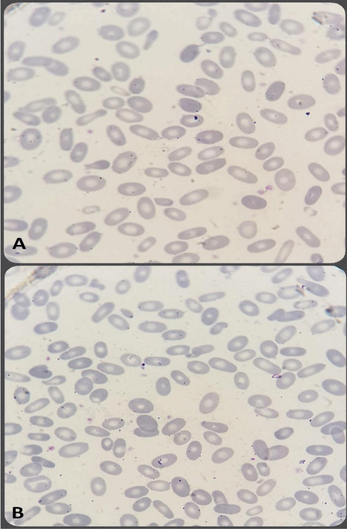
A 17-year-old patient, from a non-consanguineous marriage, the only daughter of her parents and with no particular pathological history, presented to the emergency room for acute onset dyspnea at rest, with a fever of 38°C. Evolving in the context of the deterioration of the general state. Clinical examination revealed a heart murmur, splenomegaly with mucocutaneous pallor, and failure to thrive without any other associated clinical signs. Blood culture was used to isolate Streptococcus pneumonia. Echocardiography confirmed the diagnosis of infective endocarditis. The patient was hospitalized in the cardiology department and was treated with a double antibiotic therapy consisting of amoxicillin and ceftriaxone intravenously for 6 weeks. A blood count was performed showing isolated anemia at 7g/dl of normocytic hypochromic hemoglobin. The blood smear stained by the May Grunwald Giemsa (MGG) method revealed anisopoikilocytosis made up of 70% elliptocytes (Figure 1). The hemolysis assessment was normal. Ferritin was low. Membrane protein electrophoresis was not performed due to a lack of resources. The patient received a blood transfusion and close monitoring. A family investigation based mainly on the blood smear was requested but the patient refused to cooperate.
Discussion
Hereditary Elliptocytosis (HE) is a group of disorders characterized by the presence of elliptical-shaped erythrocytes on peripheral blood film. It has a worldwide distribution but is more common in areas of endemic malaria, particularly in people of African and Mediterranean descent. [1]. The majority of HE-associated defects occur in spectrin, the major structural protein in the skeletal structure of the erythrocyte membrane. Spectrin is composed of two non-identical homologous proteins, alpha, and beta spectrin, encoded by separate genes [2].
HE and its related disorders are characterized by clinical, biochemical, and genetic heterogeneity. Manifestations range from asymptomatic carrier states to severe transfusion-dependent hemolytic anemia. In the healthy carrier, only detailed studies of the thermal stability of the red blood cell membrane as well as a detailed analysis of the protein composition of the cytoskeleton can highlight the anomaly. Mild forms include well-compensated hemolysis, without anemia, but which may be complicated by episodes of acute hemolysis, following a viral infection that stimulates the macrophage monocyte system (infectious mononucleosis, cytomegalovirus, hepatitis) [2]. In the chronic hemolytic form, there is frank hemolysis, anemia with a hemoglobin concentration between 8 and 10g/dl, hyperbilirubinemia, and splenomegaly. A particular entity is a neonatal and infantile poikilocytosis or pyropoikilocytosis. This is a very early presentation, with severe hemolysis responsible for neonatal jaundice often requiring exchange transfusion [1,2]. The reading of the blood smear is an important element of the diagnosis because usually, HE is clinically latent, it is characterized by the presence of more than 30% of elliptocytes [3]. In a normal blood smear and secondary elliptocytosis associated with deficiency anemia or certain myelodysplastic syndromes, less than 5% of elliptocytes are found [3]. HE is an autosomal dominant disease that most often affects black people. The genetic abnormality is variable. In approximately 70% of cases, it is a mutation located in the SPTA1 gene coding for the alpha chain of spectrin leading to a point of weakness in the mesh constituting the erythrocyte skeleton. The remaining cases (25-30%) result from mutations in the EPB41 gene encoding protein 4.1R, which normally binds to actin and the spectrin beta chain. In very rare cases, the mutation is located on the SPTB gene coding for the spectrin beta chain [4].
Conclusion
HE is an easily diagnosed red blood cell membrane disease. An only careful reading of the blood smear provides a decisive element in the diagnostic process. HE is a very rare pathology whose precise frequency is difficult to estimate given the great clinical heterogeneity and the frequency of asymptomatic forms.
- Llaudet-Planas E, Vives-Corrons JL, Rizzuto V, Gómez-Ramírez P, Sevilla Navarro J, Coll Sibina MT, García-Bernal M, Ruiz Llobet A, Badell I, Velasco-Puyó P, Dapena JL, Mañú-Pereira MM. Osmotic gradient ektacytometry: A valuable screening test for hereditary spherocytosis and other red blood cell membrane disorders. Int J Lab Hematol. 2018 Feb;40(1):94-102. doi: 10.1111/ijlh.12746. Epub 2017 Oct 10. PMID: 29024480.
- Soderquist C, Bagg A. Hereditary elliptocytosis. Blood. 2013 Apr 18;121(16):3066. doi: 10.1182/blood-2012-09-457788. PMID: 23729040.
- Gallagher PG. Hereditary elliptocytosis: spectrin and protein 4.1R. Semin Hematol. 2004 Apr;41(2):142-64. doi: 10.1053/j.seminhematol.2004.01.003. PMID: 15071791.
- Kalfa TA. Diagnosis and clinical management of red cell membrane disorders. Hematology Am Soc Hematol Educ Program. 2021 Dec 10;2021(1):331-340. doi: 10.1182/hematology.2021000265. PMID: 34889366; PMCID: PMC8791164.

Article Alerts
Subscribe to our articles alerts and stay tuned.
 This work is licensed under a Creative Commons Attribution 4.0 International License.

 Save to Mendeley
