Journal of Dental Problems and Solutions
Evaluation of effects of Forsus Fatigue Resistance device in correction of class II division 1 malocclusion in adolescent patient: A case report
Anupama V Jain*, Anand K Patil and Roopak Naik
Cite this as
Jain AV, Patil AK, Naik R (2022) Evaluation of effects of Forsus Fatigue Resistance device in correction of class II division 1 malocclusion in adolescent patient: A case report. J Dent Probl Solut 9(2): 028-034. DOI: 10.17352/2394-8418.000114Copyright License
© 2022 Jain AV, et al. This is an open-access article distributed under the terms of the Creative Commons Attribution License, which permits unrestricted use, distribution, and reproduction in any medium, provided the original author and source are credited.Introduction: Class II malocclusion is one of the most prevalent malocclusions. The Class II malocclusions are caused due to forwardly placed maxilla, the backward position of the mandible, or a combination of both these factors. This disparity in skeletal base growth and position can be corrected during growth spurts using functional and fixed functional appliances.
Description: An adolescent boy with Class II division 1 malocclusion, retrusive mandible, and increased overjet was treated with a pre-adjusted edgewise appliance (0.022-slot Gemini 3M -MBT prescription) along with a fixed functional appliance, Forsus TM Fatigue Resistant Device. The Skeletal age of the patient assessment using Hand wrist radiograph and CVM showed a major part of the adolescent growth spurt to be completed. Pre-treatment and post-functional cephalograms were traced and superimposed to compare changes in the skeletal base and dental structures.
Result: The Class II molar and canine relationships were corrected to class I and the mandible showed forward positioning leading to correction for the skeletal base to class I. The facial profile showed marked improvement to an orthognathic pleasing profile.
Conclusion: The purpose of this case report is to emphasize on use of fixed functional appliances in the treatment of adolescents with skeletal base discrepancies like Class II division 1 malocclusion. Intervention with fixed functional appliances at the appropriate skeletal age can prevent the need for extractions or other surgical procedures that may be needed to correct the malocclusion.
Introduction
Class II malocclusions are one of the most common problems that one comes across in orthodontic practise [1]. The facial aesthetics and functional status of the patients are adversely affected due to the malocclusion. However, within class II, there is large variability in aetiology and maxillomandibular relationships. The malocclusion could be due to a retrusive mandible, protrusive maxilla, or both [2]. According to McNamara, the most common characteristic of class II malocclusion is mandibular retrognathia rather than maxillary protrusion [3,4]. The dentoalveolar components can also have a discrepancy and could be the underlying cause of the malocclusion. Diagnosing the case correctly and identifying the aetiology of the malocclusion is essential for proper treatment planning.
The treatment modality is decided to depend upon the aetiology, clinical findings, and reporting age of the patients to the orthodontist. The options for correction of the malocclusion range from functional appliance for growth modulation, fixed orthodontic treatment with extractions in most cases, and orthognathic surgery [4].
Functional appliances like the twin block are used in the adolescent growth spurt stage and are found to be effective in the correction of the disparity in the jaw base relationship [5,6]. However, the majority of patients will have crossed their adolescent spurt and hence report late for treatment. In situations where patients are in the last phase of their growth spurt, removable functional appliances not is a good treatment option as they rely heavily on patient cooperation and need a longer duration of appliance wear [6,7]. Fixed functional appliances are used to correct the class II by stimulating the mandibular growth by repositioning the mandible forward and hence achieving pleasing facial aesthetics [7,8]. Fixed functional appliances which need minimal patient cooperation are indicated in such non-growing patients [4,9].
One of the fixed functional appliances is the Forsus™ Fatigue Resistant Device (FFRD, 3M Unitek, Monrovia, Calif) [10]. This system is used in conjunction with fixed multibracket appliances and consists of a push rod that acts on the cuspid or first bicuspid, which is inserted into a nickel-titanium cylinder/coil, springs, and exerts an approximate force of 200 grams when activated. The force level can be modified by the clinician. The FRD appliance acts by restraining sagittal maxillary growth, stimulating mandibular growth, inducing mesial movement of the mandibular arch, and distal movement of the maxillary arch [4,10,11-13].
This case report presents how FFRD can be used in the treatment of a Class II division 1 patient who had a retrusive mandible. The patient had a convex facial profile with deep mentolabial sulcus and increased overjet.
Case presentation
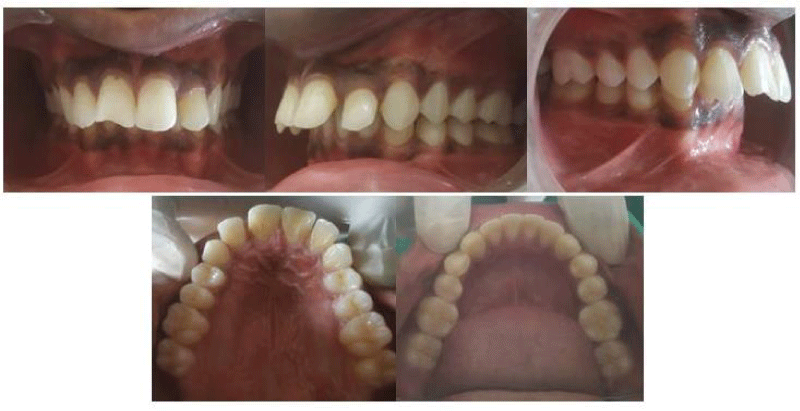
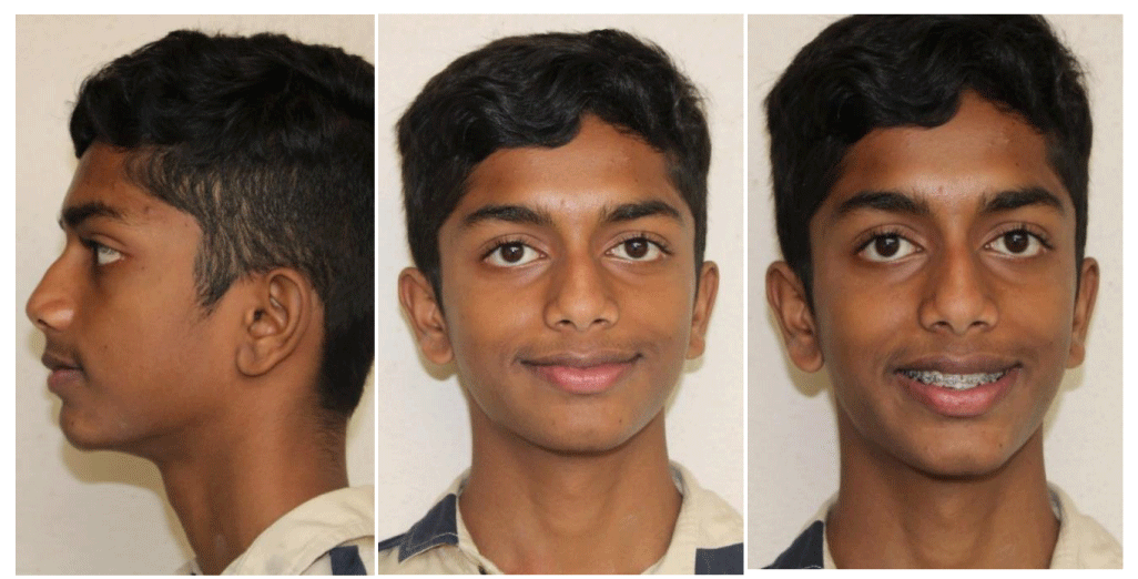
A 14-year-old boy reported to the Department of Orthodontics with a chief complaint of forwardly placed upper front teeth, convex facial profile inability to close the lips, and breathing through the mouth, especially during sleep. Extraoral clinical examination indicated a convex profile with a retrusive chin and deep mentolabial sulcus. No skeletal asymmetry was observed (Figure 1). The patient did not give a history of any habits like thumb or lip sucking. The intraoral examination (Figure 2) showed cusp-to-cusp canine and class II molar relationships on the right side and ended on the molar relationship in the left side segments with severe overjet 10mm and deep bite 6mm. The maxillary arch was constricted and the maxillary intercanine width (dentoalveolar) was decreased.
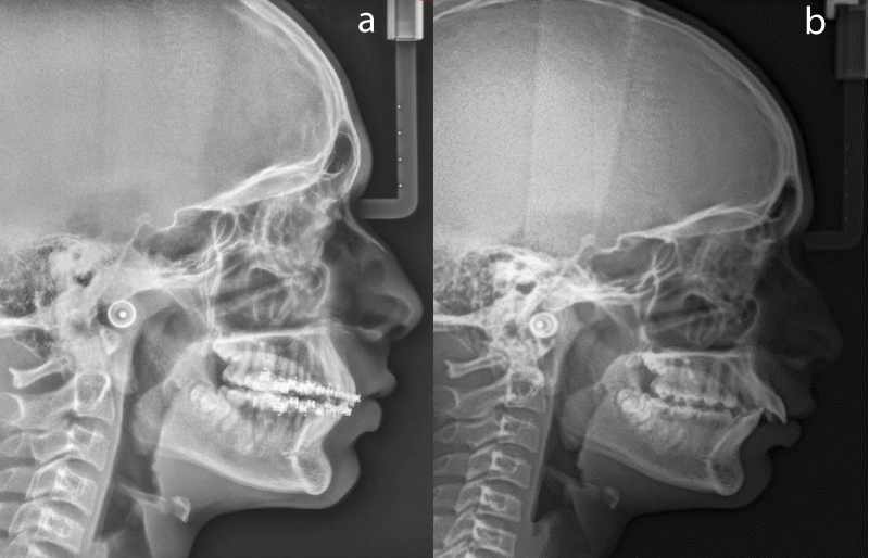
Examination of the lateral cephalometric radiograph (Figure 3a) indicated a normally positioned maxilla (SNA: 820 ), a retrognathic mandible (SNB: 760), skeletal class II malocclusion (ANB: 60), and horizontal growth pattern (Sn-Go-Gn: 320). The upper incisors were proclined (U1-NA: 320,8mm) and the lower incisors were relatively upright (L1 to NB:280, 5mm), though in relation to the mandibular base they were proclined (IMPA: 1040).
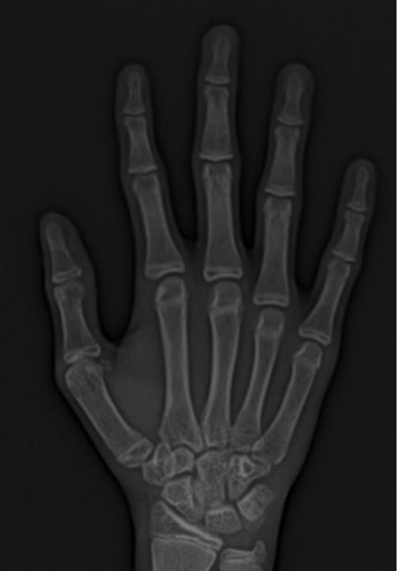
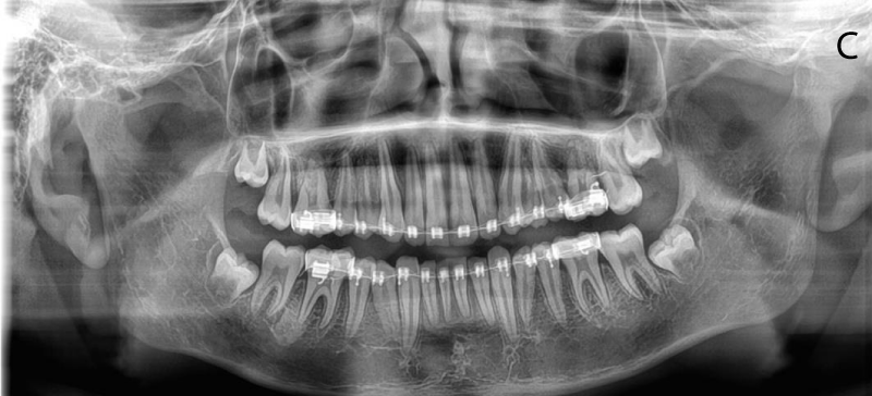
Panoramic radiographic evaluation (Figure 3a) showed the patient had the full complement of permanent teeth including the third molar tooth buds in all quadrants. Hand wrist Radiograph (Figure 3b) showed the progressive sesamoid ossification and start of fusion of epiphysis and diaphysis in most phalangeal joints MP3, PP3 indicating completion of the majority of the pubertal growth spurt. The young patient seemed to be in the last few months of a pubertal growth spurt, mostly the deceleration phase of the growth spurt.
Treatment objectives
The treatment objectives were the following:
(1) Expansion of the constricted maxillary dental arch.
(2) Correction of axial inclination of upper incisors.
(3) Closing the spaces of the maxillary and mandibular arch.
(4) Correction of severe overjet.
(5) Establishing Class I canine and molar relationships.
(6) Obtaining normal overjet and overbite and
(7) Improvement of the patient’s facial aesthetic.
Treatment plan
Visual treatment objective (VTO) (Figure 4) with the positioning of the mandible forward showed a positive, favorable improvement in facial profile and aesthetics. Considering the hand-wrist radiograph examination showing only a small percentage of growth remaining and the favorable VTO, it was decided to use a fixed functional appliance for correcting the skeletal base discrepancy.
Treatment progress
At the beginning of the orthodontic treatment, the upper and lower arches were bonded with 0.022-inch slot Gemini 3M brackets (MBT prescription). Levelling and aligning stage was started with 0.016 Niti wires, followed by 0.018 Niti, 0.017 * 0.025 Niti, 0.017 * 0.025 SS and 0.019 * 0.022 SS.
As part of the preparation of the case for FFRD appliance insertion, Figure 8 ligature ties were placed from molar to molar in both upper and lower arches in the upper arch. To minimize possible transverse side effects of the FFRD on the maxillary molars a transpalatal arch was applied in the upper arch [4] .
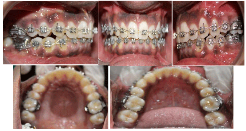
Additional buccal root torque was applied in the lower arch wire in relation to the lower anterior segment from canine to canine to limit the buccal inclination of the lower incisors [4]. FFRD appliances with a 34mm length of the rod were inserted bilaterally from the maxillary molars to the distal of the mandibular canine teeth. During treatment, the FFRD was activated using the crimp-on which was inserted on the arch wire, just distal to the lower canines where the FFRD spring attached. In 5 months, a Class I canine and molar relationships were obtained, and overjet was eliminated. The FFRD was used for 5 months duration (Figure 5).
The leveling and aligning stage prior to the FFRD application took 4 months. Active FFRD application took 5 months.
Treatment will be continued up to finishing and detailing to achieve a functional occlusal interdigitation and meet the treatment objectives mentioned above. Brackets will then be debonded. Appropriate retention protocol will be followed to maintain the skeletal, dentoalveolar, and dental corrections achieved.
Treatment results
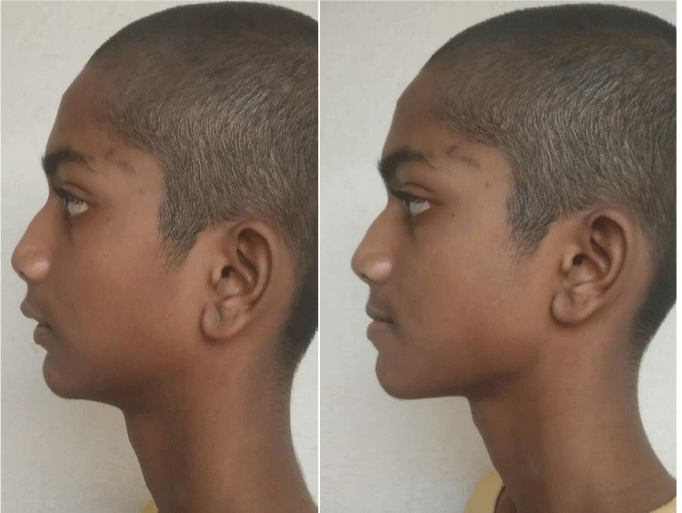
The post-fixed functional appliance intraoral photographs (Figure 6) show the correction of the class II molar relationship into the class I molar and canine relationship on both the right and left buccal segments. The ideal overjet and overbite relationship are achieved with the overjet reduced from 10 mm to 1.5 mm and overbite reduced from 6 mm to 3mm. The teeth were well aligned, and the occlusion was stable. The facial aesthetics showed marked improvement with a decrease in facial convexity and a decrease in the prominence of the mentolabial fold. The significant extra oral changes can be appreciated in post-fixed functional facial photographs (Figure 7).
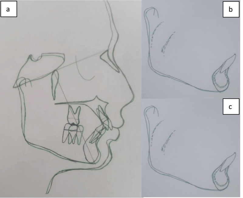
The pre-treatment and postfixed functional stage cephalometric measurements are given in Table 1. The results indicated improvement in both skeletal and dental parameters. Post-fixed functional cephalometric radiographs (Figure 8a) showed the maxillary incisor inclination was reduced with U1 to NA reducing from 32 0 to 24 0 and 8 mm to 4 mm. However mandibular incisors showed a slight increase from 280 to 320, 5 mm to 7 mm (L1 to NB). ANB angle decreased from 80 to 40. The post-fixed functional panoramic radiograph showed no alveolar bone loss or apical root resorption (Figure 8c). The maxillary base, Mandibular base, and overall cephalometric superimposition (Figure 9) showed forward and downward movement of the mandibular dentoalveolar segment and lower arch. Superimposition also shows the position of the condyle is forward and downward.
Discussion
In patients with Class II malocclusion due to mandibular retrusion, either camouflage using interarch elastics or orthognathic surgical treatment is carried out depending upon the severity of maxilla mandibular discrepancy [4,14]. The young patient had a retrognathic profile and deep bite. Conventional Extraction treatment followed by retraction of the teeth would have worsened the profile and the deep bite. Also, when the patient reported for treatment, he was in the deceleration phase of the adolescent growth spurt with the hand-wrist radiograph showing progressive ossification of the sesamoid, fusion of epiphysis, and diaphysis of the proximal phalanx of the middle finger (PP3U as per Bjork, Grave Brown method) as per the skeletal maturity indicators [15].
The cervical vertebrae C3 also shows concavity in its inferior border and shape changing to squarish from rectangular, which again indicated the completion-maturation phase of adolescent growth as per Hassel and Farman [16]. Therefore, it was decided to capitalize on whatever growth potential was left and apply non-extraction orthodontic treatment along with a fixed functional appliance to protract the mandible and the mandibular arch. The dependence on patient compliance was also reduced with the use of a fixed functional appliance system.
Aetiology of malocclusion, Rationale for use of FFRD & treatment alternatives
On intraoral observation, a constricted maxillary dental arch form with reduced intercanine width was found. A premature loss of deciduous molars and disrupted eruption pattern of permanent dentition not allowing mesial migration of the lower molars could have been the cause for the class II molar relationship. The class II molar relationship so achieved during the transition dentition could have locked the mandibular arch posteriorly and hence been the cause for the malocclusion. Further, the constricted maxillary arch also could be another reason for the retrusive position of the mandible based on the analogy of the foot and shoe relationship. The patient had a horizontal growth pattern and hence was an ideal candidate for mandibular advancement for correction of Class II malocclusion.
ForsusTM Fatigue Resistant Device is proven to be a reliable, versatile, patient-friendly, hygienic device that applied continuous consistent force levels to achieve a predictable correction of the class II malocclusion in a short duration of time by applying a mesial force on the mandibular arch and a distal force on the maxillary arch. It is also said to apply an intrusive force on maxillary molars which can decrease the posterior vertical dimension and counteract any clockwise rotation of the mandible caused due to forward positioning. In adolescent patients, who are at the end of or have just finished their pubertal growth spurts the changes are more dentoalveolar in nature.
Considering the advantages of treatment with FFRD like short treatment duration, mechanism of action, reduced dependence on patient compliance, and other advantages mentioned previously, it is best indicated in patients who have just completed their pubertal growth spurt. Hence fixed functional orthodontic treatment with correction of maxillary arch form and dentoalveolar expansion along with FFRD for forwarding positioning of the mandible and the mandibular arch was the treatment plan decided.
ForsusTM Fatigue Resistant Device can lead to favourable dentoalveolar changes and stimulates mandibular growth in patients at or before the peak phase of pubertal growth [17,18]. However, it is observed that for the patients in the post-pubertal period, mostly dental changes are encountered [19,20]. In this reported case, the correction of class II was achieved by the increase of the SNB angle by nearly 20 (2 degrees) indicating slight forward mandibular displacement. Maxillary dentoalveolar changes (Table 1) with correction of upper anterior proclination are seen. The mandibular dentoalveolar changes (Table 1) show the mesial movement of the lower dentoalveolar arch with some amount of proclination of the lower anterior despite applying additional buccal root torque to the archwire.
Minimal residual growth of the patient during orthodontic treatment could be the main contributor to the nature of these changes. Maxillary teeth especially the anterior segment demonstrated distal movement and the first molars showed very slight intrusive and mesial movement. Mandibular first molars showed mesial movement and, lower incisors exhibited proclination. The correction of the overjet was achieved by both retroclination of the upper incisors and protrusion of the lower incisors. Even with the anchorage mechanics used [4], mandibular incisors were proclined by 40. An increase in the mandibular incisor inclination is a similar common finding [20,21] of fixed functional appliances as shown by the other studies [22-24]. Li, et al. [25]. Concluded that in the correction of skeletal class II malocclusion using FFRD skeletal and dentoalveolar effects played differential roles. An evaluation by George, et al. [26] of corrected class II malocclusions showed the proportion of dentoalveolar versus skeletal contribution towards overjet and the molar correction was 87% and 13% respectively, hence they found mainly dentoalveolar effects contributed to the correction
Aslan, et al. [25-27] demonstrated the use of temporary anchorage devices(TADS) to engage the appliance to eliminate this side effect of the FRD appliance. Another technique that can be adopted is the use of large dimension mandibular rectangular archwires which fill the bracket slot more fully and incorporate of addition of negative torque in the lower incisor region [4] Zitouni and Acar [28] followed up with patients in whom the correction of class II correction with FFRD was done and the treatment results were stable for a 2-year follow-up period. Another advantage of FFRD was reported by Aslan, etal. [29], that Class II correction using FFRD provided an increase in space available for the mandibular third molars in the retromolar region. The use of FFRD also was found to have an effect on the hyoid bone position and improve the pharyngeal airway in patients with skeletal class II malocclusions and hence improved the overall quality of life of such patients [30,31].
Conclusion
Use of Forsus Fatigue Resistant Device (FFRD) as an adjunct along with fixed orthodontic treatment in an adolescent patient for correction mandibular retrusion and Class II division 1 skeletal bases resulted in favourable and prominent changes in the facial profile and dentition. Treatment with FFRD helps in the correction of the malocclusion without extractions and in a shorter period. Careful assessment of skeletal age at the time of treatment planning can help in identifying the appropriate time for FFRD application on a case-to-case basis.
Author contribution
AVJ: Conception/design of the study, Treatment/execution of the study design, Data acquisition, analysis, or interpretation. AKP: Final approval of the article/ Critical revision of the article. RDN: Writing the article/Critical revision of the article.
- Proffit WR, Fields HW Jr, Moray LJ. Prevalence of malocclusion and orthodontic treatment need in the United States: estimates from the NHANES III survey. Int J Adult Orthodon Orthognath Surg. 1998;13(2):97-106. PMID: 9743642..
- Alarashi M, Franchi L, Marinelli A, Defraia E. Morphometric analysis of the transverse dentoskeletal features of class II malocclusion in the mixed dentition. Angle Orthod. 2003 Feb;73(1):21-5. doi: 10.1043/0003-3219(2003)073<0021:MAOTTD>2.0.CO;2. PMID: 12607851..
- McNamara JA Jr. Components of class II malocclusion in children 8-10 years of age. Angle Orthod. 1981 Jul;51(3):177-202. doi: 10.1043/0003-3219(1981)051<0177:COCIMI>2.0.CO;2. PMID: 7023290..
- Atik E, Kocadereli I. Treatment of Class II Division 2 Malocclusion Using the Forsus Fatigue Resistance Device and 5-Year Follow-Up. Case Rep Dent. 2016;2016:3168312. doi: 10.1155/2016/3168312. Epub 2016 Feb 29. PMID: 27034855; PMCID: PMC4789427..
- Nelson C, Harkness M, Herbison P. Mandibular changes during functional appliance treatment. Am J Orthod Dentofacial Orthop. 1993 Aug;104(2):153-61. doi: 10.1016/S0889-5406(05)81005-4. PMID: 8338068..
- Cozza P, Baccetti T, Franchi L, De Toffol L, McNamara JA Jr. Mandibular changes produced by functional appliances in Class II malocclusion: a systematic review. Am J Orthod Dentofacial Orthop. 2006 May;129(5):599.e1-12; discussion e1-6. doi: 10.1016/j.ajodo.2005.11.010. PMID: 16679196..
- Dalci O, Altug AT, Memikoglu UT. Treatment effects of a twin-force bite corrector versus an activator in comparison with an untreated Class II sample: a preliminary report. Aust Orthod J. 2014 May;30(1):45-53. PMID: 24968645..
- Franchi L, Alvetro L, Giuntini V, Masucci C, Defraia E, Baccetti T. Effectiveness of comprehensive fixed appliance treatment used with the Forsus Fatigue Resistant Device in Class II patients. Angle Orthod. 2011 Jul;81(4):678-83. doi: 10.2319/102710-629.1. Epub 2011 Feb 7. PMID: 21299410; PMCID: PMC8919739..
- Zymperdikas VF, Koretsi V, Papageorgiou SN, Papadopoulos MA. Treatment effects of fixed functional appliances in patients with Class II malocclusion: a systematic review and meta-analysis. Eur J Orthod. 2016 Apr;38(2):113-26. doi: 10.1093/ejo/cjv034. Epub 2015 May 19. PMID: 25995359; PMCID: PMC4914762..
- Vogt W. The Forsus Fatigue Resistant Device. J Clin Orthod. 2006 Jun;40(6):368-77; quiz 358. PMID: 16804253..
- Gunay EA, Arun T, Nalbantgil D. Evaluation of the Immediate Dentofacial Changes in Late Adolescent Patients Treated with the Forsus(™) FRD. Eur J Dent. 2011 Oct;5(4):423-32. PMID: 22589581; PMCID: PMC3352683..
- Jones G, Buschang PH, Kim KB, Oliver DR. Class II non-extraction patients treated with the Forsus Fatigue Resistant Device versus intermaxillary elastics. Angle Orthod. 2008 Mar;78(2):332-8. doi: 10.2319/030607-115.1. PMID: 18251605..
- Heinig N, Göz G. Clinical application and effects of the Forsus spring. A study of a new Herbst hybrid. J Orofac Orthop. 2001 Nov;62(6):436-50. English, German. doi: 10.1007/s00056-001-0053-6. PMID: 11765707..
- Aras I, Pasaoglu A. Class II subdivision treatment with the Forsus Fatigue Resistant Device vs intermaxillary elastics. Angle Orthod. 2017 May;87(3):371-376. doi: 10.2319/070216-518.1. Epub 2016 Oct 13. PMID: 27762602; PMCID: PMC8381987..
- Björk A, Helm S. Prediction of the age of maximum puberal growth in body height. Angle Orthod. 1967 Apr;37(2):134-43. doi: 10.1043/0003-3219(1967)037<0134:POTAOM>2.0.CO;2. PMID: 4290545..
- Hassel B, Farman AG. Skeletal maturation evaluation using cervical vertebrae. Am J Orthod Dentofacial Orthop. 1995 Jan;107(1):58-66. doi: 10.1016/s0889-5406(95)70157-5. Erratum in: Am J Orthod Dentofacial Orthop 1995 Jun;107(6):19. PMID: 7817962..
- Bock NC, von Bremen J, Ruf S. Occlusal stability of adult Class II Division 1 treatment with the Herbst appliance. Am J Orthod Dentofacial Orthop. 2010 Aug;138(2):146-51. doi: 10.1016/j.ajodo.2008.09.031. PMID: 20691355..
- Bilgiç F, Hamamci O, Başaran G. Comparison of the effects of fixed and removable functional appliances on the skeletal and dentoalveolar structures. Aust Orthod J. 2011 Nov;27(2):110-6. PMID: 22372266..
- Mahamad IK, Neela PK, Mascarenhas R, Husain A. A comparision of Twin-block and Forsus (FRD) functional appliance--a cephalometric study. Int J Orthod Milwaukee. 2012 Fall;23(3):49-58. PMID: 23094559..
- Upadhyay M, Yadav S, Nagaraj K, Uribe F, Nanda R. Mini-implants vs fixed functional appliances for treatment of young adult Class II female patients: a prospective clinical trial. Angle Orthod. 2012 Mar;82(2):294-303. doi: 10.2319/042811-302.1. Epub 2011 Aug 26. PMID: 21867432; PMCID: PMC8867937..
- Aras A, Ada E, Saracoğlu H, Gezer NS, Aras I. Comparison of treatments with the Forsus fatigue resistant device in relation to skeletal maturity: a cephalometric and magnetic resonance imaging study. Am J Orthod Dentofacial Orthop. 2011 Nov;140(5):616-25. doi: 10.1016/j.ajodo.2010.12.018. PMID: 22051481..
- Ruf S, Pancherz H. Herbst/multibracket appliance treatment of Class II division 1 malocclusions in early and late adulthood. a prospective cephalometric study of consecutively treated subjects. Eur J Orthod. 2006 Aug;28(4):352-60. doi: 10.1093/ejo/cji116. Epub 2006 Apr 27. PMID: 16644850..
- Gao W, Li X, Bai Y. An assessment of late fixed functional treatment and the stability of Forsus appliance effects. Aust Orthod J. 2014 May;30(1):2-10. PMID: 24968640..
- Zhang R, Bai Y, Li S. Use of Forsus fatigue-resistant device in a patient with Class I malocclusion and mandibular incisor agenesis. Am J Orthod Dentofacial Orthop. 2014 Jun;145(6):817-27. doi: 10.1016/j.ajodo.2013.08.021. PMID: 24880853..
- Li H, Ren X, Hu Y, Tan L. Effects of the Forsus Fatigue-resistant Device on Skeletal Class II Malocclusion Correction. J Contemp Dent Pract. 2020 Jan 1;21(1):105-112. PMID: 32381810..
- George AS, Ganapati Durgekar S. Skeletal and dentoalveolar contributions during Class II correction with Forsus™ FRD appliances : Quantitative evaluation. J Orofac Orthop. 2022 Mar;83(2):87-98. English. doi: 10.1007/s00056-021-00297-z. Epub 2021 May 7. PMID: 33961059..
- Aslan BI, Kucukkaraca E, Turkoz C, Dincer M. Treatment effects of the Forsus Fatigue Resistant Device used with miniscrew anchorage. Angle Orthod. 2014 Jan;84(1):76-87. doi: 10.2319/032613-240.1. Epub 2013 Jun 17. Erratum in: Angle Orthod. 2014 Mar;84(2):383. PMID: 23772682; PMCID: PMC8683057..
- Zitouni M, Acar YB. Treatment outcome and long-term stability of class II correction with forsus fatigue resistant device in non-growing patients. Orthod Craniofac Res. 2021 Feb;24(1):130-136. doi: 10.1111/ocr.12416. Epub 2020 Aug 24. PMID: 32757406..
- Aslan BI, Akarslan ZZ, Karadağ Ö. Effects of Angle class II correction with the Forsus fatigue resistant device on mandibular third molars : A retrospective study. J Orofac Orthop. 2021 Nov;82(6):403-412. English. doi: 10.1007/s00056-021-00281-7. Epub 2021 Mar 5. PMID: 33666713..
- Kaur R, Garg AK, Gupta DK, Singla L, Aggarwal K. Effect of Twin Block Therapy Versus Fixed Functional Appliances on Pharyngeal Airway Space in Skeletal Class II Patients: A Prospective Cephalometric Study. Clin Ter. 2022 Jul-Aug;173(4):306-315. doi: 10.7417/CT.2022.2439. PMID: 35857047..
- Baka ZM, Fidanboy M. Pharyngeal airway, hyoid bone, and soft palate changes after Class II treatment with Twin-block and Forsus appliances during the postpeak growth period. Am J Orthod Dentofacial Orthop. 2021 Feb;159(2):148-157. doi: 10.1016/j.ajodo.2019.12.016. Epub 2020 Dec 31. Erratum in: Am J Orthod Dentofacial Orthop. 2021 Apr;159(4):411. PMID: 33388197.
Article Alerts
Subscribe to our articles alerts and stay tuned.
 This work is licensed under a Creative Commons Attribution 4.0 International License.





 Save to Mendeley
