Archives of Clinical Gastroenterology
Incarcerated Giant Hiatal Hernia conditioning hearth shock: case report
Medina Andrade Luis Angel1*, Cesar Manuel Vargas Sahagún2, Carlos Eduardo Rodriguez Rodriguez2, Pedro Leonardo Villanueva Solorzano2, Alberto Robles Méndez Hernández2, Bernardo Gutierrez Muñoz2, Valdez Hernandez Brenda Elizabeth3, Brigitte Marlene Chevillon Castillo1, Vallejo Ramirez Jose Eduardo1, Campos Cruz Alan Ranferi1 and Tolentino Gonzalez Christian Stefan4
2Universidad La Salle, Hospital Angeles Metropolitano, Mexico City, México
3Hospital General La Villa, México City, México
4General Surgery Department, General Zone Hospital #47 “Vicente Guerrero”, Mexican Institute of Social Security, México City, México
Cite this as
Luis Angel MA, Vargas Sahagún CM, Rodriguez Rodriguez CE, Villanueva Solorzano PL, Méndez Hernández AR, et al. (2018) Incarcerated Giant Hiatal Hernia conditioning hearth shock: case report. Arch Clin Gastroenterol 4(1): 012-014. DOI: 10.17352/2455-2283.000049Luis Angel MA, Vargas Sahagún CM, Rodriguez Rodriguez CE, Villanueva Solorzano PL, Méndez Hernández AR, et al. (2018) Incarcerated Giant Hiatal Hernia conditioning hearth shock: case report. Arch Clin Gastroenterol 4(1): 012-0
Case: A female patient of 80 years-old was referred to our hospital by septic shock and abdominal pain. At physical exam she refers abdominal and thoracic pain, dyspnea and occasional threw up for the last 2 days, with a background of this symptoms the last 5 years, and gastroesophageal reflux symptoms for 10 years. At admission, she referred epigastric and retrosternal pain, dyspnea, with an 02 of 75%, bowel sounds in left hemithorax, mean arterial pressure of 50mmHg with the use of norepinephrine. Laboratories do not reveal sepsis and CT scan reports a hiatal hernia of 9 cm with left hemithorax occupied by stomach, colon, and spleen. A cardiogenic shock by compression was suspected with this data and a laparotomy was scheduled. CT scan report was confirmed and the mentioned organs were reduced to abdomen without problems, both diaphragmatic pillars were sutured and a Nissen fundoplication completed. After 6 hours’ norepinephrine was suspended and 48 hours after the patient were discharged uneventfully.
Conclusion: Giant hiatal hernia must be suspected in patients with chronic abdominal and thoracic pain with reflux symptoms because the complications associated with this disease could have a mortality near 30% in case of strangulation and a scheduled surgery could be very safe in the correct moment.
Introduction
Hiatal hernia is a very frequent disease presented in the general population with an incidence of 20%, but this number is very variable according to different publications by the wide range of symptoms or the asymptomatic stage of many patients [1].
This kind of hernia is classified into 4 types. The type I and most common is a sliding hernia, where the stomach and cardia are displaced above the diaphragm. Type II hernia is a defect in the paraesophageal membrane where the gastric fundus is herniated but the gastric junction remains in position. Type III is a combination of the II and III. Type IV refers to a large defect in the paraesophageal membrane which allows herniation of stomach or other intraabdominal components, this is the less frequent and the one associated with severe complications like in the presented case [1].
Case Report
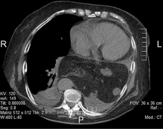
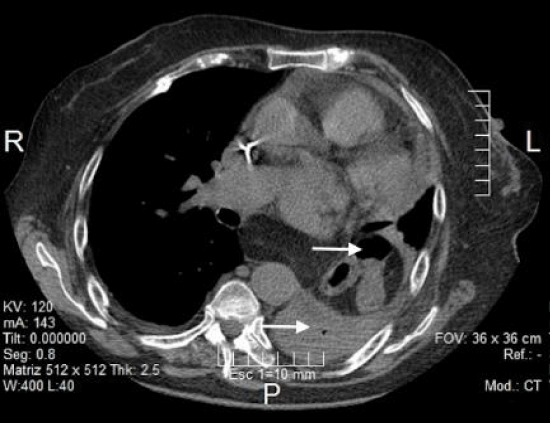
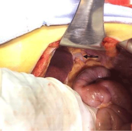
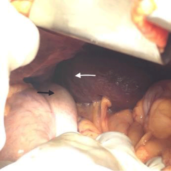
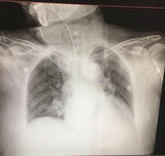
A female patient of 80 years old was referred from a second level hospital with diagnosis of septic shock and abdominal pain. She had a pathological background including diabetes mellitus 2, heart insufficiency grade 2, pulmonary chronic obstructive disease, gastroesophageal reflux for 10 years and two previous episodes of thoracic and abdominal pain associated with threw up in the last 4 weeks. At admission, patient referred intense abdominal pain mainly in epigastrium, retrosternal pain at inspiration, and have been throwing up on 6 occasions the last 8 hours. At physical exam, she had Glasgow of 15 points, with 75% basal oxygen saturation, and with supplemental oxygen by mask at 5 liters per minute with SO2 92%, with adequate right hemithorax ventilation and in the left apical zone, but with bowel sounds in the rest of left hemithorax. She maintains mean arterial pressure of 50 mmHg, with norepinephrine since the first contact hospital, heart rate of 97 beats per minute and 24 breaths per minute. In abdomen with diminished bowel sounds, pain at superficial palpation, mainly in epigastrium, without acute abdomen signs. Laboratories reported: hemoglobin of 12mg/dL, hematocrit 35%, platelets of 250000, leucocytes 8.4/mm3, neutrophils 55%, TP 14 seconds, TTP 25 seconds, glucose 135 mg/dL, creatinine 1.0 mg/dL. X rays presented an elevated left hemidiaphragm with a superimposed image in front of the hearth. CT scan was requested reporting a giant hiatal hernia of 9 cm in diameter, with stomach, transverse colon and spleen in the left thorax (Figures 1,2). Considering the laboratory findings without sepsis data and the hiatal hernia with incarcerated organs in the thorax, the diagnosis of cardiogenic shock secondary to compression was established and a laparotomy scheduled. By the cardiogenic shock with the use of norepinephrine a laparoscopic approach was discarded initially and a laparotomy was performed finding the stomach, transverse colon and spleen incarcerated in the thorax, they were reduced by a hiatal hernia of about 10 cm. of diameter without problems (Figures 3,4). A Nissen fundoplication was performed, with adequate response and remission of cardiogenic shock only six hours after surgery. A chest X-ray 24 hours after surgery shows no alteration (Figure 5). After 48 hours of hospital stay and oral feeding, the patient was discharged uneventfully. At 4 months after surgery she stills asymptomatic.
Discussion
Hiatal hernia type I is the most frequent, constituting about 95% of all hiatal hernias, and most patients with this kind of hernia are asymptomatic but may have gastroesophageal reflux. The clinical presentation of giant hiatal hernias like in the case of type IV could be very unspecific, with symptoms of gastroesophageal reflux, thoracic pain, dyspnea, exercise intolerance, or in the course of an emergency like gastric volvulus, respiratory distress or cardiac shock by compression like in the present case [2].
The presentation with dyspnea and low cardiac output have been reported only in one previous case. The first symptom can be due to reflux mediated lung fibrosis or due to compression of cardiac structures like the left atrium and coronary sinus [2].
This kind of giant hiatal hernias use to be more frequent in elderly, manifested by dyspnea and cardiac failure due to compression, with hypoxemia, hypercapnia, and respiratory distress, those symptoms will improve with supplementary oxygen and allow us to suspect the cardiogenic origin [3].
Giant hiatal hernias could present with incarcerated abdominal organs in less than 5% of the cases, but with an associated mortality near 30%, increasing this percentage by the pathologies present in the most affected population [4].
The surgical approach must be scheduled in all cases of symptomatic hiatal hernias, and in this scenario the laparoscopic approach offers the best results in terms of bleeding, hospital stay, postoperative pain, and improved quality of life compared with the open approach, turning it into the gold standard for repair [5]. The Nissen fundoplication has been accepted as the better approach and to avoid recurrence. Although this is an excellent technique in the short term, the recurrence at 10 years is estimated between 35 to 50% in most centers [6].
One of the contraindications for laparoscopic techniques is a patient in shock by the risk of cardiac arrest secondary to the decreased hearth preload during the pneumoperitoneum insufflation. For those reasons we decided not to perform the laparoscopic correction in this specific case, and patient evolved with excellent results and a hospital stay shorter than any of the reported, inclusive in some laparoscopic corrections [4-6].
Conclusion
Symptomatic hiatal hernia must be adequately studied and treated once diagnosed, especially in at-risk population like the elderly by the high rate of associated complications, and cases of incarceration by the high mortality associated, which could be avoided with a timely management.
- Dimitrios Prassas, Thomas-Marten Rolfs, Franz-Josef Schumacher (2015) Laparoscopic repair of giant hiatal hernia. A single center experience. International Journal of Surgery 20: 149-152. Link: https://goo.gl/DqHgaw
- Andrew Matar, Jad Mroue, Enrico Camporesi, Devanand Mangar (2016) Large Hiatal Hernia Compressing the Heart. The American Journal of Cardiology 117: 483-484. Link: https://goo.gl/zbTRsu
- Vincent Bunel, Pierre Mordant, Lara Ribeiro, Bruno Crestani (2017) Giant hiatal hernia: beware of the supine ICU chest X-ray!. BMJ Case Rep. Link: https://goo.gl/QL1vTG
- Ismael Mora-Guzmán, Juan Antonio del-Pozo-Jiménez, Elena Martín-Pérez (2017) A giant hiatal hernia and intrathoracic páncreas. Revista española de enfeRmedades digestivas . 109: 458-459. Link: https://goo.gl/MYyRHn
- Steve R Siegal, James P, Dolan John G Hunter (2017) Modern diagnosis and treatment of hiatal hernias. Langenbecks Arch Surg. Link: https://goo.gl/f8SKPq
- Michael Antiporda, Benjamin Veenstra, Chloe Jackson, Pujan Kandel (2018) Laparoscopic repair of giant paraesophageal hernia: are there factors associated with anatomic recurrence? Surg Endosc. Link: https://goo.gl/8Pp6VT
Article Alerts
Subscribe to our articles alerts and stay tuned.
 This work is licensed under a Creative Commons Attribution 4.0 International License.
 Save to Mendeley
