Archives of Otolaryngology and Rhinology
Adenoid cystic carcinoma of the external auditory- A Case Report
Khabti Almuhanna* and Abdullah Alfulaij
Cite this as
Almuhanna K, Alfulaij A (2018) Adenoid cystic carcinoma of the external auditory- A Case Report. Arch Otolaryngol Rhinol 4(1): 023-026. DOI: 10.17352/2455-1759.000069Background: Primary external auditory canal (EAC) malignancies are very rare with adenoid cystic carcinoma (ACC) representing approximately 5% of these tumors. There is insufficient knowledge of the natural behavior of ACC in EAC. The disease needs early detection and complete treatment because of its tendency to recur, vicinity to sensitive organs and lower response to radiation.
Case presentation: In this case study, we presented a case of young female (37 years old) with right external auditory canal (EAC) mass which was diagnosed as benign tumor, postoperative biopsy confirmed it as ACC of the EAC which is a rare case in the literature.
Conclusion: Here, we report a rare case of Adenoid Cystic Carcinoma (ACC) of External Auditory Canal (EAC) in a 37 years old Saudi female. Initial, Clinical and CT findings were suggestive of a benign cystic mass but histopathology examination confirmed that tumor was ACC of EAC. The patient underwent to radical excision of the tumor along the parotidectomy. Postsurgical recovery was uneventful with no sign of recurrence until two years of follow up.
Abbreviations
ACC: Adenoid Cystic Carcinoma; EAC: External Auditory Canal; RT: Radiotherapy
Background
Cancers of the external auditory canal (EAC) are extremely rare tumor with more than 80% being squamous cell carcinomas and adenoid cystic carcinoma (ACC) accounting for approximately 5% of all EAC [1,2] . Since 1894, only 106 cases of (ACC) involving the (EAC) have been reported in English literature [3]. Although it presents a widespread age distribution, peak incidence predominantly occurs among women, between the 5th and 6th decades of life [4]. It is a slow growing but highly invasive cancer with a high recurrence rate. Lymphatic spread to the local lymph nodes is rare, however, It is not uncommon for distant metastasis, mainly to the lungs, that may occur over the course of many years [5].
Earlier diagnosis of these tumors is of utmost importance, in view of the fact that delays in diagnosis may increases the risk of distant metastasis [5]. The propensity for perineural invasion and tendency for local recurrence are well known in ACC patients [6]. The signs and symptoms of this tumor do not always correlate with the histopathological diagnosis and subsequent clinical behavior of these tumors. Deep incisional biopsies need to be taken because superficial biopsies may frequently miss such a lesion [7].
Aggressive surgical resection with adjuvant radiotherapy is the standard treatment for local disease control [6,8]. However, there is a high risk of repeat local recurrence, because of the rarity of (ACC) of the (EAC), most of the observations drawn from various reports lack detailed comparisons of pathological findings and long-term outcome follow-up [3].
In this case study, we presented a case of young female with right external auditory canal (EAC) mass which was diagnosed as benign tumor but postoperative biopsy confirmed it as ACC of the EAC which is a rare case in the literature.
Case Presentation
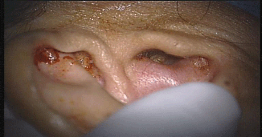
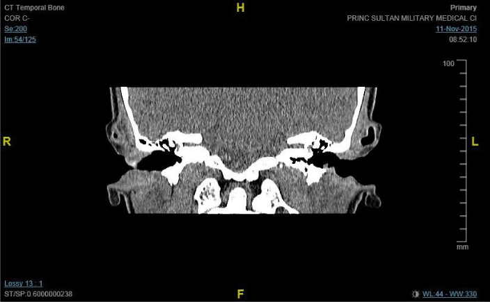
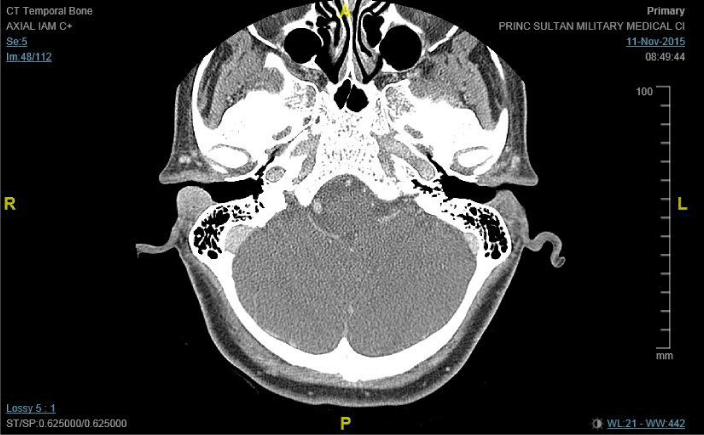
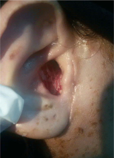
A 37-year-old female patient presented to our clinic with right external auditory canal swelling associated with on and off ear ache with no ear discharges for a duration of 10 years, it had earlier been excised three times at private clinics and histopathology reported as syringoma that was confirmed by our pathologist after reviewing the outside slides. Clinical examination revealed that the mass was occupying the concha and extended to the outer one third of the external auditory canal and causing partial obstruction of the canal (Figure 1). Pure tone audiogram showed mild conductive hearing loss at high frequency. The CT scan of temporal bone showed a well-defined faintly enhancing mass lesion measuring 8 x 10 x 13 mm which was originate from the posterior wall of the right external auditory canal, with no erosion of bone and no involvement of regional lymph node enlargement (Figure 2 A,B). Our findings were suggestive of a benign etiology such as papilloma or adenoma.
Because the patient had been followed up by hematology for low platelet and hepato-splenomegaly she had chance to do Pet scan as part of her investigations that showed right external auditory canal nodule with mild metabolic activity, likely benign. Hepato-splenomegaly had no evidence of focal or hypermetabolic lesions. Only a single small hypermetabolic left external iliac lymph node with possible reactive or inflammatory changes was observed. Her bone marrow study confirms the diagnosis of Gaucher’s disease. Based on given information the swelling (likely benign), the patient underwent simple surgery for excision of the mass, later histopathological examination revealed adenoid cystic carcinoma.
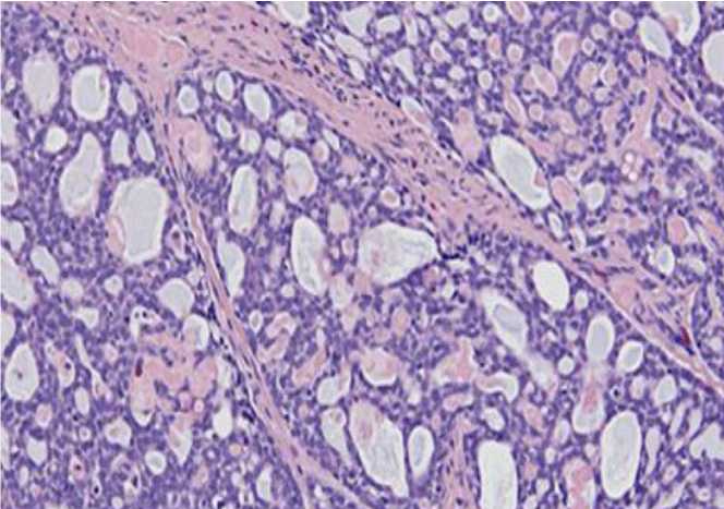
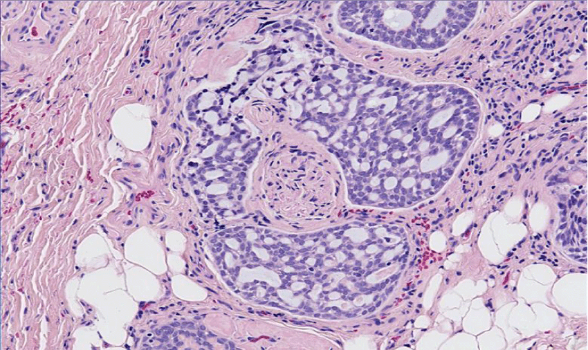
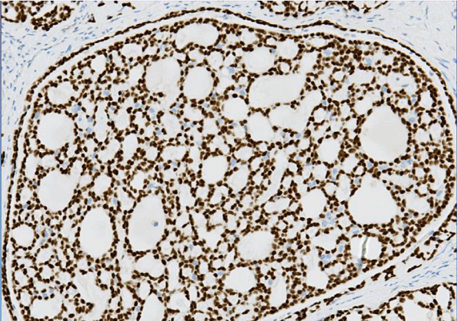
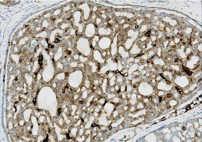
Microscopically, the tumor was composed of baseloid cells arranged in a cribriform architecture (Figure 3). The tumor infiltrated adjacent adipose tissue, skeletal muscles and very close to the elastic cartilage of the external auditory canal (Figure 4). Frequent perineural invasion was identified in this patient (Figure 5). Immunohistochemistry confirmed the diagnosis that showed strong nuclear staining in the outer baseloid cells (P63 immunohistochemistry) and membranous positivity in the inner ductal cells (CK7 immunohistochemistry) as shown in figures 6 and 7 respectively.
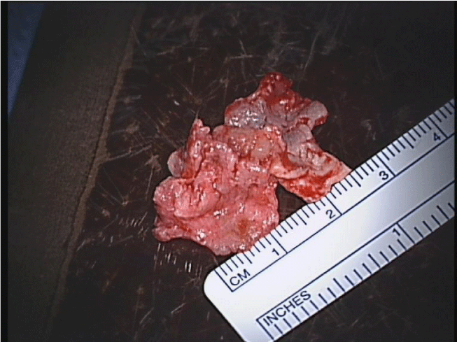
After a thorough review of histopathological and immunochemical findings patient was taken again to the operating room for more radical surgery, where we excised the tumor and most of the conchal cartilage that adjacent to the tumor we followed the tumor along the tragal cartilage and because of the extension (anterior- inferior) we had suspicion that the parotid gland was the original of the tumor so we extend our dissection to do superficial parotidectomy It has been sent for frozen section came to be negative.
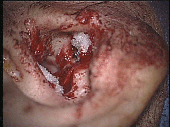
Permanent biopsy for our dissection showed clear margins and the parotid gland is negative for malignancy (Figures 8,9). After the discussion in the tumor board we elected to keep the radiotherapy as adjuvant tool. The postoperative course was uneventful and the excision site was well healed with no evidence of recurrence until the last follow up (Figure 10).
Discussion
Adenoid Cystic Carcinomas (ACC) earlier known as cylindroma seldom arise in EAC, however ACC is most common lesion of glandular origin [5]. This case of ACC involving a 37 year old female is apparently the first case of Saudi Arabia. Earlier reports clearly show that female in their forties and fifties are more prone to the AC [Choi JY 2003 4,5], our case being on the lower side of median age.
The symptoms of our case include swelling of right external auditory canal and partial hearing loss. Generally, the common clinical manifestation include otorrhea, regular pain, hearing loss and bleeding [Arshad et al 2009]. There was no discharge or bleeding in our case.
The etiology of ACC is not fully understood. ACC appears to arise from the ceremonious glands, sweat glands or ectopic salivary gland tissue [9]. In some cases, the tumor may have arisen in the adjacent parotid salivary gland and secondarily may have extended into the ear canal [10]. In our case that mass was occupying the concha and extended to the outer one third of the external auditory canal and causing partial obstruction of the canal.
Metastasis to regional lymph nodes and distant sites are well-documented so lungs and regional lymph nodes must also be evaluated with CT for metastasis. [Mitul Chaitan Bhatt 2016, 5,11,12]. Evidence of irregular bone erosion or destruction of the canal wall is indicative of malignancy [Mitul Chaitan Bhatt 2016]. However, in our study PET scan of patient showed that a faintly enhancing mass lesion was appearing from the posterior wall of the right external auditory canal, there was no erosion of bone and no involvement of regional lymph node enlargement. Only a single small hypermetabolic left external iliac lymph node with possible reactive or inflammatory changes was observed. Therefore, our findings were suggestive of a benign etiology such as papilloma or adenoma.
Histologically, our findings showed that tumor cells were composed of baseloid cells arranged in cribriform architecture and were infiltrated to adipose tissues, skeletal muscles and towards elastic cartilage of the external auditory canal and presence of strong nuclear staining in the outer baseloid cells (P63 immunohistochemistry) and membranous positivity in the inner ductal cells (CK7 immunohistochemistry). Our findings were in favor of Fliss DM 1990 and Shao-Cheng Liua 2012 [3,9]. In addition to that, in our patient we observed the frequent perineural invasion which is a classical characteristic and diagnostically helpful feature of adenoid cystic carcinoma [3,13]. Moreover, according to Nemzek WR 1998 [14], ACC is the second most common tumor associated to perineural invasion after squamous cells carcinoma. These findings confirmed the diagnosis of ACC of the EAC with perineural invasion and involvement of the tympanic portion of the temporal bone [3].
After ACC of EAC confirmation, a radical surgery was done along with the superficial parotidectomy to ensure that parotid gland is free we did not consider the Pet Scan that was requested for something else and it was not reliable result .As we discussed the case in the tumor board we elected to keep the radiotherapy as adjuvant therapy in case of recurrence, [1], although data from randomized trials is lacking, most practitioners consider such treatment to be beneficial [15-17], Al though, postoperative radiation seems to improve local control rates, the impact on ACC-specific survival is not clear [18].
Conclusion
ACC of the EAC is a rare malignant tumor. This tumor has an aggressive behavior characterized by local invasiveness and a metastatic risk of approximately 30%. There is insufficient knowledge of the natural behavior of ACC in EAC. We would like to emphasize the need for early detection of adenoid cystic carcinoma of the external auditory canal and its differentiation from other benign conditions and therefore we encourage other authors to report cases concerning diagnostic and treatment difficulties of this malignancy.
Consent form
Written informed Consent was obtained from the patient for the publication of this case report including the images.
Ethical approval
This study was approved by the Research and Ethical committee of Prince Sultan Military Medical City, Riyadh, Saudi Arabia.
Author’s contribution
Khabti Al Muhanna: Designed and supervised the study, performed clinical examination and gave final approval for the manuscript to be published. Abdullah Alfulaij: Collected demographic data, read the manuscript and helped in writing manuscript.
- Faisal D (2016) Adenoid Cystic Carcinoma of the External Auditory Canal “Rare Case Report. Glob J Oto 1: 555-567. Link: https://goo.gl/zNUJDm
- Safinaz Zainor, Hamidah Mamat, Sakina Mohd. Saad, Mohd. Razif Mohamad Yunus (2013) Adenoid cystic carcinoma of external auditor canal: A Case Report. Egyptian Journal of Ear, Nose, Throat and Allied Sciences 14: 41-44. Link: https://goo.gl/EyYchN
- Shao-Cheng Liua, Bor-Hwang Kanga, Shin Niehb, Junn-Liang Changc, Chih- Hung Wanga (2012) Adenoid cystic carcinoma of the external auditory canal. Journal of the Chinese Medical Association 75: 296- 300. Link: https://goo.gl/sqUAXt
- Waldron CA, el-Mofty SK, Gnepp DR (1988) Tumours of theintraoral minor Salivary glands: a demographic and histologic studyof 426 cases. Oral Surg Oral Med Oral Pathol 66: 323-333. Link: https://goo.gl/jNdRsm
- Dong F, Gidley PW, Ho T, Luna MA, Ginsberg LE, et al. (2008) Adenoid cystic Carcinoma of the external auditory canal. Laryngoscope 118: 1591-1596. Link: https://goo.gl/yz1w3x
- Spiro RH (1986) Salivary neoplasms: overview of a 35 year experience with 2807 Patients. Head Neck Surg 8: 177-184. Link: https://goo.gl/ig293F
- Mohammad Hussham Arshad, Umair Khalid,Shehzad Ghaffar. Adenoid cystic carcinoma of the external auditory canal. Journal of the College of Physicians and Surgeons Pakistan 19: 726-728. Link: https://goo.gl/6mzTqM
- Bhagat S, Varshney S, Bist SS, Mishra S, Aggarwal V (2012) Case Report: Adenoid Cystic Carcinoma of External Auditory Canal. Online J Health Allied Scs 11: Link: https://goo.gl/UKLbyp
- Fliss DM, Kraus M, Tovi F (1990) Adenoid cystic carcinoma of the external auditory canal. Ear Nose Throat J 69: 63545. Link: https://goo.gl/uFpbKk
- Szanto PA, Luna MA, Tortoledo ME (1984) Histologic grading of adenoid cystic carcinoma of the salivary gland. Cancer 54: 10629. Link: https://goo.gl/XWS1By
- Sridhar PS, Sharma DN, Julka PK, Rath GK (2006) Adenoid cystic carcinoma of external auditory canal with vertebral metastasis. Indian journal of Medical and Pediatric Oncology 27: 42-43. Link: https://goo.gl/RYN6JF
- Degirmeni B, Yucel A, Acar M, Albayrak R (2004) Adenoid cystic carcinoma of external auditory canal with pulmonary metastasis. Turk J Med Sci 34: 195-119. Link: https://goo.gl/Z4UZPZ
- Hicks GW (1983) Tumours arising from the glandular structures of the external auditory canal. Laryngoscope. 93: 326-340. Link: https://goo.gl/VGLNzN
- Nemzek WR, Hecht S, Gandour-Edwards R, Donald P, McKennan K. Perineural Spread of Head and Neck Tumors:How Accurate Is MR Imaging?. AJNR Am J Neuroradiol. 1998,19:701-706. Link: https://goo.gl/nh7yfP
- Garden AS, Weber RS, Morrison WH, Ang KK, Peters LJ (1995) The influence of positive margins and nerve invasion in adenoid cystic carcinoma of the head and neck treated with surgery and radiation. Int J Radiat Oncol BiolPhys 32: 619–626. Link: https://goo.gl/W3JVzV
- Balamucki CJ, Amdur RJ, Werning JW, et al. (2012) Adenoid cystic carcinoma of the head and neck. Am J Otolaryngol 33: 510–518. Link: https://goo.gl/Mcugsh
- Simpson JR, Thawley SE, Matsuba HM (1984) Adenoid cystic salivary gland carcinoma: treatment with irradiation and surgery. Radiology 151: 509–512. Link: https://goo.gl/FrPNmf
- Oplatek A, Ozer E, Agrawal A, Bapna S, Schuller DE (2010) Patterns of recurrence and survival of head and neck adenoid cystic carcinoma after definitive resection. Laryngoscope 120: 65–70. Link: https://goo.gl/Uuh6cR
- Perzin KH, Gullane P, Clairmont AG (1978) Adenoid cystic carcinomas arising in salivary glands. Cancer 42: 26582. Link: https://goo.gl/Nz89d2
- Pulec JL, Parkhill EM, Devine KD (1963) Adenoid cystic carcinoma of external auditory canal. Trans Am Acad Ophthalmol Otolaryngol 67: 673694. Link: https://goo.gl/4zNnYj
- Anna Buda Nowak, Małgorzata Swiader, Mirosława Puskulluoglu, Grzegorz Dyduch, Maciej Krupinski, Krzysztof Krzemieniecki (2015) Adenoid cystic carcinoma of the external auditory canal with metastases to lymph nodes and lungs – problematic diagnosis and treatment based on a case report. Przegląd Lekarski. Link: https://goo.gl/Tk6924
- Perzin KH, Gullane P, Conley J (1982) Adenoid cystic carcinoma of external auditory canal. A clinicopathological study of 16 cases. Cancer 50: 2873-2883. Link: https://goo.gl/shVKGk

Article Alerts
Subscribe to our articles alerts and stay tuned.
 This work is licensed under a Creative Commons Attribution 4.0 International License.

 Save to Mendeley
