Global Journal of Medical and Clinical Case Report
Cutaneous mastocytosis in an infant: Case report and clinicopathological correlation
Prithviraj Kishoresingh Solanki1*, Vijayshri Bhide2, Vijaya Gadage3, Vinay Kulkarni4 and Anil Patki4
2Consultant Pathologist, Deenanath Mangeshkar Hospital and Research Centre, Pune, India
3Consultant Hematopathologist, Deenanath Mangeshkar Hospital and Research Centre, Pune, India
4Consultant Dermatologist, Deenanath Mangeshkar Hospital and Research Centre, Pune, India
Cite this as
Solanki PK, Bhide V, Gadage V, Kulkarni V, Patki A (2021) Cutaneous mastocytosis in an infant: Case report and clinicopathological correlation. Glob J Medical Clin Case Rep 8(3): 108-111. DOI: 10.17352/2455-5282.000141Copyright
© 2021 Solanki PK, et al. This is an open-access article distributed under the terms of the Creative Commons Attribution License, which permits unrestricted use, distribution, and reproduction in any medium, provided the original author and source are credited.Mastocytosis is a disorder of clonal proliferation of the mast cells, which can be cutaneous or systemic. Abnormal mast cell infiltration comprising multifocal compact clusters or cohesive aggregates can affect one or more organ systems. Cutaneous mastocytosis is a relatively uncommon condition in the pediatric population. We report a case of 9 month infant presented with multiple papular and vesicular skin rashes since 6 months of age. On clinical examination Darier’s sign was negative. The serum tryptase levels were within normal limits. Clinical differential diagnoses were benign cephalic histiocytosis vs cutaneous mastocytosis. Skin biopsy revealed a mononuclear cell infiltrate in the papillary dermis reaching up to the dermo-epidermal junction. Toluidine blue staining highlighted the metachromatic granules. CD117, CD30 IHC stains were positive which confirmed the diagnosis of cutaneous mastocytosis. This case is presented to highlight the histomorphology and the special stains in cases of mastocytosis.
Introduction
Mastocytosis is a disorder of clonal proliferation of the mast cells, which can be cutaneous or systemic. Abnormal mast cell infiltration comprising multifocal compact clusters or cohesive aggregates can affect one or more organ systems [1]. Mastocytosis is classified mainly by pathology investigations, distribution of the lesions, and clinical manifestations. Mastocytosis can be cutaneous, in which the mast cell infiltrate remain confined to the skin. When systemic, there is the involvement of at least one extracutaneous organ, with or without evidence of skin lesions. World Health Organization classifies cutaneous mastocytosis into three sub-groups: 1) Maculopapular cutaneous mastocytosis (Previously called urticaria pigmentosa), 2) Mastocytoma, and 3) Diffuse cutaneous mastocytosis [1]. This classification is a clinical one rather than a pathologic one. The age of onset of mastocytosis is important because it has prognostic implications. The patients who present with mastocytosis before puberty has usually good prognosis [2]. We present here a case of cutaneous mastocytosis(CM) in an infant presenting with multiple skin lesions.
Case report
The patient was a 9 month male infant who was the third child of healthy nonconsanguineous parents. He was born at full-term gestation via Caesarean section to a 30-year-old mother. Since six months of age, the patient had multiple skin-colored papular and nodular lesions over the abdomen, scalp, upper and lower extremities which sometimes became red on touch or friction and showed vesiculation. The patient had at least 20 such lesions by nine months, which diminished in size but did not resolve completely. There was no family history for similar complaints or hematolymphoid neoplasms. On clinical examination, Darier’s sign was negative. There was no evidence of hepatosplenomegaly or lymphadenopathy. There were no other systemic manifestations or symptoms. Serum tryptase level done by fluoroenzyme immunoassay method at seven months of age was 5.79µg/L, which was within normal limits (<11.4µg/L). The routine hemogram was essentially within normal limits except for a mild increase in platelet count which was 503x109/L (150x109-450x109/L).
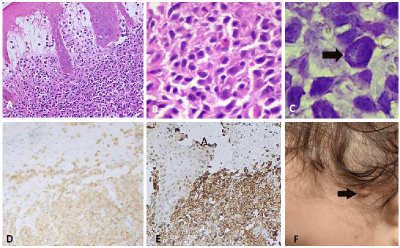
Skin biopsy of the papular lesion revealed prominent infiltration by mononuclear cells in the upper dermis reaching the dermo-epidermal junction. These cells had pale round to oval nuclei and moderate eosinophilic cytoplasm with distinct cytoplasmic borders. A few cells had characteristic peripheral cytoplasmic basophilic hue. Also seen in the dermis were lymphoid aggregates and few scattered eosinophils around the dermal blood vessels. Staining with toluidine blue stain revealed characteristic metachromatic cytoplasmic granules in a few cells. Immunohistochemical studies revealed that the mononuclear cells were positive for CD45, CD117 and CD30 and were negative for CD68, S100 protein, CD2, CD3, CD1a, CD138 and chromogranin. This immune profile was consistent with mast cells. Mib-1 labeling index was low (~1%). A diagnosis of cutaneous mastocytosis was clinically consistent with maculopapular cutaneous mastocytosis. A bone marrow aspiration was not performed Figure 1.
Discussion
Mastocytosis is a heterogeneous group of diseases characterized by the abnormal infiltration of mast cells in the skin and sometimes other organs and the simultaneous release of chemical mediators by these cells [3]. The organ most commonly involved in mastocytosis is the skin. Cutaneous mastocytosis is classified according to clinical presentation and by the onset of disease [4]. Rather than a true neoplasm, it is a hyperplastic response to abnormal stimuli. CM is a relatively uncommon condition in pediatric dermatology. Its prevalence in the USA ranged from 1: 1000 to 1: 8000 dermatology patients [5]. Accurate epidemiological data on the Indian population is not available [6].
Distribution of lesions: Most lesions are distributed over the trunk in patients with maculopapular cutaneous mastocytosis (urticaria pigmentosa) [7]. Nettleship first described the typical lesions of mastocytosis as an unusual form of urticaria. The clinical diagnostic criteria for cutaneous mastocytosis are enumerated in Table 1 [8]. A positive Darier’s sign or compatible histology is required for confirmation of the diagnosis. Although Darier’s sign is a highly specific clinical manifestation, its absence does not rule out mastocytosis as its positivity rate is variable and ranges from 88% to 92% of cases [5]. Routine hemogram findings in cutaneous mastocytosis are usually normal or may be associated with eosinophilia. Serum tryptase levels are usually obtained at the initial evaluation. Serum tryptase value of ≥20 ng/mL is one of the minor diagnostic criteria for diagnosing systemic disease based on guidelines developed in the adult population. An increased serum tryptase level is reported to help predict children at risk for episodes of severe mediator release. The serum tryptase level is a diagnostic marker and reflects the burden of (neoplastic) mast cells in mastocytosis [8]. In most cases of cutaneous mastocytosis, normal or near-normal serum tryptase levels are found. The mean tryptase level is significantly higher in patients diagnosed with diffuse cutaneous mastocytosis than in those with other forms of cutaneous mastocytosis [9]. Adult-onset mastocytosis and associated hematological manifestation are associated with activating point mutation at codon 816 of the growth factor receptor c-kit. However, this mutation seems to be lacking in most pediatric and familial mastocytosis and may represent a clonal disease that may have different mutations from that in adulthood. Activating c-kit mutation enhances mast cell proliferation and oncogenic transformation [5,10]. Clinically, SCORing index Of MAstocytosis (SCORMA) and serum tryptase levels are helpful in determining the patients’ prognosis [11].
In children, cutaneous mastocytosis follows a benign and self-limiting clinical course and rarely remains active through adolescence, posing the risk of systemic disease in adult life [2].
Histologically, there is a dermal infiltrate of mononuclear cells with abundant eosinophilic cytoplasm, distinct cytoplasmic boundaries, and large round, pale nuclei. They are usually present in the papillary dermis. Eosinophils are also often present. Edema of the papillary dermis and subepidermal vesiculation can be seen. Intraepidermal accumulation of melanin pigment is also seen in many cases [12,13]. A possible morphological differential diagnosis of cutaneous mastocytosis in the pediatric population is histiocytic diseases. These may be Langerhan cell histiocytosis or non-Langer cell histiocytosis (eg. Benign cephalic histiocytosis). Basic dyes like Giemsa, toluidine blue, Astra blue, etc, can be used to highlight the metachromatic granules of mast cells but they can be sometimes positive in histiocytes [14]. Immunohistochemical stains also aid in the diagnosis. Mature mast cells are positive for CD29, CD33, CD34, CD45, CD61, CD117, CD30 etc. Immature or neoplastic mast cells also express CD2, CD13, and CD25. Mast cells are negative for CD1, CD1a, CD8, CD10, CD17, CD19, and CD24. All normal and neoplastic mast cells co-express CD117 and tryptase [1,12,16]. Tryptase staining by IHC is a superior marker to metachromatic staining with toluidine blue, which can also stain macrophages. Aberrantly expressed IHC markers by mast cells are CD14, CD68 (Monocytic/histiocytic markers), CD63, CD203c (basophilic leukemia), CD33, MPO (myeloid), CD2 (T-cells), etc [17].
Treatment and follow-up: Generally, no treatment or only topical corticosteroid treatment is needed for cutaneous mastocytosis. Bone marrow examination is not recommended in the pediatric population with isolated cutaneous mastocytosis. In patients with only cosmetic complaints, no therapy or topical corticosteroid therapy is usually given in children older than two years. For patients of cutaneous mastocytosis with complaints of itch, redness, and swelling, avoidance of food that provokes the lesions and systemic therapy (H1 and H2 blockers) is given. In isolated cutaneous mastocytosis, annual follow-up checkups and telephone consultations once every six months are recommended [15,16].
Conclusion
The present case of mastocytosis had negative Darier’s sign and normal serum tryptase level. Skin biopsy and IHC staining aided in confirmation of diagnosis. A good clinicopathologic correlation and a high index of suspicion helped in arriving at the diagnosis.
- Campo E, Harris NL (2017) WHO Classification of Tumours of Haematopoietic and Lymphoid Tissues. United States: International Agency for Research on Cancer. Link: https://bit.ly/2YlYgJP
- Castells M, Metcalfe DD, Escribano L (2011) Diagnosis and treatment of cutaneous mastocytosis in children. Am J Clin Dermatol 12: 259-270. Link: https://bit.ly/3uI4wHu
- Valent P, Horny HP, Escribano L, Longley BJ, Li CY, et al. (2001) Diagnostic criteria and classification of mastocytosis: a consensus proposal. Leuk Res 25: 603-625. Link: https://bit.ly/3Ag4zeA
- Wolff K, Komar M, Petzelbauer P (2001) Clinical and histopathological aspects of cutaneous mastocytosis. Leuk Res 25: 519-528. Link: https://bit.ly/3Bg1rRp
- Kiszewski AE, Duran‐Mckinster C, Orozco‐Covarrubias L, Gutierrez‐Castrellon P, Ruiz‐Maldonado R (2004) Cutaneous mastocytosis in children: a clinical analysis of 71 cases. J Eur Acad Dermatol Venereol 18: 285-290. Link: https://bit.ly/3oBBurX
- Srinivas SM, Dhar S, Parikh D (2015) Mastocytosis in children. Indian Journal of Paediatric Dermatology 16: 57-63. Link: https://bit.ly/2YpCILJ
- Ben-Amitai D, Metzker A, Cohen HA (2005) Pediatric cutaneous mastocytosis: a review of 180 patients. Isr Med Assoc J 7: 320-322. Link: https://bit.ly/3DfjtUn
- Carter MC, Clayton ST, Komarow HD, Brittain EH, Scott LM, et al. (2015) Assessment of clinical findings, tryptase levels, and bone marrow histopathology in the management of pediatric mastocytosis. Journal of Allergy and Clinical Immunology 136: 1673-1679. Link: https://bit.ly/3FkVKUE
- Sperr WR, Jordan JH, Fiegl M, Escribano L, Bellas C (2002) Serum tryptase levels in patients with mastocytosis: correlation with mast cell burden and implication for defining the category of disease. Int Arch Allergy Immunol 128: 136-141. Link: https://bit.ly/3acEh2q
- Lange M, Nedoszytko B, Górska A, Żawrocki A, Sobjanek M, et al. (2012) Mastocytosis in children and adults: clinical disease heterogeneity. Arch Med Sci 8: 533-541. Link: https://bit.ly/3Bh6otc
- Heide R, van Doorn K, Mulder PG, van Toorenenbergen AW, Beishuizen A, et al. (2009) Serum tryptase and SCORMA (SCORingMAstocytosis) Index as disease severity parameters in childhood and adult cutaneous mastocytosis. Clin Exp Dermatol 34: 462-468. Link: https://bit.ly/3Fn7XIr
- Hale CS, Hamodat M (2021) Cutaneous mastocytosis. Link: https://bit.ly/3iBMDFz
- Lever WF (2009) Lever's Histopathology of the Skin. Argentina: Wolters Kluwer Health. Link: https://bit.ly/3iwZbxR
- Christopher L, John BD, Suvarna KS (2012) Bancroft's Theory and Practice of Histological Techniques. United Kingdom: Elsevier Health Sciences.
- Czarny J, Lange M, Ługowska-Umer H, Nowicki RJ (2018) Cutaneous mastocytosis treatment: strategies, limitations and perspectives. Postepy Dermatol Alergol 35: 541-545. Link: https://bit.ly/3laOv9L
- Heide R, Beishuizen A, De Groot H, Den Hollander JC, Van Doormaal JJ, et al. (2008) Mastocytosis in children: a protocol for management. Pediatr Dermatol 25: 493-500. Link: https://bit.ly/3uJUuWh
- Horny HP, Sotlar K, Valent P (2014) Mastocytosis: immunophenotypical features of the transformed mast cells are unique among hematopoietic cells.Immunology and allergy clinics of North America 34: 315-321 Link: https://bit.ly/2YgY5yV

Article Alerts
Subscribe to our articles alerts and stay tuned.
 This work is licensed under a Creative Commons Attribution 4.0 International License.

 Save to Mendeley
