Archive of Urological Research
Clinical application of PCNL in oblique supine lithotomy position for upper urinary calculi
Hai Yang Zhou#, Xin Feng Chen#, Hua Zhu, Dong Hua Gu, Xiao Dong Pan and Bing Zheng*
#These authors contributed equally to this work.
Cite this as
Zhou HY, Chen XF, Zhu H, Gu DH, Pan XD, et al. (2021) Clinical application of PCNL in oblique supine lithotomy position for upper urinary calculi. Arch Urol Res 5(1): 013-019. DOI: 10.17352/aur.000031Objective: To investigate the effect of PCNL (Percutaneous Nephrolithotomy) in the treatment of upper urinary tract calculi in oblique supine lithotomy position and prone position.
Methods: Retrospectively collected data from 184 patients and 80 patients who underwent PCNL in prone and oblique supine lithotomy position respectively in our hospital from 2016 to 2019. The intraoperative and postoperative observation indicators were compared to evaluate the advantages and disadvantages of oblique supine lithotomy position.
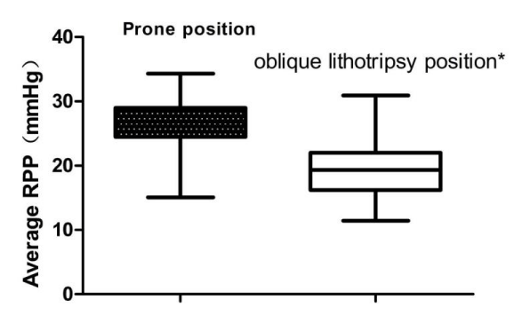
Results: A total of 264 patients were recruited, PCNL in the treatment of upper urinary tract calculi in oblique supine lithotomy position (n=80) compared to prone position (n=184) had significantly shorter operation time (49.03±12.64min, 62.14±14.82min, P=0.032), higher stone removal rate in first week (82.5%>69.0%,P=0.023), lower Hb (Hemoglobin) reduction (9.94±3.62,12.83±4.01,P<0.01) and lower possibility of fever (8.7%,25.5%,P=0.002); patients who underwent PCNL in oblique supine lithotomy position had significantly lower renal pelvis pressure(18.70 ± 4.06mmHg, 26.28 ± 3.42mmHg, P <0.01)AT the same time, PCT (Postoperative inflammatory markers) were significantly increased when intraoperative renal pelvis hypertension occurred and the duration was more than 5min
Conclusion: The oblique supine lithotomy position was associated with shorter operation time, lower renal pelvis pressure, incidence of postoperative related complications and combination of percutaneous nephroscope with ureteroscope under special condition, which is of great significance.
Abbreviations
RPP: Renal Pelvic Pressure; PCNL: Percutaneous Nephrolithotomy; ESWL: Extracorporeal Shock Wave Lithotripsy; SIRS: Systemic Inflammatory Response Syndrome; BMI: Body Mass Index; CT: Computed Tomography; PCT: Procalcitonin; Hu: Housefield unit; Hb: Hemoglobin
Introduction
In 1976, when Fernstrom and Johansson successfully removed kidney stones through percutaneous renal channels for the first time, PCNL gradually became the gold standard for the treatment of kidney stones ≥2cm in diameter [1]. The oblique supine lithotomy position was introduced by Ibarluzea In 2007 [2], compared with the traditional prone position, which confirmed with higher stone removal rate [3-5]. The purpose of this study is to compare the treatment effect of prone position and oblique supine lithotomy position in the treatment of upper urinary tract stones with PCNL by retrospective study. Related studies are helpful to evaluate the advantages of oblique supine lithotomy position in the application of PCNL. Our hospital has performed PCNL for oblique lithotripsy for upper urinary calculi since 2016, and has achieved good results. It is reported as follows.
Object
General information: Data of 264 patients who underwent PCNL surgery in our hospital from September 2016 to June 2019 were retrospectively collected, including 184 patients in the prone position group and 80 patients in the oblique supine lithotomy position group.
Inclusion criteria: (1) The patient was diagnosed with kidney stones or kidney stones combined with ureteral stones by B-ultrasound, KUB, CT or IVP; (2) Unilateral kidney stones, stone length ≥ 2cm, single or multiple, located above the junction of waist 2 ureter and renal pelvis; (3) After the application of empirical antibiotics with (or without), two consecutive urine cultures are negative.
Exclusion criteria: (1) Severe cardiopulmonary dysfunction or tumors in other organs; (2) Pregnant women; (3) Horseshoe kidney and duplication kidney; (4) A history of open kidney surgery; (5) Active urinary tract infection (the effect of sensitive antibiotic treatment is not good); (6) Coagulation dysfunction due to poor drug control; (7) The glomerular filtration rate of the affected side is <10mL/min.
The patients with urinary tract infection were treated with sensitive antibiotics according to the results of urinary culture. B ultrasound, KUB + IVP or retrograde contrast, dual energy CT were used to determine the location and size of stones.
Operative procedure
All patients underwent PCNL surgery, which was performed by the same surgical team led by the same surgeon, and also by the same urology specialist surgical nurses and anesthesiologists.
Method
Prone position PCNL
After general anesthesia, the lithotomy position was taken, and a zebra safety guide wire was placed under the cystoscope. An F6 ureteral catheter was placed along the guide wire. Select the 11th intercostal space or the 12th lower costal space, between the posterior axillary line and the scapular line, and perfuse normal saline with a 50ml syringe retrogradely through an F6 ureteral catheter, resulting in "artificial hydronephrosis". Under the guidance of B-ultrasound, the 18G renal puncture needle punctured the renal calyceal dome precisely, and the clear liquid outflow indicated that the puncture was successful (a lot of blood liquid indicated that the puncture failed, and a second puncture was needed). The "J" guide wire was placed into the renal collection system through the needle sheath, and the fascial expander was gradually expanded to F20, and the working sheath was retained. Nephroscope was entered into the renal collecting system through the channel to look for stones. After smashing the stones with holmium laser, the stones were flushed out through the perfusion fluid, and the larger stones were taken out in the stone basket. F4.8 double J tube and F16 nephrostomy tube were routinely indwelling Figure 1.
Oblique supine lithotomy position PCNL
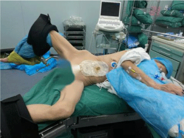
The lower torso of the supine position is turned to the opposite side, the body is turned into a healthy side by the waist cushion, the back is close to the edge of the bed, the shoulders are fixed with shoulder rests, and the cushions are placed behind the hips. Tilt back 45 ° to the operating table 3L washing liquid was placed on the abdomen to reduce the lateral displacement of the patient. The lumbar bridge was adjusted to make the waist show a folding knife position. The puncture area of the patient side was exposed. The patient side lower limb flexed knee adduction was slightly abducted and fixed on the foot rest. The healthy side lower limb flexed knee was placed on the horizontal plate of the abduction. The chest and abdomen were in oblique supine lithotomy position, while the lower limbs and buttocks were in rotation 90° lithotomy position. Under the guidance of color ultrasound, puncture was performed in the posterior line of 12 subcostal axilla, the puncture Angle was 12°~25° from the ventral Angle perpendicular to the skin, the puncture target was located in the middle or lower calyces of the affected kidney, and the renal puncture channel was established in the same prone position. F4.8 double J tube and F16 nephrostomy tube were routinely indwelling.
Intraoperative pressure monitoring of the renal pelvis
By placing the pressure sensor (Pressure Sensor (Beijing Jishiba Medical Devices Co., Ltd. ICU Medical Model 42584-05)) in the renal pelvis along the puncture sheath through the extension tube, it is fixed in the plane of the renal pelvis. After the calibration and adjustment, the invasive blood pressure measurement channel connected to the ECG monitor (Shanghai Hanfei Medical Devices Co., Ltd. Delgde State BSM3500) is connected. Data collection software was used to input the intraoperative data into the database in real time, and then SPSS was used for retrospective statistics to calculate the intraoperative mean renal pelvic pressure.
During the operation, two ureteroscopy combined with ureterolithiasis
retrograde ureteroscopy: COOK F16 ureteroscopy dilatation sheath was placed along the guide wire, and Olympus ureteroscopy was placed for lithotripsy; anterograde ureteroscopy: ureteroscopy was used to look for residual stones in the upper ureter from the calyx through the renal puncture channel. Holmium laser stone fragmentation, through the channel out or quarrying nets to remove gravel.
Observation data
Intraoperative: observe and record the operation time, intraoperative pressure of renal pelvis, Hemoglobin (Hb) changes before and after operation, hospital stay and other indicators of the two groups. KUB or CT was reviewed on the same day and one month after the operation, and every three months thereafter, no stones remained if the stones were ≤3mm. If there are significant residual stones after surgery, consider the second stage PCNL or ESWL assisted stone removal depending on the situation and the patient's intention.
Postoperative: observed the incidence of postoperative complications (including fever, blood transfusion, bleeding, difficult to control, need to undergo vascular embolization, sepsis, etc.) in the two groups; Postoperative day1 PCT changes;Postoperative stone removal rate (SFR) and secondary residual stone treatment.
Statistical analysis
SPSS 20.0 software is used for statistical analysis. The measurement data is represented by mean ± standard deviation (). The comparison between groups is performed by t-test. If the variance is not uniform, the corrected t-test (t-test) is used. Counting data are expressed by the number of cases and percentages. Chi-square test is used for comparison between groups. According to the theoretical frequency, Pearson chi-square or Fisher exact test is selected. Statistical significance was found at p <0.05.
Result
General information
In terms of average age, the prone position group was 49.6 ± 12.9 years old and the oblique supine lithotomy position group was 52.3 ± 13.4 years old (P = 0.425). The vast majority of patients in both groups were male (P = 0.196) and had no BMI. Statistical differences (P = 0.283). Stone characteristics of the two groups of patients: there were no statistical differences in preoperative eGFR, stone distribution, and degree of hydronephrosis. The average diameter of the two groups of stones was 2cm. The oblique supine lithotomy position group: 21.24 ± 4.11mm, the prone position group: 20.85 ± 4.30mm, P = 0.499); (Table 1 for details).
Intraoperative and postoperative data
There was no significant difference between the two groups in intraoperative channel establishment time and puncture channel number (P>0.05). The operation time in the oblique supine lithotomy position was significantly lower than that in the prone group (49.03±12.64 in the oblique supine lithotomy position and 62.14±14.82 in the prone group, P=0.012). There was no significant difference in postoperative complications except for the patients with postoperative fever (>38.50C) in the prone position group and the oblique lithotomy group (7/80,47/184,P<0.01). The length of postoperative hospital stay was lower in the oblique ankylotomy group than in the prone position group, and the difference was statistically significant (6.32±2.54, 8.94±3.02, P=0.02). The Renal Pelvis Pressure (RPP) in the oblique supine lithotomy position was significantly lower than that in the prone position (18.70±4.06mmHg, 26.28±3.42mmHg, P <0.01); The Hb reduction after operation in prone position wsa higher (9.94 ± 3.62g / L, 12.83± 4.01, P <0.01); On the first week after the operation, the stone removal rate in the group with oblique supine lithotomy position was significantly higher than that in the group with prone lithotomy, with statistical difference (82.5% vs 69.0%, P=0.023); Patients with residual stones were treated with ESWL (23/80,59/184,P>0.05) or two-stage percutaneous nephrolithotomy>5/80,24/184,P>0.05. Finally, there was no significant difference in stone clearance rate between the two groups after 1 month (Table 2 for details).
Renal pelvis pressure
The RPP transient ≥30mmHg occurred in two groups were 152 and 32 cases, and ≥40mmHg were 115 cases and 28 cases, respectively. In both groups, the RPP was ≥30mmHg during the operation, and the total time was> 5min. There were 135 and 22 cases, respectively. There were 109 cases and 17 cases with total time> 5min (Table 3 for details).
Renal pelvis hypertension and Postoperative day1 PCT (procalcitonin) change
Considering intraoperative renal pelvis hypertension alone, there was no statistically significant difference in postoperative day1 PCT changes between patients in the high pressure group (RPP>30mmHg or>40mmHg Group) and the low pressure group (RPP>30mmHg Group, P>0.05, RPP>40mmHg Group, P>0.05). ; When the total duration of intraoperative renal pelvis hypertension (≥30mmHg) ≥5min, the postoperative day1 PCT was significantly higher than that of patients with transient renal pelvis hypertension (total duration <5min) (RPP>30mmHg Group, P <0.01,RPP>40mmHg Group,P<0.001) (Table 4 for details).
Discussion
Although the incidence of complications of PCNL has been greatly reduced compared with traditional open surgery, there are still risks of fever, sepsis, renal hemorrhage, rupture of pleura and lung, and intestinal injury. Sirs [6] has been reported in 28% of PCNL patients. Closely related factors include the presence of infectious stones, operation time, and stone load, etc. As a controllable risk factor, RPP has gradually been valued by urologists. Under normal physiological conditions, the RPP fluctuates from 1.47 to 4.11 mmHg [7].In some special cases, high pressure of renal pelvis may cause serious consequences of death [8]. Therefore, it is of great significance to control PCNL intraoperative renal pelvic pressure. In this study, the proportion of mean pyelic pressure and pyelic hypertension under oblique obviotomy was lower than that in prone position, with statistical significance.
Recent studies have shown that intraoperative RPPs above 30mmHg are closely related to complications caused by pelvic venous reflux [9], especially when patients are complicated by urinary tract infections. High RPP can cause tears in the pyloric dome (that is, the weak anatomical part of the puncture path), increasing the risk of bleeding. At the same time, high RPP will be absorbed through the renal pelvis and lymphatic reflux, and hemodynamic changes cause circulation Overload, when combined with urinary tract infection, bacteria and endotoxin enter the blood, causing postoperative fever and sepsis. Moreover, in vitro study showed that the pressure of renal pelvis above 30mmhg (3.99kpa) for more than 50s would significantly increase the postoperative fever rate [10]. In this study, the intraoperative renal pelvis hypertension the total time was less than 5min; Only when the intraoperative renal pelvis hypertension with total time is more than 5min, the difference of postoperative fever between the two groups is statistically significant, and the persistent high pressure of the renal pelvis is the key factor leading to the reflux of the renal pelvis vein and the postoperative complications.
There was no significant difference in postoperative complications except the number of postoperative fever (>38.50C) patients. Related studies have shown that PCT, as an inflammatory index of early elevation (the first day after operation), has a better predictive effect on the development tendency of sepsis [11-13]. When intraoperative pyel hypertension (≥30 mmHg or 40 mmHg) and duration of more than 5 min, the PCT value of the first day after operation was significantly lower than that of the prone group; When the duration of renal pelvis hypertension was less than 5 min, there was no significant difference in PCT value between the two groups. Considering that transient renal pelvis hypertension was not enough to cause sufficient perfusion fluid reflux, the immune system could still resist after operation.
Factors that affect the pressure of the renal pelvis during the operation include the number of puncture channels, the accuracy of the positioning of the renal pelvis system, the position during the operation, the lithotripsy method, and the inner diameter of the scope sheath. In this study, the RPP was significantly lower in the oblique supine lithotomy position than in the prone position and was statistically significant. The following factors may be considered: under the oblique anterotomy, the puncture channel is usually lower than the renal pelvis plane, which is conducive to the outflow of intraoperative lavage; Relevant studies have shown that distal ureteral obstruction may lead to a sharp increase in intraoperative renal pelvic pressure [14], resulting in renal unit damage and reduction. Therefore, maintaining the patency of the lower urinary tract through intraoperative retrograde ureteroscopy can avoid the accumulation of lithotripsy in the lower urinary tract during percutaneous nephroscopy, resulting in high secondary RPP. The lack of abdominal organ compression reduces renal pelvic pressure at the oblique anteriorite position;
At the same time, the early control of intraoperative perfusion through the syringe has great randomness and uncertainty. Excessive perfusion flow will cause renal pelvis hypertension and bacterial reflux such as the circulatory system [9,15], resulting in urinary sepsis and renal impairment [16]. Or the intraoperative perfusion pressure can be adjusted by adjusting the height of 3L bag of perfusion fluid. Although the stability of intraoperative perfusion pressure is ensured to some extent, there is still a lack of specific quantitative indicators, and the effect fluctuates greatly before and after. In this study, the perfusion pump was used to precisely control the low-pressure perfusion water flow, to ensure the stability of the pressure of the renal pelvis during the operation, and to minimize the transient high pressure of the renal pelvis during the operation.Meanwhile, low intraoperative RPP should also be considered, that is, insufficient perfusion leads to unclear intraoperative visual field and lack of compression effect leads to increased intraoperative blood loss. In this study, the RPP at the oblique avulsion position was lower than that in the traditional prone position.
Intraoperative percutaneous nephroscope combined with ureteroscope in special cases
Studies have shown that the single stone removal rate for staghorn stones treated with PCNL is only 56% [17]. Multistage PCNL is associated with an increased risk of related complications [18,19]. Soft ureteroscopes have been proven to be safe and effective in treating upper urinary calculi ≤ 2 cm. A meta-analysis showed that the soft mirror can also achieve satisfactory results in the treatment of urinary calculi > 2cm [20]. Conventional prone position cannot be used to treat ureteral calculi in one stage, especially when contralateral ureteral calculi are combined. However, oblique anteropsis can be used to treat ureteral calculi in one stage by retrograde ureteroscopy without changing body position. At the same time, retrograde ureteroscopy can actively expand the ureter to avoid the probability of "stone street" forming in the lower segment of ureter after the operation. Retrograde ureteroscopy can be used when the percutaneous passage is not well established. When there are cast stones in the renal pelvis and the establishment of artificial hydronephrosis is not smooth, the retrograde ureteroscopy can be first used to crush to a small volume and partially assist the establishment of puncture channels.Intraoperative combination of percutaneous nephroscope and ureteroscope can verify the residual stones with each other, the surgern can remove them promptly during the operation, and increase the Stone Free Rate (SFR)after operation Figure 2.
The operation time is directly related to the incidence of postoperative complications. The longer the operation time, hypothermia, increased pelvic venous return, the effects of the circulatory system, and the risk of anesthesia may occur. In this study, the operation time of the oblique supine lithotomy position was significantly lower than that of the prone position. It was considered that the oblique supine lithotomy position was maintained during the operation without the change of body position; the puncture channel was often lower than the horizontal plane of the renal pelvis, and the gravity effect + irrigation during the operation was convenient for the removal of gravel, which improved the efficiency of stone removal in disguise. Ureteral calculi can be treated simultaneously by the combination of two mirrors (ureteroscopy and nephroscope) at the oblique invert site. ureteroscope can be used to observe the conditions of the renal pelvis and calyx first, to guide the establishment of puncture channels in percutaneous nephroscopy, and even to observe the puncture process directly, reducing the probability of puncture failure Figure 3.
With the maturity of PCNL technology and the continuous update of equipment, combined with the rich surgical experience of PCNL, postoperative complications can be reduced.It is not difficult to find that the group of oblique supine lithotomy position is at the level of low-pressure perfusion, and the incidence of complications above grade II is only (10/ 80, 12.5%), which reflects the safety of oblique supine lithotomy position from the side. With the rise of ultra-micro percutaneous nephroscope (SMP) in recent years, the narrower endoscopic sheath will lead to the increase of the pressure of the renal pelvis during the operation. In combination with the characteristics of low pressure of the renal pelvis in this position, it can better reflect the advantages of oblique supine lithotomy position.
Deficiencies
The sample size in this study is small; Since the incisive-puncture channel under the oblique anaposyte is far away, it is necessary to grasp the vascular area of the kidney more accurately in the process of establishing the channel. Real-time three-dimensional color ultrasound during the operation is needed to improve the vascular identification rate and thus reduce the probability of intraoperative bleeding. At the same time, the puncture channel is longer, and the scope of the lens swing is limited during operation. A small amplitude of the lens swing may cause the puncture point to deviate from the predetermined target. In addition, compared with the prone position of abdominal cushions and compression of abdominal organs, kidney mobility in the oblique supine lithotomy position is greater than that of the prone position, which makes it difficult to accurately locate the entire puncture, and it requires more accumulation of puncture experience, extending the learning curve. Real-time color Doppler ultrasound tracking is needed to clarify the differences in renal activity during different positions to guide the establishment of percutaneous channels during the operation.
Conclusion
Oblique supine lithotomy position can reduce the pressure of renal pelvis during operation and improve the rate of postoperative stone removal (SFR). For special cases, especially the complicated upper urinary calculi of renal calculi with ureteral calculi, it can realize the combination of double mirror (percutaneous nephroscope + ureteroscope) lithotripsy, significantly reduce the operation time, thus, reduce the amount of blood loss during operation and reduce the incidence of postoperative complications.
Ethical standard
Ethical and regulatory approvals were sought and obtained from the Second Affiliated Hospital of Nantong University, China.
The research project was funded by the Health and Family Planning Commission of Nantong City, Jiangsu Province with a major scientific fund number of WKZL2018022. /p>
Authors’ contribution
HaiYang Zhou1#: Project development, Data Collection, Data analysis,Manuscript writing
XinFeng Chen2#: Project development, Data Collection, ata analysis
Bing Zheng*: Project development,Surgical operation, Data analysis,Manuscript editing
Hua Zhu3: Data Collection, Data analysis
Donghua Gu4 :Data Collection, Data analysis
Xiaodong Pan 5: Data Collection, Data analysis
- Fernstrom I, Johansson B (1976) Percutaneous pyelolithotomy. A new extraction technique. Scand J Urol Nephrol 10: 257-259. Link: https://bit.ly/3gwd5jk
- Ibarluzea G, Scoffone CM, Cracco CM, Poggio M, Porpiglia F, et al. (2007)Supine Valdivia and modified lithotomy position for simultaneous anterograde and retrograde endourological access. BJU Int 100: 233-236. Link: https://bit.ly/32u5xWh
- Guanghua P, Leming S, Donghua X, Huang J, Zhong Y, et al. (2018)Suctioning flexible ureteroscopic lithotripsy in the oblique supine lithotomy position and supine lithotomy position: a comparative retrospective study. Minerva Urol Nefrol 70: 612-616. Link: https://bit.ly/3n3bP8D
- Leng S, Xie D, Zhong Y, Huang M(2018)Combined Single-Tract of Minimally Percutaneous Nephrolithotomy and Flexible Ureteroscopy for Staghorn Calculi in Oblique Supine Lithotomy Position. Surg Innov 25: 22-27. Link: https://bit.ly/3gnknpN
- Al-Dessoukey AA, Moussa AS, Abdelbary AM, Zayed A, Abdallah R, et al.(2014 Percutaneous nephrolithotomy in the oblique supine lithotomy position and prone position: a comparative study. J Endourol 28: 1058-1063. Link: https://bit.ly/2QjXDfJ
- Lojanapiwat B, Kitirattrakarn P(2011)Role of preoperative and intraoperative factors in mediating infection complication following percutaneous nephrolithotomy. Urol Int 86: 448-452. Link: https://bit.ly/3sBpODQ
- Djurhuus JC, Frokjaer J, Munch Jorgensen T, Knudsen L, Pham T, et al. (1990)Regulation of renal pelvic pressure by diuresis and micturition. Am J Physiol 259: R637- R644. Link: https://bit.ly/2P4MhM3
- Özlem T, Hüsnü T, Ilker Ü, Voyvoda N, Şerifoğlu I(2013)Changes in renal Doppler ultrasonographic parameters in patients managed with rigid ureteroscopy. Acta Radiol 54: 327-332. Link: https://bit.ly/3apdel2
- Wen Z, Guohua Z, Kaijun W, Li X, Chen W, et al. (2008)Does a smaller tract in percutaneous nephrolithotomy contribute to high renal pelvic pressure and postoperative fever?. J Endourol 22: 2147-2151. Link: https://bit.ly/3glgUIl
- Loftus CJ, Hinck B, Makovey I, Sivalingam S, Monga M, et al.(2018)Mini Versus Standard Percutaneous Nephrolithotomy: The Impact of Sheath Size on Intrarenal Pelvic Pressure and Infectious Complications in a Porcine Model. J Endourol 32: 350-353. Link: https://bit.ly/3dx2RgU
- Mat-Nor MB, Md Ralib A, Abdulah NZ, Pickering JW 2016)The diagnostic ability of procalcitonin and interleukin-6 to differentiate infectious from noninfectious systemic inflammatory response syndrome and to predict mortality. J Crit Care 33: 245-251. Link: https://bit.ly/3tAuX0r
- Qi T, Lai C, Li Y, Chen X, Jin X(2021)The predictive and diagnostic ability of IL-6 for postoperative urosepsis in patients undergoing percutaneous nephrolithotomy. Urolithiasis. Link: https://bit.ly/3glnuhW
- Takahashi W, Nakada TA, Yazaki M, Oda S(2016)Interleukin-6 Levels Act as a Diagnostic Marker for Infection and a Prognostic Marker in Patients with Organ Dysfunction in Intensive Care Units. Shock 46: 254-260. Link: https://bit.ly/32vswjG
- Suki W, Eknoyan G, Rector FC, Seldin DW(1996)Patterns of nephron perfusion in acute and chronic hydronephrosis. J Clin Invest 45: 122-131. Link: https://bit.ly/3v4I22l
- Troxel SA, Low RK(2002)Renal intrapelvic pressure during percutaneous nephrolithotomy and its correlation with the development of postoperative fever. J Urol 168: 1348-13151. Link: https://bit.ly/3dvdS2a
- Christian S, Sero A, Dalibor P, Popiolek M, Celia A, et al. (2018)Worldwide Use of Antiretropulsive Techniques: Observations from the Clinical Research Office of the Endourological Society Ureteroscopy Global Study. J Endourol 32: 297-303. Link: https://bit.ly/3n3f7sG
- Sachin P, Apul G, Rahul Y, El-Nahas R, et al.(2012)Factors affecting stone-free rate and complications of percutaneous nephrolithotomy for treatment of staghorn stone. Urology 80: 960-961. Link: https://bit.ly/3nabf9q
- Senocak C, Ozbek R, Bozkurt OF, Unsal A(2018)Predictive factors of bleeding among pediatric patients undergoing percutaneous nephrolithotomy. Urolithiasis 46: 383-389. Link: https://bit.ly/3akGkC3
- Ramón de Fata F, Pérez D, Resel-Folkersma L, Galán JA, Serrano A, et al. (2013)Analysis of the factors affecting blood loss in percutaneous nephrolithotomy: a registry of the Spanish Association of Urology in the supine position. Actas Urol Esp 37: 527-532. Link: https://bit.ly/3dxuM0h
- Aboumarzouk OM, Monga M, Kata SG, Traxer O, Somani BK(2012) Flexible ureteroscopy and laser lithotripsy for stones >2 cm: a systematic review and meta-analysis. J Endourol 26: 1257-1263. Link: https://bit.ly/32umVtX
Article Alerts
Subscribe to our articles alerts and stay tuned.
 This work is licensed under a Creative Commons Attribution 4.0 International License.


 Save to Mendeley
