Open Journal of Orthopedics and Rheumatology
Chondrosarcoma of the right iliopubic branch incidentally discovered: About a case
Youness Mokhchani1,2*, Abdelhay Rabbah1, Abderrafia Rachdi1,2, Jalal Boukhriss1,2, Bouchaib Chafry1,2 and Mustapha Boussouga1,2
2Faculty of Medicine and Pharmacy, Mohammed V University -Rabat- 10000, Morocco
Cite this as
Mokhchani Y, Rabbah A, Rachdi A, Boukhriss J, Chafry B, et al. (2022) Chondrosarcoma of the right iliopubic branch incidentally discovered: About a case. Open J Orthop Rheumatol 7(1): 008-010. DOI: 10.17352/ojor.000044Copyright License
© 2022 Mokhchani Y, et al. This is an open-access article distributed under the terms of the Creative Commons Attribution License, which permits unrestricted use, distribution, and reproduction in any medium, provided the original author and source are credited.Chondrosarcoma is the most common primary malignant bone tumor after myeloma and osteosarcoma, ranking third. It may be in second place in terms of frequency after osteosarcoma if we consider myeloma as a tumor of bone marrow cells rather than a bone tumor because it is a hematopoietic derivation [1]. Its incidence is 1-4 cases/100,000 in Europe [2,3]. There is a male predominance reported in the literature with a sex ratio of 2 according to Evans [4]. The age of onset is generally between 40 and 70 years [5] with an average age of 43 years according to J. Douglas [6]. The entire skeleton can be affected, but the sites most frequently affected are the pelvis in 24 to 38%, followed by the proximal femur in 16 to 27%, the ribs in 8% of cases, the proximal humerus in 9%, and the distal femur in 6% of cases [5]. Spinal localization remains rare (1 to 7%), as well as the involvement of the hands and feet (1 to 3%), the latter occurring mainly in the context of enchondromatosis or Ollier's disease [7].
We report a case of chondrosarcoma developing at the expense of the right iliopubic branch, incidentally discovered during a radiological assessment.
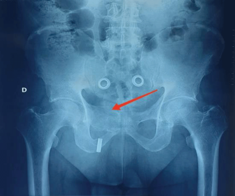
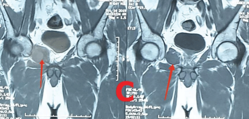
This is a 64-year-old patient, who consults for pain in the right hip occurring after a domestic accident (following a slip in the bathroom) which led to the forced separation of the thighs. In addition, the clinical examination found pain during mobilization of the hip and especially on palpation of the right groin region. In view of these clinical data, an X-ray of the pelvis was performed, which revealed an image of osteolysis located at the level of the right iliopubic branch, blowing the cortical without breaking them (Figure A). Magnetic Resonance Imaging (MRI) revealed a large polylobed tumor mass with a cartilaginous cuff with endopelvic extension very suggestive of chondrosarcoma (Figures B,C). The indication for a surgical biopsy was retained because of the radiological aspects of the tumor which are in favor of malignancy. The anatomopathological study of the biopsy specimen found elements in favor of well-differentiated chondrosarcoma of low grade of malignancy (grade I of O'NEAL and ACKERMAN). The extension assessment was without anomaly.
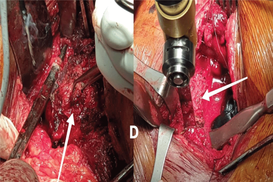
An extra-marginal resection of the tumor was performed in collaboration with a visceral surgeon. The approach was centered on the region of the right groin and directed downwards and inwards to become elective on the tumor. The dissection was careful to avoid any lesion of the femorocutaneous nerve and the external iliac vessels on the lateral side and the right spermatic cord on the medial side. Resection removed the right iliopubic branch after the protection of the endopelvic organs with a retractor (Figure D). The closure was carried out plane by plane on the suction Redon drain after placement of a wall reinforcement plate (Figure E).
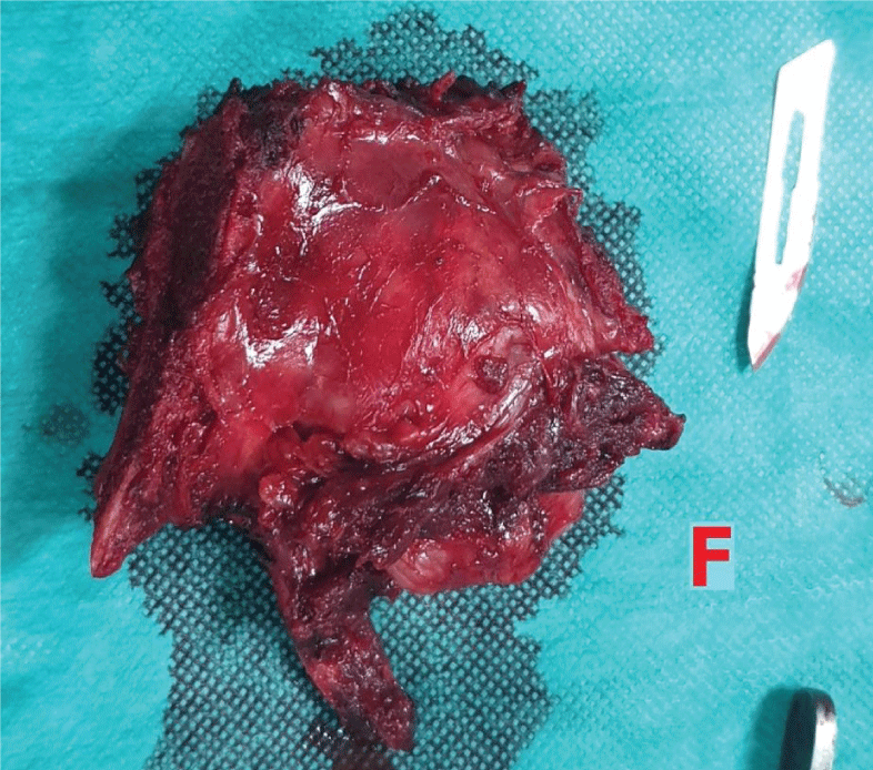
The anatomo-pathological study of the excision piece found healthy resection limits (Figure F,G), passing at least 2 cm from the caudal and left and right lateral resection margins, classified "R0" by the anatomopathologist , according to the “R” classification of the FNCLCC (National Federation of Centers for the Fight Against Cancer) [8-10]. The postoperative follow-up was uneventful, the patient's verticalization was authorized but pressure on the right lower limb was deferred to 1 month. Clinical and radiological controls after two years of follow-up did not find any recurrence.
Chondrosarcoma is a tumor that generally occurs in the pelvis [11], it is radio-chemo-resistant, its management is often surgical. The pelvis remains a difficult location since there are no major anatomical barriers to tumor extension, which means that these sarcomas produce a large painful extra-skeletal mass without other symptoms [12]. Recurrence and systemic spread often lead to poor results, and depend on several prognostic factors, such as the size of the tumor, its location, its histological grade, as well as the quality of the surgical intervention (surgical margin obtained) [11-13]. For our patient, the rather early fortuitous discovery of the tumour, its low histological grade, as well as its good extraction, were factors behind the good evolution.
- Multiple Myéloma : MSD Manuel – professional version.
- Hardes J Chondrosarcoma of bone: diagnosis and therapy. Arne Streit buerger, Georg Gosheger, Department of Orthopaedics, University Hospital of Münster, Germany.
- Rozeman LB, Hogendoorn PC, Bovée JV. Diagnosis and prognosis of chondrosarcoma of bone. Expert Rev Mol Diagn. 2002 Sep;2(5):461-72. doi: 10.1586/14737159.2.5.461. PMID: 12271817.
- Evans HL, Ayala AG, Romsdahl MM. Prognostic factors in chondrosarcoma of bone: a clinicopathologic analysis with emphasis on histologic grading. Cancer. 1977 Aug;40(2):818-31. doi: 10.1002/1097-0142(197708)40:2<818::aid-cncr2820400234>3.0.co;2-b. PMID: 890662.
- Lee FY, Mankin HJ, Fondren G, Gebhardt MC, Springfield DS, Rosenberg AE, Jennings LC. Chondrosarcoma of bone: an assessment of outcome. J Bone Joint Surg Am. 1999 Mar;81(3):326-38. doi: 10.2106/00004623-199903000-00004. PMID: 10199270.
- Pritchard DJ, Lunke RJ, Taylor WF, Dahlin DC, Medley BE. Chondrosarcoma: a clinicopathologic and statistical analysis. Cancer. 1980 Jan 1;45(1):149-57. doi: 10.1002/1097-0142(19800101)45:1<149::aid-cncr2820450125>3.0.co;2-a. PMID: 7350999.
- Ucla E, Tomeno B, Forest M. Facteurs du pronostic tumoral dans les chondrosarcomes de l'appareil locomoteur. A propos de 180 cas [Tumor prognostic factors in chondrosarcoma of the locomotor system. Apropos of 180 cases]. Rev Chir Orthop Reparatrice Appar Mot. 1991;77(5):301-11. French. PMID: 1836882.
- Enneking WF, Spanier SS, Goodman MA. A system for the surgical staging of musculoskeletal sarcoma. Clin Orthop Relat Res. 1980 Nov-Dec;(153):106-20. PMID: 7449206.
- Wittekind C, Compton CC, Greene FL, Sobin LH. TNM residual tumor classification revisited. Cancer. 2002 May 1;94(9):2511-6. doi: 10.1002/cncr.10492. PMID: 12015777.
- Tomeno B. In Forest M., Tomeno B., Vanel D. Diagnosis of tumors and pseudotumoral lesionsof bone and joint.
- Ozaki T, Hillmann A, Lindner N, Blasius S, Winkelmann W. Chondrosarcoma of the pelvis. Clin Orthop Relat Res. 1997 Apr;(337):226-39. doi: 10.1097/00003086-199704000-00025. PMID: 9137194.
- Pritchard DJ, Lunke RJ, Taylor WF, Dahlin DC, Medley BE. Chondrosarcoma: a clinicopathologic and statistical analysis. Cancer. 1980 Jan 1;45(1):149-57. doi: 10.1002/1097-0142(19800101)45:1<149::aid-cncr2820450125>3.0.co;2-a. PMID: 7350999.
- Sheth DS, Yasko AW, Johnson ME, Ayala AG, Murray JA, Romsdahl MM. Chondrosarcoma of the pelvis. Prognostic factors for 67 patients treated with definitive surgery. Cancer. 1996 Aug 15;78(4):745-50. doi: 10.1002/(SICI)1097-0142(19960815)78:4<745::AID-CNCR9>3.0.CO;2-D. PMID: 8756367.
Article Alerts
Subscribe to our articles alerts and stay tuned.
 This work is licensed under a Creative Commons Attribution 4.0 International License.


 Save to Mendeley
