Open Journal of Trauma
Biomaterials for the rehabilitation of bone and teeth tissues from the sequelae of oral trauma
Mayra de la C Pérez Álvarez1,2* and Amisel Almirall La Serna2,3
2Professor, UNESCO Chair of Biomaterials, Biomaterials Center, University of Havana, Cuba
3Senior Researcher, Biomaterials Center, University of Havana, Cuba
Cite this as
Pérez Álvarez MC, La Serna AA (2020) Biomaterials for the rehabilitation of bone and teeth tissues from the sequelae of oral trauma. Open J Trauma 4(1): 047-048. DOI: 10.17352/ojt.000032Diagnosis and treatment for traumatic dental injuries are very complex due to the multiple trauma entities represented by six luxation types and nine fracture types affecting both the primary and permanent dentitions. When further considering that fracture and luxation injuries are often combined, and can involve either dentitions, a great number of trauma scenarios may result [1]. Classifications, epidemiological, clinical and radiographic studies are considered very important to evaluate the particular case in each patient in order to correctly plan and conduct the treatments of these injuries. Therefore, even though oral-facial trauma has been extensively investigated, in vivo clinical essays are frequently needed to understand and improve the healing processes after trauma and to determine the best treatment in each case [2,3].
Traumatic dental injuries can affect soft tissues, dental organs and bone and the resulting damage depends on the severity of the injury and time elapse, among other factors [4]. Pulp tissue is one of the most commonly affected, with the outcome of pulp necrosis which could, in time, cause an osteolytic injury in the bone tissue surrounding the tooth apex.
The tortuous evolution of these teeth in endodontic treatments has made common the use of intrachannel medication with calcium hydroxide, claiming that it is essential to control infection within the necrotic system of the dental canal and favors periapical repair. This is possible due to the calcium hydroxide antibacterial action and its ability to induce the formation of hard tissue in bone and teeth. Its use also generates a mechanical apical healing barrier, seals the duct system and reduces the edema. The main inconvenient when using this material is the high degradation rate, which leads to less time of therapeutic action and therefore to a limited degree of reparation of the tissue in some pathologies [5,6].
Nowadays new methods and biomaterials for periapical lesions, root fractures and false dental canal are under study. The continuous search for biomaterials for reparation and regeneration of periapical tissues, with or without surgery, has brought as a consequence the increase and evolution of these materials. The improvement of qualities such as the bioactive ability to stimulate the mineralization of teeth, the affinity for growth factors and the property to provide a favorable environment for cells in order to promote the cicatrization of periapical tissue are among the main goals of research. In order to improve the results of the endodontic treatment the combination of different materials has been analyzed.
The Biomaterials Center of the University of Havana, Cuba, together with other international institutions, the University of Granada, Spain and the Leduc Dental Products Factory in Montevideo, Uruguay, have studied the live response of combinations of biomaterials for the endodontic treatment of traumatized teeth, including hydroxyapatite and zinc oxide.
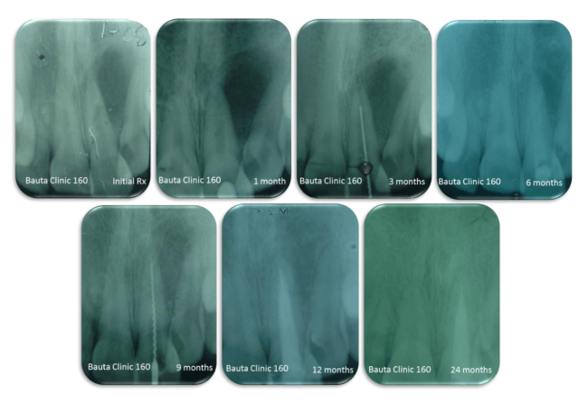
Although in clinical practice the technique of filling the root canal with calcium hydroxide is common, our group has been working in the combined use of calcium hydroxide with fine-grained hydroxyapatite (less than 0.1). A clinical study involving 11 patients with periapical lesions, which had been indicated tooth extraction in most of the cases, were treated with the intrecanal application of the material after removing the internal content of the lesion by root canal via. The clinical and radiographic responses in bone repair of periapical osteolytic lesions were evaluated and the effectiveness of treatment after 360 days reached a success rate of 91,66% [7]. The radiographic evolution of one of the patients successfully treated with this combination of biomaterials is shown in Figure 1.
The success of the procedure may be attributed to the bioactive property of hydroxyapatite to promote the mineralization of hard tissues when in contact. In this case the stability of hydroxyapatite also helps to increase the time of therapeutic action of the calcium hydroxide providing a stable support which acts as a drug release system and provides a barrier for biological fluids to access the site and dissolve the calcium hydroxide. Another advantage of this combined material is the permanence of the filler in the conduct, which extends the periods among consultations and therefore, reduces their number.
Some other materials could also been used in order to improve the therapeutic action of calcium hydroxide. Among them, zinc oxide has shown promising results and some of the encouraging experiences achieved have been reported. The remineralization ability at different root dentin regions of a hydroxyapatite-based cement containing zinc oxide was evaluated in vitro. The results showed that the mineralization occurred in all regions of the root dentin and therefore this material can be used as an endodontic sealer [8]. Another study revealed that this material also has an improved long-term sealing ability [9].
The purpose of the improvement of the biomaterials used to treat periapical lesions thought root channels should not only solve the endodontic problem, but also improve the long-term conservation of the tooth. Today at the Biomaterial Center of the University of Havana, other combinations of biomaterials are being studied for this purpose, looking for research projects to start the phase of clinical evaluation in humans, with controlled clinical trials. The final objective will be to have a product, in the medium term, with better results and superior properties for clinical use than the biomaterials that we use today.
As a conclusion, the materials studied can successfully be applied for intracanal medication to treat bone defects in periapical endodontic procedures.
- Andreasen JO, Lauridsen E, Gerds TA, Ahrensburg SS (2012) Dental Trauma Guide: a source of evidence-based treatment guidelines for dental trauma. Dent Traumatol 28: 142-147. Link: https://bit.ly/36xHOI5
- Andreasen JO, Andersson L (2011) Critical considerations when planning experimental in vivo studies in dental traumatology. Dent Traumatol 27: 275-280. Link: https://bit.ly/3jB3XJa
- Andersson L, Andreasen JO (2011) Important considerations for designing and reporting epidemiologic and clinical studies in dental traumatology. Dent Traumatol 27: 269-274. Link: https://bit.ly/2GFYous
- Cohen DS, Blanco L, Prigione C, Anaise C (2020) Traumatismos De Alto Impacto En Pacientes Con Tratamiento Ortodóncico. Rev Cir Infantil 24-34. Link: https://bit.ly/30A6hbG
- Soares IJ, Cantarini C, Miraglia Cantarini JP, Goldberg F (2018) Empleo del MTA en la obturación de perforaciones radiculares de origen iatrogénico. Rev Asoc Odontol Argent 106: 127-135. Link: https://bit.ly/3liE9lk
- Velazque Rojas L, Simões-Nogueira A, Sampaio do Vale I, Tiegui Neto V, Barreto Gonçales AG, et al. (2014) Enucleación de quiste periapical simultáneo a la obturación del sistema de conductos radiculares. Rev Cubana Estomatol 51. Link: https://bit.ly/30DixIv
- Pérez Álvarez MC, Cruz YM, Delgado JA, Almirall A, Rodriguez Hernández JA, et al. (2016) Repair of periapiacal lesions Using a Combination of Hydroxyapatite and Calcium Hydroxide. Oral Health and Dentistry Jouranal 1: 30-38 Link: https://bit.ly/34u5LNz
- Toledano M, Muñoz-Soto E, Aguilera FS, Osorio E, Pérez-Álvarez MC, et al. (2019) The mineralizing effect of root dentin sealeron based on zinc oxide modified hydroxyapatite. Clinical Oral Investigations 24: 285-299. Link: https://bit.ly/34r1Tgl
- Toledano M, Muñoz-Soto E, Aguilera FS, Osorio E, González-Rodríguez MP, et al. (2019) A zinc oxide-modified hydroxyapatite-based cement favored sealing ability in endodontically treated teeth. J Dent 88: 103-162. Link: https://bit.ly/3ntZ7jp
Article Alerts
Subscribe to our articles alerts and stay tuned.
 This work is licensed under a Creative Commons Attribution 4.0 International License.


 Save to Mendeley
