Archives of Clinical Gastroenterology
Bezoars: An unusual cause of intestinal obstruction in children?
Hemonta KR Dutta1, Mauchumi Baruah2* and Debasish Borbora3
2MD, Associate Professor, Department of Physiology, Assam Medical College & Hospital, Dibrugarh, Assam, India
3PhD, Assistant Professor, Gauhati University, Assam, India
Cite this as
: Dutta HK, Baruah M, Borbora D (2021) Bezoars: An unusual cause of intestinal obstruction in children. Arch Clin Gastroenterol 7(2): 049-052. DOI: 10.17352/2455-2283.000098Intestinal obstruction in children are mostly caused by congenital anomalies of the gastrointestinal tract. Ingested foreign bodies can cause GI disturbance including obstruction. Bezoars are concretions of human or vegetable fibers that accumulate in the gastrointestinal tract. The most common type of bezoar in human is the trichobezoar, which is mostly made of hair, followed by bezoars made of vegetable or fruit fiber (phytobezoars), milk curd (lactobezoars), or any indigestible material. Intestinal obstruction due to bezoars is an uncommon cause of intestinal obstruction among children and adolescents. We present seven cases of intestinal obstruction in children and young adults caused by bezoars between 2012 and 2019. The age ranged from 11 months to 16 years. There were four females to three males. One adolescent girl had trichobezoars, another had tricho-phytobezoars, three had phytobezoars and two had lactobezoars. The presentations were epigastric discomfort, nausea, intestinal obstruction, chronic malnutrition, recurrent vomiting and abdominal distension. One adolescent girl with Rapunzel syndrome, required psychiatric treatment following removal of the trichobezoar. All the children except one required surgery. A child with lactobezoar improved with conservative treatment and passed the bezoar after enema. All the operated patients had uneventful recovery. The mean follow up was 83.5 months.
Introduction
Bezoars are concretions of human or vegetable fibers that accumulate in the gastrointestinal tract. Although bezoars are most commonly found in the stomach, they can be found anywhere in the gastrointestinal (GI) tract [1]. The most common type of bezoar in human is the trichobezoar, which is mostly made of hair, followed by bezoars made of vegetable or fruit fiber (phytobezoars), milk curd (lactobezoars), or any indigestible material. Intestinal obstruction in children is mostly caused by congenital anomalies of the gastrointestinal tract. Bezoars is an uncommon cause of intestinal obstruction among children and adolescents. We present seven cases of intestinal obstruction in children and young adults caused by bezoars between 2012 and 2019.
Patients & methods
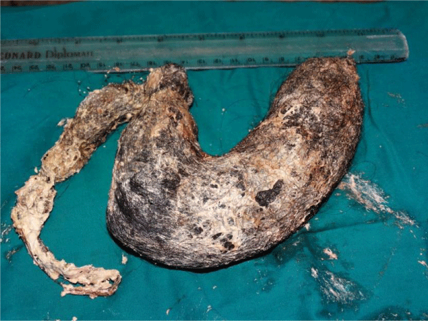
Seven children & adolescents presenting with intestinal obstruction and other GI disorders between 2012 and 2019 are presented. There were three males and four females (Table 1). Age ranged from 11 months to 16 years (median= 88 months; mean = 96.3 months). The common presenting symptoms were epigastric discomfort, nausea, recurrent vomiting, abdominal distension, constipation and bilious vomiting. One adolescent girl had history of trichotillomania (pulling out her own hair) and trichophagia (eating her hair) (Figure 1). Another 13 year old girl had trichophytobezoar, but she gave no history of trichophagia. Both these girls had poor nutritional status and required psychiatric treatment following surgery. Three patients had phytobezoar and two had lactobezoars. Five patients presented with features of acute intestinal obstruction. Six patients needed surgery and all of them had uneventful recovery. One patient improved with conservative treatment and had passed the bezoar rectally after an enema. The mean follow up was 83.5 months.
Discussion
Bezoars are defined as aggregates of undigested or inedible material found anywhere in the gastrointestinal tract (GIT) and most commonly found in the stomach [1]. Plant fiber, hair. and medication bezoars have all been described. Charak, the ancient Indian physician described bezoars in his treatise ‘Charaka Samhita’ in the second and third centuries BC. In ancient times, bezoars were thought to have medicinal and magical and they were considered antidotes to a variety of poisons and diseases [2,3]. Schonborn [4] first published a description of the surgical removal of bezoars in the 19th century
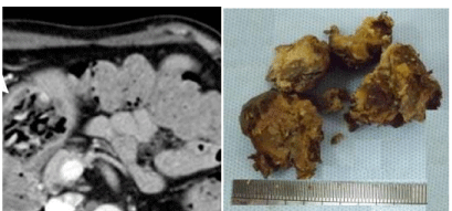
According to the compositions, bezoars are classified into four types: Phytobezoars are composed of indigestible food particles that are found in vegetable or fruit fibers, trichobezoars are composed of a conglomeration of hair and food particles, lactobezoars are composed of milk protein, or pharmacobezoars are concretions of various medications [5]. Phytobezoars are the most common type of bezoars, accounting for approximately 40% of all reported bezoars. In our series of seven cases, three patients had phytobezoars and one had trichophytobezoar. Of these four cases, three had undigested vegetables and fruit fibres and seeds (Figure 2). One patient (patient no.3, Figure 3) had phytobezoars composed of Bhimkal banana seeds, a particular variety of banana commonly seen in the state of Assam, India. The large, undigested seeds of Bhimkal clumps together forming a mass and cause intestinal obstruction and colic. This is similar to bezoars caused by persimmon seeds. The skin of unripe persimmons contains high concentrations of the persimmon tannin. Upon reaction with stomach acid, persimmon tannin is believed to polymerize and form a conglomerate in which cellulose, hemicelluloses, and various proteins are accumulated [6]. Two young children had lactobezoars, one of them improved with conservative treatment and passed the bezoar in pieces after a glycerine enema.
An altered gastric physiology, such as impaired gastric emptying or reduced acid production, is a well-known cause of bezoars. Bezoars are usually caused by previous gastric operations, such as vagotomy or partial gastrectomy, and can also be caused by gastroparesis or a gastric outlet obstruction [7]. The initial presentation of a bezoar often depends on its composition. Lactobezoars may present in premature infants or newborns with symptoms of feeding intolerance, abdominal distension, irritability, and/or vomiting. Physical examination may reveal a palpable abdominal mass in these patients. Simpsons [8] noted that pharmacobezoars can present with symptoms of gastric outlet obstruction, and can develop toxic symptoms because of their pharmacologic properties. Trichobezoars can take time to form—sometimes up to several years—and these bezoars may first present with subtle symptoms such as nausea or early satiety. However, as trichobezoars grow in size, they may present with epigastric pain, gastric outlet obstruction, ulceration, GI bleeding, and occasionally perforation [9]. One of our patient had a type of trichobezoar called Rapunzel syndrome, which is characterized by presence of hairballs or hair-like fibers which occupies the stomach and extends upto the small bowel. This results from chewing and swallowing hair or any other indigestible materials (trichophagia), often associated with hair-pulling disorder (trichotillomania, an impulse control disorder involving a compulsive urge to pull out one’s hair) in young girls [10-12]. The syndrome is named after the long-haired girl ‘Rapunzel’ in the fairy tale by the Brothers Grimm [13]. The patient in our series presented with poor nutritional status, nausea and recurrent abdominal pain. On exploration, the bezoar completely filled up the stomach and extended across the duodenum upto proximal few centimeters of jejunum. She required psychiatric treatment after removal of the bezoar. Another adolescent girl had trichophytobezoar, but she did not give any history of trichotillomania or trichophagia. However, she was provided psychiatric counseling following successful removal of the bezoar. Phytobezoars usually form more rapidly than trichobezoars.
Diagnosis of bezoars need obtaining a thorough history and clinical examination. Inquiry into the patient’s diet, food habit and medication is important. In a quite patient with thin abdominal muscles, a lump may be palpable. Halitosis may signify the presence of putrefying material in the stomach, and clinicians may observe patches of alopecia in patients with trichobezoars similar to those seen in individuals with trichotillomania. Investigation of such patients include x-ray abdomen in erect posture (in cases of instestinal obstruction) and ultrasonography (USG). Detection rate of trichobezoars by USG was reported to be around 88% [14]. Upper gastrointestinal series can demonstrate a filling defect in the stomach, however, this procedure has the risk of causing obstruction or perforation. CT scan demonstrates heterogeneous masses containing trapped air bubbles or homogenous mottled appearance in the region of stomach or intestine [14]. For gastric bezoars, endoscopy remains the investigation of choice [15].
Treatment of bezoars involve surgical removal of the bezoar and prevention of recurrence. Although surgical removal has been considered the standard treatment option for gastrointestinal bezoar, increasing use of endoscopy for diagnostic and therapeutic purposes has been reported with a success rate ranging from 50% to as high as 100% [16-18]. Apart from surgical removal, a wide range of therapeutic options have been used, including acetylcysteine, papain, metoclopramide, cellulase, and even instillation of Coca-Cola [17-23]. However, adverse effect of some of these therapies have been reported [24]. Blam and Lichtenstein [25] reported a new endoscopic technique of removal of gastric phytobezoars via suction through a large-channel endoscope. The authors found this technique to be a safe, effective, and rapid method of removing large gastric phytobezoars. Although endoscopic therapy has a role in some types of bezoars and may be the procedure of choice in terms of fragmentation and retrieval, surgical removal should be considered in patients who fail medical therapy or who have complications such as significant bleeding, obstruction, and/or perforation. For patients with Rapunzel syndrome, long-term follow-up with upper GI endoscopy or USG abdomen and psychiatric follow-up with psychotherapy and cognitive behavioural therapy are critical for prevention of recurrence [26].
- Kuhn BR, Mezoff AG (2011) Bezoars. In: Wyllie R, Hyams JS, Kay M, editors. Pediatric Gastrointestinal and Liver Disease. 4th ed. Philadelphia, Pennsylvania: Elsevier Saunders 319–322.
- Williams RS (1986) The fascinating history of bezoars. Med J Aust 145: 613–614. Link: https://bit.ly/3izaRRC
- Eng K, Kay M (2012) Gastrointestinal Bezoars: History and Current Treatment Paradigm. Gastroenterol Hepatol (N Y) 8: 776–778. Link: https://bit.ly/3xeYY7u
- Schonborn B (1883) Eine durch gastrotomie entfernte Haargeschwulst aus dem Magen eines jungen Mädchens. Arch klin Chir 29: 609.
- Sanders M (2004) Bezoars: from mystical charms to medical and nutritional management. Practical Gastroenterology 28: 37–50. Link: https://bit.ly/3xfk7y6
- Krausz MM, Moriel EZ, Ayalon A, Pode D, Durst AL (1986) Surgical aspects of gastrointestinal persimmon phytobezoar treatment. Am J Surg152: 526–530. Link: https://bit.ly/3pGsgcm
- Zamir D, Goldblum C, Linova L, Polychuck I, Reitblat T, et al. (2004) Phytobezoars and trichobezoars: a 10-year experience. J Clin Gastroenterol 38: 873–876. Link: https://bit.ly/3iyVokn
- Simpson SE (2011) Pharmacobezoars described and demystified. Clin Toxicol (Phila) 49: 72–89. Link: https://bit.ly/3wkDsy8
- Ciampa A, Moore BE, Listerud RG, Kydd D, Kim RD (2003) Giant trichophytobezoar in a pediatric patient with trichotillomania. Pediatr Radiol 33: 219–220. Link: https://bit.ly/3zgYPSF
- Sah DE, Koo J, Price VH (2008) Trichotillomania. Dermatol Ther 21: 13–21. Link: https://bit.ly/3gp4Bcr
- Ventura DE, Herbella FA, Schettini ST, Delmonte C (2005) "Rapunzel syndrome with a fatal outcome in a neglected child". J Pediatr Surg 40: 1665–1667. Link: https://bit.ly/3wdUei5
- Dutta HK (2020) Rapunzel Syndrome: A Case Report and Literature review. Annals of Short Reports 3: 1050. Link: https://bit.ly/3gnXxNb
- Grimm Brothers (1994-99) Rapunzel. Translated by Godwin-Jones R. Richmond, Virginia Commonwealth University Department of Foreign Languages.
- Ripolles T, Garcia-Aguayo J, Martinez MJ, Gil P (2001) Gastrointestinal Bezoars: sonographic and CT characteristics. Am J Roentgenol 177: 65–69. Link: https://bit.ly/3vc9ACv
- Wang YG, Seitz U, Li ZL, Soehendra N, Qiao XA (1998) Endoscopic management of huge bezoars. Endoscopy 30: 371-374. Link: https://bit.ly/3gdCiyM
- Lee SJ, Cheon GJ, Oh WS, et al. (2010) Clinical characteristics of gastric bezoars. Korean J Helicobacter Up Gastrointest Res 10: 49–54.
- Kement M, Ozlem N, Colak E, Kesmer S, Gezen C, et al. (2012) Synergistic effect of multiple predisposing risk factors on the development of bezoars. World J Gastroenterol 18: 960–964. Link: https://bit.ly/3vk7y3u
- Erzurumlu K, Malazgirt Z, Bektas A, Dervisoglu A, Polat C, et al. (2005) Gastrointestinal bezoars: a retrospective analysis of 34 cases. World J Gastroenterol 11: 1813–1817. Link: https://bit.ly/3vk7y3u
- Schlang HA (1970) Acetylcysteine in removal of bezoar. JAMA 214: 1329. Link: https://bit.ly/3geI9UA
- Kramer SJ, Pochapin MB (2012) Gastric phytobezoar dissolution with ingestion of diet coke and cellulase. Gastroenterol Hepatol (N Y) 8: 770–772. Link: https://bit.ly/3wdUQUV
- Pollard HB, Block GE (1968) Rapid dissolution of phytobezoar by cellulase enzyme. Am J Surg 116: 933–936. Link: https://bit.ly/357bvxA
- Holloway WD, Lee SP, Nicholson GI (1980) The composition and dissolution of phytobezoars. Arch Pathol Lab Med 104: 159-161. Link: https://bit.ly/2SpKmmR
- Ladas SD, Triantafyllou K, Tzathas C, Tassios P, Rokkas T, et al. (2002) Gastric phytobezoars may be treated by nasogastric coca-cola lavage. Eur J Gastroenterol Hepatol 14: 801–803. Link: https://bit.ly/2U0giP9
- Zarling EJ, Moeller DD (1981) Bezoar therapy: complication using Adolph’s meat tenderizer and alternatives from literature review. Arch Intern Med 141: 1669–1670. Link: https://bit.ly/3iChORI
- Blam ME, Lichtenstein GR (2000) A new endoscopic technique for the removal of gastric phytobezoars. Gastrointest Endosc 52: 404–408. Link: https://bit.ly/3vncFQt
- Miltenberger RG, Rapp JT, Long ES (2001) Habit Reversal Treatment Manual for Trichotilomania. 170–195. Link: https://bit.ly/3izDwFS
Article Alerts
Subscribe to our articles alerts and stay tuned.
 This work is licensed under a Creative Commons Attribution 4.0 International License.


 Save to Mendeley
