International Journal of Dermatology and Clinical Research
Acquiring accidental aspergillosis
Mitchell S Davis1*, Lydia Shedlofsky2 and Christine C Lin3
2HonorHealth, Scottsdale, Arizona, USA
3HonorHealth/Affiliated Dermatology, Scottsdale, Arizona, USA
Cite this as
Davis MS, Lydia Shedlofsky DO, Lin CC (2021) Acquiring accidental aspergillosis. Int J Dermatol Clin Res 7(1): 007-009. DOI: 10.17352/2455-8605.000041Copyright
© 2021 Davis MS, et al. This is an open-access article distributed under the terms of the Creative Commons Attribution License, which permits unrestricted use, distribution, and reproduction in any medium, provided the original author and source are credited.Aspergillus is an all-pervasive mold with the potential to cause severe invasive infections in the immunocompromised. A rare cutaneous manifestation of Aspergillus infection, primary cutaneous aspergillosis (PCA), occurs in just 1-5% of invasive aspergillosis cases. Prompt treatment is indicated as PCA may progress to a disseminated state. We present a unique case of an immunocompetent individual diagnosed with PCA two weeks after trauma and subsequent surgery.
Introduction
Aspergilli are ubiquitous fungal genera that include more than 350 species across various habitats. These environments include household dust, plants, common soil, and building material, with estimates that humans inhale thousands of Aspergillus spores daily [1-3]. Fortunately, the pathogenic subspecies are few and include A. fumigatus, A. flavus, A. terreus, and A. ustus. When these Aspergillus species directly inoculate a host and the infection manifests in the skin, the condition is known as Primary Cutaneous Aspergillosis (PCA). This cutaneous variant of Aspergillus infection primarily affects patients with compromised immune function, and a concomitant break in skin integrity is often required [2]. Cases in the immunocompetent population are sporadic, with most cases involving traumatic inoculation or recent surgery [2].
PCA typically manifests with macules, papules, nodules, and plaques near a site of disrupted skin integrity (e.g., venipuncture for a blood test) [4]. The most commonly described initial lesions are red or violet papules or plaques [5-7]. Blood or pus-filled vesicles may develop within these lesions and eventually ulcerate or produce a darkly pigmented crust [5]. The most common locations for PCA lesions to develop are on the head and extremities.
If PCA is suspected on presentation and then confirmed with biopsy and staining, prompt treatment is necessary to prevent disseminated aspergillosis. This report demonstrates an exceptionally uncommon case of an immunocompetent patient with PCA.
Case
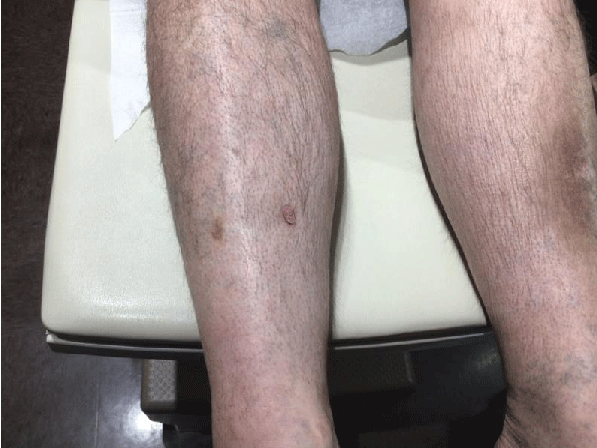
A 55-year-old male with a past medical history significant for squamous cell carcinoma presented to an outpatient dermatology clinic complaining of a non-healing wound. He experienced trauma and subsequent surgery to the left leg 14 weeks prior. Upon physical exam, a left, anterior, mid-tibial prominence with central ulceration was noted with induration and four centimeters of surrounding erythema (Figure 1). Given the differential diagnosis for a non-healing wound (i.e., non-melanoma skin cancer, bacterial infection, fungal infection, etc.), wound culture was ordered and the patient was prescribed ten days of doxycycline monohydrate. Informed consent was signed prior to obtaining a sample for wound culture.
Culture results revealed Aspergillus. Additional infectious workup and labs were unremarkable, with the exception of leukocytosis(18.9). The patient was referred to surgery for consultation; however, it was determined that the patient was not a candidate for surgery as the infection was limited to superficial soft tissue.
Approximately one month after his visit to the dermatology clinic, the patient called the after-hours emergency number with a high fever requesting advice. At the on-call doctor’s recommendation, he presented to the emergency department. Chest imaging revealed a right basilar interstitial consolidation and pleural effusion consistent with pneumonia. Urinalysis showed trace leukocyte esterase, white blood cells (30-50), and few bacteria in the urine. Complete blood count revealed leukocytosis (17.6). The patient declined the need for admission and was discharged with azithromycin and an albuterol inhaler. The patient was instructed to follow up with dermatology.
The patient was seen by dermatology and prescribed itraconazole. Two weeks later, the patient was seen in the clinic for follow-up. The wound appeared to have responded very well to itraconazole with no residual ulceration (Figure 2).
Discussion
Primary cutaneous aspergillosis (PCA) is an uncommon opportunistic fungal infection that typically occurs at sites of skin injury (e.g., intravenous access sites, burns, surgery) [2]. In cases of invasive aspergillosis, the rate of cutaneous involvement is reported to range from 1-5%.1 PCA may present with macules, papules, nodules, plaques, and hemorrhagic bullae [4-7]. If PCA is suspected, skin biopsy with hematoxylin and eosin are the first steps in diagnosis. This is typically followed by special stains such as the methenamine silver and Periodic acid–Schiff stain, which may show findings characteristic of PCA. Findings include septate hyphae and acute-angled branching [2,4]. The detection of galactomannan antigen in serum may also aid in diagnosis and determine the possible hematological extension of disease; this antigen is a component of the Aspergillus cell wall [8].
Aspergillus infection has been associated with multiple disorders characterized by immune system suppression [9]. Known risk factors in indults include diabetes, HIV, long-term steroid use, and malignancy (particularly hematological) [4,10]. In children, leukemia, and lymphomas are the most common diseases associated with PCA [10]. Both the innate immune system (e.g., neutrophils, macrophages) and adaptive immune system (e.g., T-cell immunity) are involved in the host’s defense against Aspergillus [9]. In an immunocompetent host, macrophages destroy Aspergillus conidia, while hyphae are damaged by monocytes and polymorphonuclear leukocytes [4]. Deficiencies in any component of the host’s innate and adaptive immune system increase the risk of fungal infection [11]. It is exceptionally uncommon for PCA to affect immunocompetent individuals [2].
Currently in the literature, there is a paucity of reported cases of PCA in the immunocompetent population. Avkan-Oguz et al. and Van Burik et al. reference nine cases of posttraumatic or postoperative PCA in immunocompetent patients [2,12]. The majority of the reported cases involved surgical wounds or trauma to the lower extremities or hands sustained during farming or traffic accidents [2,12]. Clinical presentation of these cases varied widely and included dermatologic findings such as erythematous papules, nodules, plaques, and hemorrhagic bullae [12,13]. Treatment regimens included azole antifungals, amphotericin, and surgical debridement [2,12].
Our patient’s case was similar to several of the cases reported in the literature in that our patient was diagnosed with PCA after leg trauma and subsequent surgery. Additionally, as with several reported cases of PCA, our patient’s infection responded well to an azole antibiotic. The dark, musky-colored prominence with central ulceration on our patient’s leg may be considered by some to be a unique presentation of PCA. However, a clinician needs to recognize the wide variability in the presentation of this cutaneous fungal infection. Lastly, it is important to recognize that cases of PCA in the immunocompetent population generally have better outcomes when compared to those patients in the immunocompromised population [14].
Conclusion
We report a case of primary cutaneous aspergillosis after lower extremity trauma in an immunocompetent individual. The clinical manifestations of PCA are variable and clinicians must acknowledge the possibility of PCA infection in their immunocompetent patients. Prompt diagnosis and management are crucial to prevent PCA from progressing to disseminated aspergillosis. Management typically includes several weeks of an azole antifungal and possibly surgical debridement.
- Bernardeschi C, Foulet F, Ingen-Housz-Oro S, Ortonne N, Sitbon K, et al. (2015) Cutaneous Invasive Aspergillosis: Retrospective Multicenter Study of the French Invasive-Aspergillosis Registry and Literature Review. Medicine (Baltimore) 94: e1018. Link: https://bit.ly/3cFCmEY
- Avkan-Oğuz V, Çelik M, Satoglu IS, Ergon MC, Açan AE (2020) Primary Cutaneous Aspergillosis in Immunocompetent Adults: Three Cases and a Review of the Literature. Cureus 12: e6600. Link: https://bit.ly/3cCpXl7
- Swarnagowr BN, Sangeetha S (2014) Disseminated Aspergillosis in an Autopsy Case of Young Male. J Evol Med Dent Sci 3: 6382-6385. Link: https://bit.ly/3nHBcyJ
- Mada PK, Koppel DAS, Al Shaarani M, Chandranesan ASJ (2020) Primary cutaneous Aspergillus fumigatus infection in immunocompetent host. BMJ Case Rep 13: e233020. Link: https://bit.ly/3xdiDFU
- Kishore M, Gupta P, Bhardwaj M (2018) Cytomorphological Diagnosis of Isolated Cutaneous Aspergillosis in an Immunocompetent Host. Indian Dermatol Online J 9: 206-208. Link: https://bit.ly/3rhP59p
- Liu X, Yang J, Ma W (2017) Primary cutaneous aspergillosis caused by Aspergillus.fumigatus in an immunocompetent patient: A case report. Medicine (Baltimore) 96: e8916. Link: https://bit.ly/3HLJLR8
- Pincelli MS, García JP, Sangueza OP, Sangueza M (2021) Primary Cutaneous Aspergillosis in Immunocompetent Patients Due to Aspergillus niger: A Report of 4 Cases. Am J Dermatopathol 43: 506-509. Link: https://bit.ly/3xjhlJt
- Fontaine T, Latgé JP (2020) Galactomannan Produced by Aspergillus fumigatus: An Update on the Structure, Biosynthesis and Biological Functions of an Emblematic Fungal Biomarker. J Fungi Basel Switz 6: E283. Link: https://bit.ly/3xhlzBf
- Ben-Ami R, Lewis RE, Kontoyiannis DP (2010) Enemy of the (immunosuppressed) state: an update on the pathogenesis of Aspergillus fumigatus infection. Br J Haematol 150: 406-417. Link: https://bit.ly/3r3nLvm
- Torrelo A, Hernández-Martín A, Scaglione C, Madero L, Colmenero I, et al. (2007) Primary cutaneous aspergillosis in a leukemic child. Actas Dermosifiliogr 98: 276-278. Link: https://bit.ly/3DKQo3X
- Segal BH, DeCarlo ES, Kwon-Chung KJ, Malech HL, Gallin JI, et al. (1998) Aspergillus nidulans infection in chronic granulomatous disease. Medicine (Baltimore) 77: 345-354. Link: https://bit.ly/3xdVaEH
- van Burik JAH, Colven R, Spach DH (1998) Cutaneous Aspergillosis. J Clin Microbiol 36: 3115-3121. Link: https://bit.ly/3CKXrYQ
- Avkan-Oğuz V, Çelik M, Satoglu IS, Ergon MC, Açan AE (2020) Primary Cutaneous Aspergillosis in Immunocompetent Adults: Three Cases and a Review of the Literature. Cureus 12: e6600. Link: https://bit.ly/3cCpXl7
- Pfeiffer CD, Fine JP, Safdar N (2006) Diagnosis of Invasive Aspergillosis Using a Galactomannan Assay: A Meta-Analysis. Clin Infect Dis 42:1417-1727. Link: https://bit.ly/3xegPMV
Article Alerts
Subscribe to our articles alerts and stay tuned.
 This work is licensed under a Creative Commons Attribution 4.0 International License.


 Save to Mendeley
