Archive of Urological Research
A Band from the “Blue”
Padmanabha Vijayan*
Cite this as
Vijayan P (2023) A Band from the “Blue”. Arch Urol Res 7(1): 001-003. DOI: 10.17352/aur.000042Copyright
© 2023 Vijayan P. This is an open-access article distributed under the terms of the Creative Commons Attribution License, which permits unrestricted use, distribution, and reproduction in any medium, provided the original author and source are credited.Several lesions of the bladder are incidental findings and a few of them are seen against a background of bladder outlet obstruction. Tuberculosis of the bladder is associated with characteristic lesions and changes. Presented here is a never-seen-before bladder lesion in a patient presenting with symptoms of Bladder Outlet Obstruction due to an enlarged tuberculous prostate, and no evidence of tuberculous cystitis.
Introduction
Tuberculosis of the lower urinary tract though common in developing countries may manifest with fever, loss of weight, and lower urinary symptoms such as frequency, urgency, hematuria, hemospermia, obstructed ureters, contracted bladder, etc. Tuberculosis involving the prostate is rare but has been encountered and reported from time to time. The presence of a Y-shaped band in the lumen of the urinary bladder has never been reported in the available medical literature. Moreover, it had remained symptomless until its accidental discovery by sonologic imaging.
Case report
A 64-year-old male presented with a poor urinary stream, delay in initiating voiding, difficulty’s and need to strain to void for 5 years. His symptoms were now sufficiently bothersome for him to seek medical attention. He had lost about 5 kilograms of weight in the past 6 months. He had no other constitutional symptoms, and his past medical history was non-contributory, as was his personal history.
Physical examination revealed an emaciated male with painless distension of the bladder seen above the pubis. There were no other abdominal findings. DRE revealed a normal-sized prostate, and a distended bladder was palpable rectally. No other abnormalities were found on a review of his other systems.
His hematology and biochemistry profiles were normal. The urine examination was normal. At Uroflowmetry, he voided 224 ml with a Peak flow of 5.4 ml/s and a Mean flow of 3.3 ml/s, leaving behind a large residue.
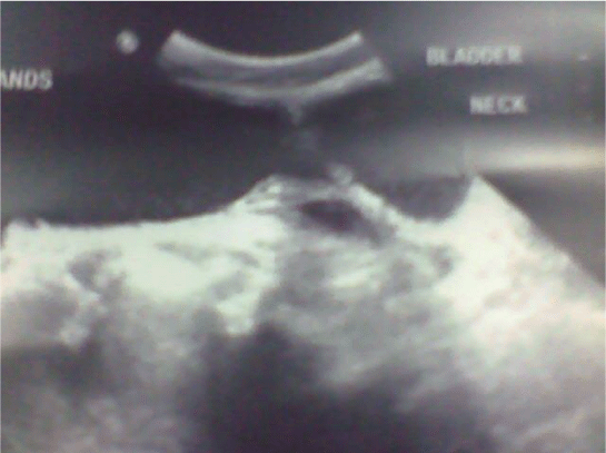
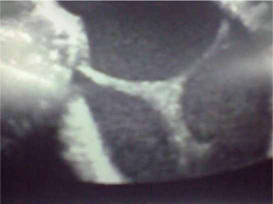
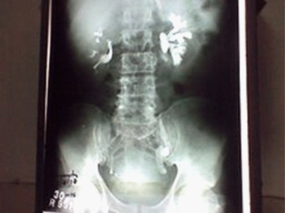
Ultrasonography showed a normal right kidney, with no ureteric dilatation. The left kidney showed hydronephrosis, with hydro ureter all the way to the ureterovesical junction. A Y-shaped band of isoechoic tissue was seen in the posterior wall, with the stem at the bladder neck, and the branches extending to each lateral wall (Figures 1,2). No other abnormality of any viscera or structure was found. Excretory urography revealed good renal function on both sides, with left hydroureteronephrosis (Figure 3). The bladder was well distended, with filling defects seen at the bladder neck, and high on the right lateral wall. Some irregularity of the left lateral wall of the bladder was also noted at its junction with the dome.
On cystoscopy, the band was found to be beginning at the posterior lip of the bladder neck and extending to the lateral walls. The prostate showed asymmetric enlargement, with occluding lateral lobes and the left lobe extending across the midline considerably. No other structural abnormality was found. The urine showed turbidity. The endoscopic view of the band was not recorded, as there was no facility for the same. The bladder capacity was 350 ml.
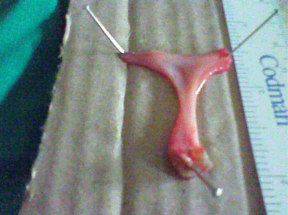
The limbs of the band in the bladder were endoscopically detached from the bladder and the tissue was removed intact (Figure 4). Resection of the left lobe of the prostate was accompanied by the expulsion of copious, thick purulent fluid. The resection was completed, and the patient voided with the improved flow after catheter removal 72 hours later.
Histopathology of the “band” showed a fibrous core covered with transitional normal epithelium. No other tissue was found. The prostate showed tubercular granulomata. The patient received Anti-tubercular chemotherapy for 12 months and improved clinically with a gain in weight. He continued to void to his satisfaction. He has been followed up for 2 years as of date.
Discussion
Tuberculous ulcers in the bladder are rare but when present are superficial with a central area surrounded by raised granulation. They are close to ureteric orifices but as the disease progresses may appear in any part of the bladder. Progressively the inflammation involves the muscle, which gets replaced by fibrous tissue. The fibrosis around the ureteric orifice may contract and produce a stricture or become withdrawn, rigid, and dilated assuming a golf hole appearance. The rigid lower ureter may reflux. The mucosal lesions when healed have a stellate appearance caused by fibrous tissue meeting at a central point – the site of the initial area of infection.
Tuberculosis of the prostate is rare and is mostly diagnosed by a pathologist or is found during TURP [1]. Rarely cavitations may occur leading to fistulae [2].
The aetiology of this bladder band is difficult to ascertain. Acquired conditions that produce bands are known to do so following the resolution of inflammation, and due to the resultant fibrosis, on the surfaces of organs and structures – intestine, pleura, pericardium, etc. The histology of the band showed neither granulomata nor any muscle tissue. The transitional epithelial lining over the band was completely intact.
One possible explanation is an embryological origin. The fusion of the trigone with the vesicourethral canal may have resulted in a structure like the one seen – much like the mountain ranges seen geologically when landmasses have moved from different locations to eventually fuse. The presence of a fibrous core may represent remnants of primitive mesoderm over which normal epithelium has grown.
However, it is difficult to answer two important questions – why has this not been seen before, and why did this condition remain quiescent for six-and-a-half decades? It is our postulation that since there was no obstruction to the flow of urine from the ureters or out of the bladder, this remained quiescent and was only picked up when ultrasonography was performed for some other, unrelated condition.
Embryologically, the urinary bladder is derived partly from the ectodermal cloaca and partly from the caudal ends of the mesonephric ducts. After separation from the cloaca, the ventral part of the latter becomes divided into 3 parts.
- Cranial - vesicourethral canal continuous with allantoic duct into this part mesonephric ducts open.
- Middle narrow channel –the pelvic portion
- Caudal – deep phallic section closed externally by a urogenital membrane.a
The 2nd and 3rd parts together, constitute the urogenital sinus. The ureter and mesonephric ducts come to open separately into the vesicourethral part. The termination of the mesonephric duct then moves caudally to open into that part which will form the prostatic urethra. The mesonephric ducts contribute to the formation of the trigone and dorsal walls of the prostatic urethra. The remainder of the vesicourethral part forms the body of the bladder and part of the prostatic urethra.
Furthermore [3], the mesonephric ducts drain into the urogenital sinus, and the epithelia fuse. The ureteral bud evaginates from the mesonephric duct. The mesonephric duct beyond the ureteric bud dilates as a common excretory duct – the precursor of hemitrigone. The right and left common excretory ducts get absorbed into the urogenital sinus. The mesenchyme of the common excretory ducts migrates toward the midline. As the common excretory ducts get absorbed into the urogenital sinus, the orifices of the ducts move caudally. The continued growth of the epithelium and mesoderm of the absorbed excretory ducts separate the ureteric orifices and establish the framework of the primitive trigone.
Complete duplication of the urinary bladder in the coronal plane with no associated abnormalities has been reported [4]. In this particular case, it may be postulated that at this stage of the development of the primitive trigone, it got split in the coronal plane resulting in two trigonal plates- a superficial and a deep one. The superficial plate remained as a Y-shaped band and the deep one as the proper trigone.
It is believed that the presence of a tuberculous lesion of the prostate may not be in any way related to the Y-band, which may indeed be a red herring. The anatomy and the shape of this band are perplexing.
- Sporer A, Auerbach O. Tuberculosis of prostate. Urology. 1978 Apr;11(4):362-5. doi: 10.1016/0090-4295(78)90232-7. PMID: 664142.
- Patoir G, Spy E, Cordier R. Trois cas de fistules vésico- ou uréthro-rectales tuberculeuses [3 cases of tubercular vesico- or urethrorectal fistula]. J Urol Nephrol (Paris). 1969 Mar;75(3):210-7. French. PMID: 5815312.
- Hamilton WJ, Mossman HW (Eds). The Urogenital system In Human Embryology, Prenatal development of form and function, New York Macmillan Press Ltd. 1976; 377.
- Oğuzkurt P, Ozalevli SS, Alkan M, Kayaselcuk F, Hiçsönmez A. Unusual case of bladder duplication: complete duplication in coronal plane with single urethra and no associated anomalies. Urology. 2006 Nov;68(5):1121.e1-3. doi: 10.1016/j.urology.2006.06.002. PMID: 17113907.
Article Alerts
Subscribe to our articles alerts and stay tuned.
 This work is licensed under a Creative Commons Attribution 4.0 International License.


 Save to Mendeley
