Open Journal of Trauma
Gamma angle, a measurement tool of the femoral head angular translation in adults hips with cam or mix form of impingement femoroacetabulare
Vladimir Andjelkovic1*, Zoran Andjelkovic2 and Dušan Stojanovic2
2Department of Orthopedics and Traumatology, General Hospital Leskovac, Serbia
Cite this as
Andjelkovic V, Andjelkovic Z, Stojanovic D (2019) Gamma angle, a measurement tool of the femoral head angular translation in adults hips with cam or mix form of impingement femoroacetabulare. Open J Trauma 3(1): 012-017. DOI: 10.17352/ojt.000021Femoral head translation leads to the cam deformity development. It is formed on the femoral head-neck junction. Cam deformity produces femoroacetabular impingement. There are no particular techniques for femoral head translation assessment. The offset index is the most regularly applied. It quantifies the relation between the femoral head and neck junction. We introduce the original method to test the femoral head translation.
The purpose of this survey was to draw, measure and to test gamma angle rates. Tested groups were subjects with cam and mix form of femoroacetabular impingement. We compare gamma angle values with the offset index. Gamma angle role in femoral head angular inclination measurement, we considered.
Material: We measured the gamma angle on the preoperative X-rays of the hips. 51 subjects with mixed and cam form of femoroacetabular impingement we analyzed. Standardized preoperative anteroposterior and profile X-ryas we managed.
Method: Two femoral neck axes we drew. The angle they made we named gamma angle. We assumed this angle measure femoral head angular inclination. Gamma angle higher than 3° was pathological. We calculated and tabulated data.
The results: Gamma angles mean was 6,30° on the AP and 5,97° on the profile X-rays. The gamma angle sensitivity of 90,32% was on AP X-rays. On the other X-rays, sensitivity was smaller: 60-85%. Specificity, positive and negative predictive values were over 90%. We established a high negative correlation between the offset index and gamma angle values.
Conclusion: Gamma angle measured the angular inclination of the femoral head on the hips X-rays. This angle might be an appropriate tool. One can apply this tool in people with the closed proximal femoral epiphysis.
Introduction
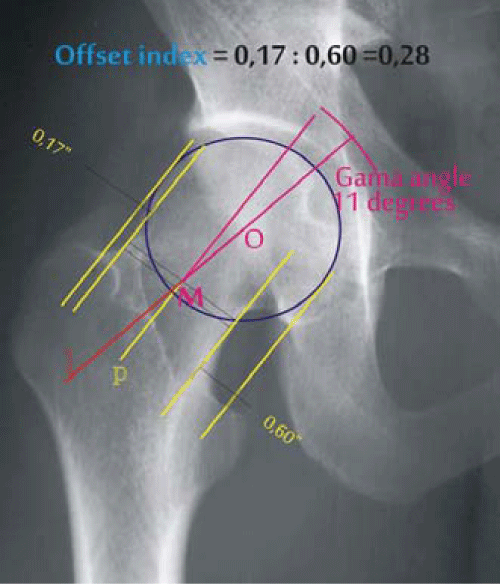
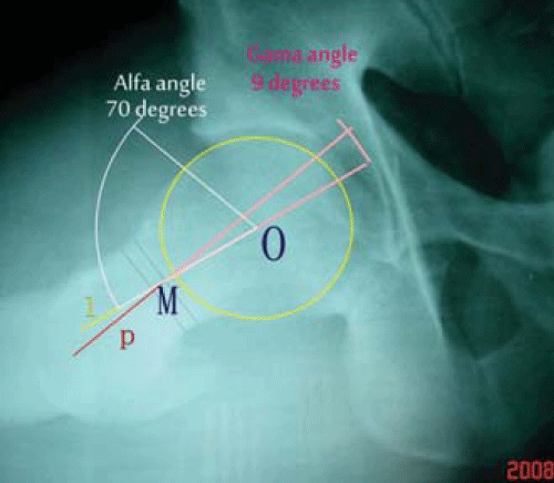
Femoroacetabular impingement (FAI) causes groin pain in young people. Its principal causes are morphological differences in the proximal femur and/or acetabulum. While the hip moves, femoral neck strikes on the acetabulum margin. This leads in labral and labrum adjacent cartilage lesion of the anterosuperior acetabulum. These lesions present early arthritic changes of the hip [1-10]. Murray (2) assumed that ‘’femoral head tilt’’ is a cause of hip arthritis. This tilt originated from mild adolescent femoral head epiphysiolysis. Harris described femoral head tilt as a ” pistol grip” deformity [5,8]. Ganz introduced the theory of hip arthritis development during femoroacetabular impingement. He distinguished three morphotypes of FAI: cam, mixed and the pincer form [1,9-11]. Etiology of the cam form of FAI is not definite. It is a secondary osteochondral bone hillock or cam deformity. It is localized in superior, anterior or both, anterosuperior femoral head-neck junction. Many authors speculate that adolescents femoral head translation being an underlying etiology. It induces cam deformity development [12-17]. The femoral head center rests on, or tight around the femoral neck axis. In some proximal femur pathology, this center is faraway from the femoral neck axis [18]. We used offset indexes to study the relationship between the femoral head and neck. They measure femoral head translation on the femoral neck (Figures 1,2). The offset indexes normal values are 0,80-1,20 [18-20]. Goodman [21], and Albert [22] measured femoral head translation. They used the femoral neck axis and femoral head epiphysis. Their name for this angle was ‘’femoral head tilt angle’’. Some authors suggested the angle created between two femoral neck axes [23,24]. They named it gamma angle. This angle could be the measure of femoral head angular inclination. We wondered if it was feasible to measure the gamma angle in the adult’s hips. This hips should have the cam and mixed form of FAI. Standardized anteroposterior and profile X-rays we could manage. Study question was if this angle is femoral head angular inclination measurement tool. We hypothesized that it was conceivable in the adult’s hips to draw and measure the gamma angle that tests the femoral head angular inclination.
The purpose of the survey was to draw, measure and to test gamma angle. To correlate the gamma angle with offset index values. To assess the gamma angle role in the femoral head angular inclination measurement.
Material
We have used two data series in this survey. The original data set we downloaded from the report of V. Andjelkovic [24], with his agreement. From this report, we have used gamma angle values. This author measured this angle on the hips X-rays. He used two types of standardized X-rays of the hips. One was anteroposterior (AP) X-ray. The second was profile Dunn-Ripstein-Mȕller (DRM90) X-rays. The X-rays he made on the adult’s asymptomatic individuals. We used the gamma angles mean, standard deviation and confidence interval. This encouraged us to determine gamma angles upper and smaller limits in the adult hips (Table 1). We added and subtracted three standard deviations to the Mean to achieve the gamma angle limits. For AP X-rays it was: -1,61° To achieve the purpose of this survey, we used preoperative X-rays of the hips. The standardized anteroposterior (AP) was the first one. Profile Dunn Ripstein Mueller in 90 degrees of hip flexion (DRM90) was the second [23-26]. We did preoperative X-rays. We did images of subjects with the cam and mix form of FAI. In these X-rays, we drew two neck axes. First was” literature gold standard” or 3 points femoral neck axis. We spotted it with letter l. This line always contains femoral head center O (Figures 1,2). The second was femoral neck inner third, two parallel lines neck axis. We spotted it with letter p [24,26-28]. If the femoral head center (O) lied out of the p-axis than these axes formed sharp angel lMp. We named this angle, gamma angle (γ), (Figures 1,2). When the axes l and p overlapped, the femoral head had an anatomical position. Femoral head center (O) moves from its anatomical position when the femoral head scrolls. The femoral neck axis p creates an acute angle gamma with axis l. This angle is the femoral head angular inclination on the femoral neck inner third. If the femoral head center (O) exists below the axis p, an inferior femoral head inclination appeared. If the femoral center O lies above the axis p we had the superior femoral head inclination. Posterior position of the femoral head center on the axis p showed posterior femoral head inclination. Anterior position of femoral head center on the axis p was the anterior femoral head inclination. We used the offset indexes to correlate the gamma angle values. These indexes are the standard parameter to measure femoral head translation [4,8,23,24,28]. We drew and measured offset indexes on both femoral neck axes. On the axis l we named it to offset index-l (OFI-l)). On the axis p, we named it to offset index-p (OFI-p) (Figures 1,2). We prepared and tabulated data series. The Kolmogorov-Smirnoff test verified the data distribution normality. To assess the Mean, we used the paired two-tailed t-tests. The Pearson correlation coefficient we used to measure correlation power. Contingency tables 4 x 4 we used to analyze the sensitivity, specificity, positive and negative predictive gamma angle value. Power of the trial was set at 80% with a beta error of 0,20. Conclusion error of 5%, where p<0,05 value rejects the hypothesis. We used the” SPSS 20 for Windows” program to evaluate data. The Corell DrawX7 program we used to process hips X-rays. We used the first data set (Table 1) from the study reported in 2018. [24]. This data set is a shyness of the gamma angle data. He measured this angle by accident in adults asymptomatic hips. Gamma angle 0° was detected in 85 hips from142 AP hips X-rays. In these hips, the p and l femoral neck axis overlapped. Gamma angle was 1° in 39 hips and 17 hips had gamma angle 2°. Only one hip had 3° gamma angle. Overall Mean was 0.542° (99% confidence interval was 0.387-0.697). In 144 DRM90 hips X-rays, 96 hips had 0° gamma angle value. Gamma angle was 1°in 27 hips. In 19 hips this angle was 2°. In 2 hips gamma angle value 3° (Mean: 0.507; 99% of a confidence interval 0.343-0.671). We added and subtracted 3 standard deviations to the Mean. This is how we came to the gamma angle values limits of -3° < γ < +3°. This humbled the gamma angle must not reach 3° in any angle on the femoral neck axis l. We gathered the second data set from the preoperative hips X-rays of the operated subjects. In 8 hips of mix form FAI, gamma angle was fewer than 3° on the AP hips X-rays (Tables 2-4). In these hips on DRM90 X-rays gamma angle was bigger than 3°. Gamma angle was bigger than 3° in 12 hips. On AP X-rays in only 3 hips. In 9 hips on AP and DRM90 hips X-rays: Mean: 5,08° (95% confidence interval: 4,30°- 6,23°). In the same group, the gamma angle was fewer than 3° in three hips on DRM90 hips X-rays. On AP X-rays in these hips, gamma angle was higher than 3°. In 17 hips gamma angle was higher than 3° (in 8 hips only on DRM90 hips X-rays). In 9 hips in both X-rays gamma angle was higher than 3° (Mean: 4,29°; 95% confidence intervals: 3,41°- 5,16°). Gamma angle was fewer than 3°in three hips on AP X-rays of a cam form of FAI. In this group, the gamma angle was higher than 3° on DRM90 hips X-rays. In 28 hips, gamma angle was higher than 3° in 9 hips on AP X-rays. The other of 18 hips had gamma angle higher than 3°on both hips X-rays. Theirs Mean was: 6,82 ° (95% confidence interval: 6,06 -7,57°). On DRM90X-rays gamma angle was fewer than 3° in eight hips. In these hips, gamma angle was higher than 3° on AP hips X-rays. From 22 hips gamma angle was higher than 3° in 3 hips on DRM90 X-rays. In 19 hips, gamma angle was higher than 3° on both hips X-rays: Mean of 7,27° (95% confidence interval: 5,89 - 8,00°). Together 40 hips (23 cams, 17 mixes from FAI),had pathological gamma angle value on AP hips X-rays.Theirs Mean was 6,30 (95% confidence interval: 5,52°- 7,07°). In 39 hips(17 cams,12 mix form FAI), on the DRM90 hips X-rays was pathological. Theirs Mean was 5,97(95% confidence interval: 5,12°- 6,81°) (Tables 2,3). Gamma angle had a high sensitivity on the AP hips X-rays in cam form FAI (90,32%). On the other X-rays, sensitivity was lower: 60-85%. Gamma angle specificity was significant on hips X-rays (98, 63% - 99,30%). A significant positive predictive values gamma angle had on AP: 89, 47% and 97,56% DRM90 hips X-rays. Negative predictive values were on AP: 92, 81% and 97,96% on DUM90 hips X-rays (Table 4). We measured the offset index using l and p femoral neck axes in 51 operated hips. In 40 AP hips X-rays in line l offset index had Mean (l) ≈ 0,55. In 41 hips in line p offset index had Mean (p)≈ 0,54. Offset index l in 50 DRM90 hips X-rays was Mean (l) ≈ 0,62. Offset index p was fewer than 0,80 (Mean (p)≈ 0,57). Gamma angle and offsets index on the AP hips X-rays had a significant negative correlation. On the line l it was: ρ = - 0,913 (p<0,05). Correlation on the line p femoral neck axis, was ρ = - 0,957 (p< 0,05). A significant negative correlation had gamma angle and offset index on the DRM90 hips X-rays. It was on the line l: ρ = - 0,939 (p<0,05). On the line p correlation coefficient was ρ= - 0,932 (p< 0,05) (Table 5). We drew and measured gamma angle values in adult persons with the FAI. Gamma angle is the quantitative angular distance between the two femoral neck axes. This distance measures the femoral head inclination on the femoral neck inner third. We set the angle gamma and its upper and lower limits in asymptomatic persons. This provided us to measure the femoral head angular inclination pathological values. We assumed that this inclination was the femoral head tilt in people. Gamma angle drawing and measuring on hips X-rays weren’t demanding technique. The suggested technique has its disadvantages. The cam deformity sometimes involves the femoral neck inner third. It was problematic to detect the marginal spots on the femoral neck that specify two parallel lines. In such situations, we had to lateralize two parallel lines as much as cam deformity demands. This extends the distance between inner parallel line middle and the femoral head center. This makes OM distance longer reducing gamma angle values. We didn’t check the reliability and reproducibility of the gamma angle. We notice that the femoral neck has a compound three-dimensional anatomy [29,30]. The recommended method simplifies femoral neck axes drawing and gamma angle measuring. Despite these disadvantages the recommended tool has its stand. It measures femoral head angular inclination in adults diseased hips. This can aid in detecting the etiology of the femoral head and neck pathology. We observed the value of the gamma angle, measured in the newer report [24]. This encouraged us to determine gamma angle limits in asymptomatic adults hips (γ±3°). We decided that any value that exceeds these limits has pathological interest. The gamma angle had sensibility range 60-90% for taken X-rays. Detected lower rate of gamma angle sensitivity was incorrect-lower. In one hip the gamma angle was fewer than 3° in one plane. In the new plane on the same hip, it was over 3°. This meant that the gamma angle was higher than 3° at the list in one plane in all tested hips. This produced the strong gamma angle sensitivity in disclosure of the femoral head angular inclination. We detected gamma angle high specificity (98-99,30%) If the femoral head angular inclination does not occur. The strength of the gamma angle to predict femoral head angular inclination was significant (PPV: 89-97, 56%). Gamma angle prediction of non-existent disease was also extremely significant (NPV: 92-97,96%). We measured the offset indexes and correlated them with the gamma angle. Both tested femoral head tilt. We calculated the significant negative correlation of both. This suggested the significant statistical correlation of the gamma angle and offset indexes. Gamma angle measures and quantify femoral head angular inclination on the femoral neck inner third. Murray (2) was the first who measured femoral head translation. He determined the vertical distance between the femoral head center and the femoral neck axis. His axis connected middles to the intertrochanteric line and the femoral neck inner third line. In patients with before existed hips arthritis, he used the AP hips X-rays. Femoral head translation distance from the neck axis he reported in millimetres. Murray couldn’t apply the method to the profile hips X-rays. This method quantified femoral head translation and suggested it as a cause of hip arthritis. Goodman (21) measured femoral head translation too. He used femoral neck axis and femoral head epiphysis in the adults’. Proximal femur cadaver bones and the X-rays of the same he tested. He didn’t define his method. Goodman used femoral neck axis and a femoral head epiphysis line in adult hips. His line of the epiphysis was invisible on the hips X-rays. Albert et al. on the nuclear magnetic resonance imaging, measured femoral head inclination. He determined the angle between the femoral neck axis and the proximal femoral epiphysis line [22]. In adolescents, Southwick measured slipping of the femoral head epiphysis before its closure. He measured the head-shaft angle between the epiphysis line and femoral diaphysis [31]. Muggier presented two femoral neck-lines in drawing femoral neck axis. He measured femoral head translation using an index. This index presented a vertical distance between the femoral head center and the’ real femoral neck axis’’. This idea mimics Murray’s method [32]. Andjelkovic Z. As suggested on the gamma angle existence. This angle he drew on cadaveric femora and radiographic images of these femora. He tested the two parallel lines role in drawing the femoral neck axis [23]. Andjelkovic V measured gamma angle in adults asymptomatic hips. Gamma angle of less than 3°was founded. He proposed gamma angle measuring in symptomatic adults hips. This study proposes the measurement method for femoral head inclination. Further studies are necessary to check this method in the larger groups of the patients. We recommend intra-observer and inter-observer evaluation of the results. our data requires standardization the method of two parallel line femoral neck axis drawing. Comparison of the gamma angle values on the pre- and postoperative hips X-rays is necessary. Gamma and alpha angle relationships could give an answer of cam deformity development in the femoral head-neck junction. We presented a new method to draw, test and measure gamma angle on the hips X-rays. We applied this method to the persons with the cam and mixed form of femoroacetabular impingement. This angle could be a proper tool to measure femoral head angular inclination in the adults.Method
Results
Discussion
Conclusion
- Ganz R, Parvizi J, Beck M, Leunig M, Notzli H, et al. (2003) Femoroacetabularimpingement: a cause for osteoarthritis of the hip. Clin Orthop Relat Res 417: 112-120. Link: http://bit.ly/2KKykhh
- Murray RO (1965) The aetiology of primary osteoarthritis of the hip. Br J Radiol 38: 810-824. Link: http://bit.ly/33YsHnk
- Resnick D (1976) The ‘tilt deformity’ of the femoral head in osteoarthritis of the hip: a poor indicator of the previous epiphysiolysis. Clin Radiol 27: 355-363. Link: http://bit.ly/2Hwoz4r
- Tanzer M, Noiseux N (2004) Osseous abnormalities and early osteoarthritis: the role of hip impingement. Clin Orthop 429: 170-177. Link: http://bit.ly/2KVthtc
- Harris WH (1986) Etiology of osteoarthritis of the hip. Clin Orthop Relat Res 213: 20-33. Link: http://bit.ly/2ZkLNoa
- Ito K, Kahlnor M, Leunig M, Ganz R (2004) Hip morphology influences the pattern of femoral- acetabular impingement. Clin Orthop 429: 262-271.
- Murgier J, Espié A, Bayle-Iniguez X, Cavaignac E, Chiron P (2013) Frequency of radiographic signs of slipped capital femoral epiphysiolysis sequelae in hip arthroplasty candidates for coxarthrosis. Orthop Traumatol Surg Res 99: 791-797. Link: http://bit.ly/31X9kt1
- Aronson J (1986) Osteoarthritis of the young adult hip: etiology and treatment. Instr Course Lect 35: 119-128. Link: http://bit.ly/31P5jqc
- Ganz R, Leunig M, Leunig-Ganz K, Harris WH (2008) The etiology of osteoarthritis of the hip: an integrated mechanical concept. Clin Orthop Relat Res 466: 264-272. Link: http://bit.ly/2zgl3Gs
- Siebenrock KA, Wahab KH, Werlen S, Kalhor M, Leunig M, et al. (2004) Abnormal extension of the femoral head epiphysis as a cause of cam impingement. Clin Orthop Relat Res 418: 54-60. Link: http://bit.ly/2ZgmYWf
- Ito K, Minka MA, Leunig M, Werlen S, Ganz R (2001) Femoroacetabular impingement and the cam-effect: AMRI based quantitative study of the femoral head-neck offset. J Bone Joint Surg Br 83: 171-176. Link: http://bit.ly/2zaRRRd
- Lutken P (1961) Bone-bridge formation between the greater trochanter and the femoral head: a normal variation of the pattern of the ossification in the upper end of the femur in adolescence. Acta Orthop Scand 31: 209-215.
- Morgan JD, Somerville EW (1960) Normal and abnormal growth at the upper end of the femur. J Bone Joint Surg Br 42-B: 264-272. Link: http://bit.ly/2Z5PkYc
- Carsen S, Moroz PJ, Rakhra K, Ward LM, Dunlap H, et al. (2014) Beaule´ PE.The otto franc award. On the etiology of the cam deformity: A cross-sectional pediatric MRI study. Clin Orthop Relat Res 472: 430-436. Link: http://bit.ly/2TOOitC
- Siebenrock KA., Behning A, Mamisch TC, Schwab JM (2013) Growth plate alteration precedes cam-type deformity in elite basketball players. Clin Orthop Relat Res 471: 1084-1091. Link: http://bit.ly/2HiA3YT
- Angel JL (1964) The reaction area of the femoral neck. Clin Orthop 32: 130-142. Link: http://bit.ly/2zdTRbK
- Odgers PNB (1931) Two details about the neck of the femur: (1) the eminentia, (2) the empre inte. J Anat 65: 352-362. Link: http://bit.ly/2Mt5bJy
- Toogood PA, Skalak A, Cooperman DR (2009) Proximal femoral anatomy in the normal human population. Clin Orthop Relat Res 467: 876-885. Link: http://bit.ly/30th0Ty
- Nötzli HP, Wyss TF, Stoecklin CH, Schmid MR, Treiber K, et al. (2002) The contour of the femoral head/neck junction as a predictor for the risk of anterior impingement. J Bone Joint Surg Br 84: 556-560. Link: http://bit.ly/33PUKFt
- Eijer H, Leunig M, Mahomed MN, Ganz R. Anterior femoral head-neck offset: a method for measurement. Hip Int. 2001; 11: 37–41.
- Goodman DA, Feighan JE, Smith AD, Latimer B, Buly RL, et al. (1997) Subclinical slipped capital femoral epiphysis. Relationship to osteoarthrosis of the hip. J Bone Joint Surg Am 79: 1489-1497. Link: http://bit.ly/2zeDo6R
- Albers CE, Steppacher SD, Haefeli PC, Werlen S, Hanke MS, et al. (2015) Twelve Percent of Hips with a Primary Cam Deformity Exhibit a Slip-like Morphology Resembling Sequelae of Slipped Capital Femoral Epiphysis. Clin Orthop Relat Res 473: 1212-1223. Link: http://bit.ly/2KN7Sna
- Andjelković Z, Mladenović D, Vukasinović Z, Arsić S, Mitković M, et al. (2014) Contribution to the method for determining femoral neck axis. Srp Arh Celok Lek 142: 178-183. Link: http://bit.ly/2ZibqSc
- Andjelkovic V, Andjelkovic Z, Stojanovic D (2018) Femoral Neck Axis Drawing with Two Parallel Lines in Asymptomatic Adults. Archives of Radiology 1: 22-30. Link: http://bit.ly/30yvUaM
- Tönnis D (1987) General Radiography of the Hip Joint. In: Tönnis D, ed. Congenital dysplasia and dislocation of the hip in children and adults. Berlin: Springer-Verlag 100-142. Link: http://bit.ly/2NmU8RV
- Tannast M, Siebenrock KA, Anderson SE (2007) Femoroacetabular Impingement: Radiographic diagnosis – what the radiologist should know. Am J Roentgenol 88: 1540-1552. Link: http://bit.ly/2ZfF0ff
- Dunn DM (1952) Anteversion of the neck of the femur: a method of measurement. J Bone Joint Surg Br 34: 181-186. Link: http://bit.ly/2L1FOLT
- Andjelković Z, Mladenović D (2013) Measuring the osteochondral connection of the femoral head and neck in patients with impingement femoroacetabular by determining the angle of two alpha in lateral and anteroposterior hip radiographic images. Vojnosanit Pregl 70: 259-266. Link: http://bit.ly/33Qo52w
- Bonneau N, Libourel PA, Simonis C, Puymerail L, Baylac M, et al. (2012) A three-dimensional axis for the study of femoral neck orientation. J Anat 221: 465-476. Link: http://bit.ly/2Z4CjxO
- Atkins PR, Shin Y, Agrawal P, Elhabian SY, Whitaker RT, et al. (2019) Which Two-dimensional Radiographic Measurements of Cam Femoroacetabular Impingement Best Describe the Three-dimensional Shape of the Proximal Femur? Clin Orthop Relat Res 477: 242-253. Link: http://bit.ly/33NZJGv
- Southwick WO (1967) Osteotomy through the Lesser Trochanter for Slipped Capital Femoral Epiphysis. J Bone Joint Surg Am 49: 807-835. Link: http://bit.ly/2Z9MHVe
- Murgier J, Reina N, Cavaignac E, Espié A, Bayle-Iniguez X, et al. (2014) The frequency of sequelae of slipped upper femoral epiphysis in cam-type femoroacetabular impingement. Bone Joint J 96-B: 724-729. Link: http://bit.ly/2Zh5qgO
Article Alerts
Subscribe to our articles alerts and stay tuned.
 This work is licensed under a Creative Commons Attribution 4.0 International License.


 Save to Mendeley
