Open Journal of Trauma
Giant Arachnoid Cyst of Interhemispheric Fissure with Bilateral Extension across Midline Presenting with Seizure after Motor Vehicle Accident: A Serious Acute Post-Traumatic Complications and Management Review
Guru Dutta Satyarthee*
Cite this as
Satyarthee GD (2017) Giant Arachnoid Cyst of Interhemispheric Fissure with Bilateral Extension across Midline Presenting with Seizure after Motor Vehicle Accident: A Serious Acute Post-Traumatic Complications and Management Review. Open J Trauma 1(2): 026-028. DOI: 10.17352/ojt.000006Intracranial arachnoid cyst (IHAC) is considered as rare congenital lesions. Usually the arachnoid cyst remains asymptomatic and incidentally picked -up on routine cranial imaging. Mostly, the intracranial arachnoid cyst is located in the sylvan fissure, cerebello-pontine angle, or suprasellar region. However, arachnoid cyst located in the interhemispheric fissure is regarded as extremely uncommon, however observed only in children due to clinical symptoms, however, extremely rare in adult. Till date only fifteen such cases are reported in the western literature. Most of reported cysts were associated with extension from interhemispheric fissure to either one side across the midline. To the best of author’s knowledge, current case is unique and represent first case of giant IHAC in the western literature, with gigantic size associated with extension across the midline bilaterally and occurring in a relatively younger age and presenting with seizure, occurred two hours following trauma, also necessitated cystoperitoneal shunt surgery for associated raised intracranial pressure and noticed headache relief and control of seizure on antiepileptic medication noticed in the follow-up period.
Introduction
Intracranial arachnoid cyst is a cystic spaces, filled with liquid material like cerebrospinal fluids, located in the craniospinal compartment. IHAC is rare developmental lesion, constitutes about 1% of all intracranial lesions [1,2]. In the brain, usually located in the cisternal spaces and larger intracranial intracerebral fissures eg. Sylvan fissure, cerebellopontine angle, or suprasellar cisternal area. However, interhemispheric fissure arachnoid cyst is extremely uncommon, and majority reported in children and typically associated with corpus callosum agenesis [3,4]. However IHAC is extremely rare in adults and only fifteen cases are reported till date. In adults population classically it is not associated with corpus callosum agenesis unlike to paediatric cases. IAC mostly reported in sixth or seventh decades of life except two reported cases by [1,2], reported one case each, noticed in fifth decade of life. Usually it remains asymptomatic and picked on routine imaging. However, asymptomatic or large incidental intracranial arachnoid cyst may show acute deterioration in the neurological status following trauma due to acute hemorrhage into cyst or acute enlargement of arachnoid cyst and may even require emergency treatment and investigated with cranial computed tomography scan [5-8]. Our patient developed seizure after sustaining trauma, could have been caused by very small hemorrhage or acute enlargement or aggravation of hydrocephalus and increased mass effect [9-11]. Management, diagnosis and pertinent literature is briefly discussed.
Case Illustration
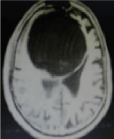
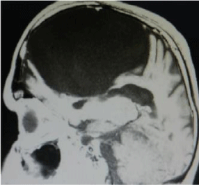
A 45-year-old male presented to with complaints of repeated episodes of generalized tonic clonic seizure for last three years, first episode occured two hours after sustaining head injury following motor vehicle accident and labile mood, suddenly used to start cry or laughing without any reason or proper situation or provocation causing severe social embarrassment. On Examination, he was conscious, with mild impairment of recent memory with episodes of unprovocative laugher and cry, with visual acuity of 6/18 on both sides and fundi showed papilloedema, positive palmomental reflex and rest of neurological examination were within normal limit. MRI brain revealed a giant cystic mass lesion, located in the interhemispheric fissure showing hypointense signal on T1W image (Figure 1) and oval shaped, well-defined outlines and smooth-walled, located in midline in the interhemispheric fissure causing displacement of medial part of both adjoining cerebral hemispheres with compressed lateral ventricles and displaying hyperintense signal on T2W image (Figure 2), sagittal section T1W magnetic resonance images revealed compression and distortion and inferiorly displacement of corpus callosum (Figure 3). On contrast administration, no enhancement of cyst wall or content was noticed. His seizure was controlled with phenytoin 300 mg daily dosages, however, he also needed cystoperitoneal shunt surgery for progressive headache during follow-up. At last follow-up his seizure were controlled on antiepileptic medication and headache also subsided.
Discussion
Interhemispheric fissure arachnoid cyst is characterized by presence of a cystic spaces filled with cerebrospinal fluids like content , located in the various parts of the craniospinal compartment, mostly in the intracranial compartment, being uncommon in the spinal region. In the intracranial compartment, IHAC is usually located in the cisternal spaces and larger intracranial intracerebral fissures. IHAC is considered extremely rare and originates in the arachnoid space located between two cerebral hemispheres, as classically not associated with agenesis of corpus in elderly age [1]. Mostly IHAC have unilateral lateralization to, barring one case- a 58 -year male reported by Hirohata et al. who also had bilateral extension of IHAC into adjacent cerebral hemisphere causing displacement, distortion and compression causing mass effect [3]. To the beset of knowledge of authors, current case is unique first case in literature, of giant size, with bilateral extension. The differential diagnosis includes neuroepithelial cysts, ependymal cysts and colloid cysts located in interhemispheric fissure [2,4].
IHSC may get symptomatic due to larger size as our case giant size, may present with seizure, headache, limb weakness and neuropsychological dysfunction [5,8].
De K et al. observed arachnoid cyst is intra-arachnoidal cerebrospinal fluid collections that are usually asymptomatic, but may become acutely symptomatic because of haemorrhage and enlargement cyst, which may even follow minor head trauma. As current case also sustained injury and presented with seizure, however CT scan head did not reveal presence of fresh hemorrhage in the arachnoid cyst, but hemorrhage might have been subtle to be pocked on CT scam and more so CT scan was carried about after ten days following motor vehicle accident. After trauma, these cyst may present with seizure. De K et al. reported of a child, who presented with headaches after minor head trauma and CT scan head revealed presence of arachnoid cyst with associated evidence of haemorrhage, which was managed surgically and author further cautioned headache, behavioral or cognitive changes and ataxia are more commonly associated than vomiting, visual disturbances or seizures and these symptoms following minor head trauma necessitate cranial CT scan [8].
Surgical management options include cystoperitonreal shunt or microsurgical excision or endoscopic fenestration. However minimum invasive surgery should be option of choice and currently endoscopic fenestration is considered as treatment of choice [1,3,4,7-10]. Each procedure can have better outcome. Even, cystoperitonreal shunting for symptomatic arachnoid cyst also be successful surgical option in the resource constraint centre with lacking endoscopic facility [5,6].
The mechanism of epileptogenesis in arachnoid cyst-related epilepsy is unknown. However, compression of the adjoining neurovascular structures of brain may be possible mechanism. Okada and colleagues analyzed the relationships among seizures, regional cerebral blood perfusion and volume of arachnoids cysts. The volume of arachnoid cysts in patients without epilepsy was significantly larger comparing to patients with epilepsy. And patients with epilepsy demonstrated hypoperfusion on SPECT study [11].
Conclusion
Every patients with known asymptomatic or incidental large intracranial arachnoid cyst should try to avoid trauma as these cases are relatively more vulnerable for acute deterioration in the neurological status following the minor trauma and may develop hemorrhage into cyst or acute enlargement of arachnoid cyst is also known and may even and need to be investigated with fresh cranial computed tomography scan as few of these cases may need emergency treatment. Even in the older age, possibility of arachnoid cyst should be considered as one of differentials in cases presenting with raised intracranial pressure or seizure.
- Jakubiak P, Dunsmore RH, Beckett RS (1968) Supratentorial brain cysts. J Neurosurg 28: 129-136. Link: https://goo.gl/6nyRKy
- Matsuda M, Hirai O, Munemitsu H, Kawamura J, Matsubayashi K (1982) Arachnoid cysts: Report of two adult cases in the interhemispheric fissure and over the cerebral convexity. Neurol Med Chir (Tokyo) 22: 71-76. Link: https://goo.gl/YJmD3v
- Hirohata M, Matsuo H, Miyagi J, Kajihara K, Shigemori M, (1992) Interhemispheric arachnoid cyst; Report of three cases. No Shinkei Geka 20: 701-705. Link: https://goo.gl/Bwl2oS
- Watanabe M, Kameyama S, Takeda N, Tanaka R (2004) Two cases of symptomatic interhemispheric arachnoid cyst in the elderly. Surg Neurol 42: 346-351. Link: https://goo.gl/O7Et94
- Kotil K, Balci N, Bilge T (2007) Intracranial symptomatic giant arachnoid cyst of the interhemispheric fissure presenting with frontal lobe syndrome. Turk Neurosurg 17: 147-151. Link: https://goo.gl/PR5sOC
- Satyarthee GD, Mahapatra AK (2015) Is subduro-peritoneal shunt surgery the first or last resort in managing subdural effusion developing after supratentorial tumor surgery in infancy? J Pediatr Neurosci 10: 83-85. Link: https://goo.gl/SxOXoW
- Satyarthee GD, Mahapatra AK (2002) Seizure as presentation of pituitary adenomas: series of four cases. Neurosciences Today 6: 167-169.
- De K, Berry K, Denniston S (2002) Haemorrhage into an arachnoid cyst: a serious complication of minor head trauma. Emerg Med J 19: 365-366. Link: https://goo.gl/VXQ6b5
- Pasricha R, Satyarthee Guru Dutta, Bhawani Shankar S (2016) Seizures following spinal dysraphism – surgery: A Series of Four Cases with Literature Review. American Journal of Clinical Neurology and Neurosurgery 2: 41-44. Link: https://goo.gl/zV7zGc
- Raheja A, Satyarthee GD, Mahapatra AK (2015) Chronic subdural hematoma development in accelerated phase of chronic myeloid leukemia presenting with seizure and rapid progression course with fatal outcome. Roman Neurosurgery 29: 199-202. Link: https://goo.gl/5m1Jvt
- Okada Y, Hamano K, Iwasaki N, Horigome Y, Enomoto T, et al. (1998) Epilepsy accompanied by intracranial arachnoid cysts: Studies on volume and regional cerebral blood perfusion using MRI and SPECT. J Epilepsy 11: 195-201. Link: https://goo.gl/NWPWjA
Article Alerts
Subscribe to our articles alerts and stay tuned.
 This work is licensed under a Creative Commons Attribution 4.0 International License.


 Save to Mendeley
