Open Journal of Trauma
Impact of Clavicular Shortening after Midclavicular Fracture: A Retrospective Series
Axel Jubel1*, Gereon Schiffer2, Jonas Andermahr3 and Christoph Faymonville4
2Orthopedic Trauma-Department, Vinzenz-Pallotti-Hospital, Bergisch Gladbach, Germany
3Orthopaedic Trauma-Department, Hospital of Mechernich, Germany
4Orthopedic Trauma-Department, University Hospital of Cologne, Germany
Cite this as
Jubel A, Schiffer G, Andermahr J, Faymonville C (2017) Impact of Clavicular Shortening after Midclavicular Fracture: A Retrospective Series. Open J Trauma 1(1): 014-019. DOI: 10.17352/ojt.000004Background: Clavicular shortening often occurs after midclavicular fractures and its impact on functional outcomes has thus far been evaluated solely by radiographic and surgeon-based measures, with divergent findings.
The goal of this study was to evaluate shoulder function and disability after midclavicular fractures in relation to shortening and compare it with that of healthy individuals and individuals with nonunion.
Methods: Seventy-one adult patients (38±14 years) with midclavicular fractures that had been treated nonoperatively were reviewed retrospectively after a mean follow up of 28±15 months. The primary outcome variables were Disabilities of the Arm, Shoulder, and Hand, Constant–Murley, and Cologne clavicle scores. Range of motion was calculated as the difference in degrees between the injured and uninjured sides. Control cohorts of 35 healthy adults and 28 persons with nonunion were assembled.
Results: Average shortening was 1.2±0.75 cm. Patients with clavicular shortening of >2 cm (Group 3) had significantly more pain, greater loss of mobility and lower Constant–Murley scores than patients with shortening < 1 cm (Group 1) and healthy controls. Shortening deformity of more than 2 cm associated with Disabilities of the Arm, Shoulder, and Hand, Constant–Murley, and Cologne clavicle equivalent to those of subjects with nonunion. Shortening deformity of more than 2 cm is functionally equivalent to nonunion.
Conclusions: Shortening deformities after clavicular fractures in adults greatly impact functional outcomes. Patients perceive a shortening deformity of ≥ 2cm as conferring significant disability. These findings suggest that the goal of therapy for diaphyseal clavicular fractures should be restoration of anatomical length of the clavicle.
Introduction
With a yearly incidence of 20–30 per 100,000, clavicular fractures are one of the most frequent injuries of the adult skeleton [1]. Midclavicular fractures lead to a typical deformity: the lateral fragment displaces caudally, anteriorly and medially, leading to angulation and overall shortening of the clavicle.
Because good results with minimal functional deficits following nonoperative treatment of clavicular fractures have been reported in the past, it has been standard to manage this injury nonoperatively and allow healing in the resulting deformed position [2]. However, some studies have suggested a high incidence of symptomatic non- and mal-union after nonoperative treatment of displaced fractures in adults [3]. Symptomatic patients typically have marked displacement at the fracture site characterized by shortening [4,5].
Thus, whether shortening deformity after diaphyseal clavicular fracture influences shoulder function has been debated in published reports [6-9]. An increasing number of studies have reported that such shortening deformity is associated with greater disability of the shoulder [10-13], whereas other studies have reported no such association [2,14,15].
The goal of this retrospective study was to evaluate the influence of clavicular shortening after midclavicular fractures on clinical outcomes in patients treated nonoperatively.
Patients and Methods
The previously collected data of 189 patients with a history of clavicular fracture were retrospectively evaluated with a focus on shortening deformities and a new clavicular fracture scoring system validated [16].
Inclusion criteria for this analysis were isolated midshaft clavicular fracture, nonoperative treatment, and bony union.
Exclusion criteria included: follow-up < 12 months, operative treatment, age < 16 years, presence of acute injury or acute or chronic disease of the ipsi- or contra-lateral shoulder girdle, polytrauma, previous history of a contralateral clavicular fracture, refracture, and pathologic fracture.
Among the cohort of 189 patients 108 had been treated operatively and were therefore excluded, leaving 71 patients with complete data and a mean follow-up of 28 months enrolled in this study. These data were analyzed with a focus on shortening deformities. Control cohorts of 35 adult patients with healthy shoulders and 24 subjects with nonunion were assembled. Table 1 gives summarized relevant variables of the investigated patients and control groups.
Evaluation
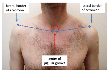
The data sets for each patient included: difference in length of clavicles, length being measured from the center of the jugular groove to the lateral border of acromion (Figure 1) and clavicular shortening being defined as the difference between the affected and unaffected sides [16]; subjective assessment of pain severity on a visual analog scale (VAS 0–100 points) [17]; range of motion (ROM) of the shoulder joint using the neutral-zero method; patient’s rating of ROM by VAS 1–6 (German school grades), and Constant–Murley [18], Disabilities of the Arm, Shoulder, and Hand (DASH) [19], and Cologne clavicle scores (CCS) [16].
DASH is a responsive, validated, and reliable patient-oriented outcome measure for assessing disability of the upper extremity: the higher the DASH-score the greater the disability, 100 points indicating a completely disabled extremity and 0 points a “perfect” extremity.
The subjective variables of pain and activities of daily living account for 35% of the total Constant–Murley shoulder score, whereas the objective variables of ROM and power account for the other 65%. To make these variables age-independent, a relative Constant–Murley score was calculated from the Constant–Murley score and expressed as a percentage of the score of the uninjured side. Thus, the best relative Constant–Murley score is 100 %. Higher scores denote better function and greater satisfaction whereas lower scores denote greater disability. The CCS [16], is a patient-oriented instrument for assessing outcomes of midclavicular fractures. It comprises six categories: three objective (clavicular shortening, ROM anteflexion, pain (VAS score, 0–100) and three corresponding subjective items (asymmetry of shoulder (VAS), limitations in daily activity (VAS), and limitations in sports or heavy physical work (VAS). A final (seventh) category is radiographic assessment according to the Nordqvist criteria [2], which are as follows: bony healing with displacement of the fragments less than the shaft width and angulation less than 30°; healing with deformity with fragment displacement more than shaft width and angulation >30°, and nonunion. The final score is determined by adding the individual values for each item. The following ranges were used to evaluate final outcomes: 0–3 points = very-good, 4–8 = good, 9–14 = moderate, and 15–24 = poor. DASH, CS and CC scores were the primary outcome variables.
ROM was calculated as the difference in degrees between the injured and uninjured sides.
For detailed analysis, the data sets were divided into the following three groups based on the magnitude of shortening (Table 1): Group 1 = shoulder shortening (∆L) ≤ 0.5 cm, Group 2 = 0.5 cm < ∆L < 2 cm, Group 3 = ∆L ≥ 2 cm. There were two groups of controls: Group 4 = nonunited fractures and Group 5 = healthy individuals.
IRB approval: The study was approved by the IRB of the University Hospital of Cologne, number 16-219.
Statistics: The collected data were assessed using the statistics program IBM® SPSS® (release 21.0, 1989–2012). Descriptive statistical tests and the Mann–Whitney-U test were used. The level of significance was set at (**) p < 0.001 and (*) p < 0.05.
Results
The mean duration of follow-up of the entire cohort was 28 ± 15 months. Patients with shortening of ∆L ≥ 2 cm (Group 3, Table 1) were significantly older (average age of 48 years; p < 0.05) than those with less shortening (average age of 36 years). Patients in the clavicular nonunion control group (average age 43 years) tended to be older, but this difference was not statistically significant (p = 0.11).
Clavicular shortening
The frequency of various degrees of shoulder shortening (∆L) is depicted in Table 1. Average shortening was 1.2 ± 0.75 cm (mean 1.0 cm, range 0–3 cm). Average shortening in Groups 2 and 3 was 1.9 ± 0.1 cm (mean 2.0 cm) and 2.8 ± 0.2 cm (mean 3.0 cm), respectively. In the healthy control group, one individual had a length difference of 0.5 cm (Table 1).
Pain
Sixteen of 21 patients (76%) in Group 1 (∆L ≤ 0.5 cm), 18/33 (55%) in Group 2 (0.5 cm < ∆L < 2 cm), 7/17 (41%) in Group 3 (∆L ≥ 2 cm), and 7/24 (28%) in Group 4 (nonunion) were completely pain-free.
The average subjective pain scores were 6 ± 2.5 points (mean 0, range 0–35) in Group 1, 17 ± 4 points (mean 0, range 0–80) in Group 2, 21 ± 6 points (mean 20, range 0–70) in Group 3, and 28 ± 5 points (mean 21, range 0–90) in Group 4.
The average subjective pain scores of Groups 2 and 3 differed significantly from those of the healthy control group (Group 5) and Group 1 (all p < 0.001).
There was no significant difference between Group 2 and Group 3 (p = 0.475) or between Group 3 and Group 4 (p = 0.475).
Range of motion
Limitations in ROM were largest in Group 3 and the nonunion control group. The average deficit of ROM in active flexion was 3°± 1.7° in Group 1, 6°± 2° in Group 2, 17°± 6° in Group 3, and 23°± 6.5° in Group 4, respectively. The differences in ROM between Groups 2 or 3 and healthy controls are significant (both p < 0.001).
Patient rating of deficit in ROM (VAS 1–6)
The average deficit of ROM in active flexion was rated by patients’ VAS scores as 1.5 ± 0.6 in Group 1, 2.2 ± 0.2 in Group 2, 2.5 ± 0.3 in Group 3 and 3.3 ± 0.3 in Group 4, respectively.
There were no significant differences in these ratings between Groups 2 and 3 and Groups 3 and 4. The ratings of Group 2 and 3 were significantly different from those of Group 5 (healthy individuals) (p < 0.001) and Group 1 (∆L ≤ 0.5 cm) (p < 0.001).
Constant–Murley scores: Figure 2 shows relative Constant–Murley scores in box plot form. The mean values were 95% in Group 1, 90% in Group 2 (0.5 cm < ∆L < 2 cm), 78% in Group 3 (∆L ≥ 2 cm), and 75% in Group 4 (nonunion). The values for Groups 2 and 3 are significantly lower than those of healthy controls and in Group 1 (p < 0.001). There were no significant differences between Groups 3 (∆L ≥ 2 cm) and 4 (nonunion), in pain scores, ROM, or Constant–Murley scores.
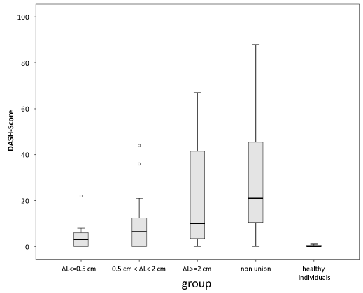
DASH scores: Figure 3 shows DASH scores in box plot form. The average values were 5.5 ± 2 points in Group 1 (∆L ≤ 0.5 cm), 8.9 ± 2.3 points in Group 2 (0.5 cm < ∆L < 2 cm), 21 ± 6.4 points in Group 3 (∆L ≥ 2 cm), and 29 ± 5 points in Group 4 (nonunion). The values for Groups 2 and 3 are significantly higher than those of healthy controls and in Group 1 (p < 0.001).
There were no significant differences between Groups 3 (∆L ≥ 2cm) and 4 (nonunion) in pain scores, ROM, or Constant–Murley, CCS, and DASH scores.
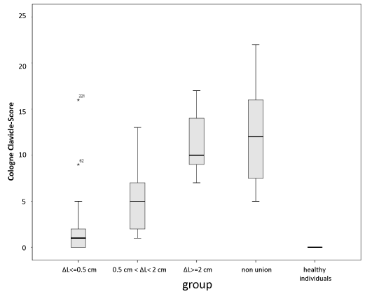
Cologne clavicle scores: Figure 4 depicts CCS in box plot form, low values indicating a good result and 0 points being the best. The mean values were 1.3 ± 0.6 points in Group 1 (∆L ≤ 0.5 cm), 5.1 ± 0.7 points in Group 2 (0.5 cm < ∆L < 2 cm), 12 ± 0.9 points in Group 3 (∆L ≥ 2 cm), and 12.1 ± 1 points in Group 4 (nonunion). The values for Groups 2 and 3 are significantly higher than those of healthy controls and in Group 1 (p < 0.001).
Discussion
From a biomechanical point of view, one of the responsibilities of the clavicle is to hold the glenohumeral joint lateral to the trunk of the body and thereby ensure freedom of the joint [20]. Additionally, the clavicle serves as a stable muscle origin [20]. Although shoulder asymmetry after clavicular fractures is a complex three-dimensional problem, in symptomatic patients shortening of the medio-lateral length of the clavicle is frequently the most characteristic finding [5,6,10,11,21]; it is also sometimes present in asymptomatic patients. There is increasing evidence that patients can have substantial dissatisfaction following clavicular malunion because of symptoms including pain, weakness, and easy fatigability [4,5,10,11,22].
McKee et al.’s working group [5], reported on a series of 15 patients who showed consolidated bony healing on radiographs yet were not pain-free an average 20 months post-trauma. These patients had an average clavicular shortening of 2.9 cm. In similar investigations, Chan et al. [23], found an average shortening of 2–3 cm, and Bosch et al. [4] found shortening of 1.6 cm. However, these studies comprised highly specific patient cohorts composed exclusively of patients who had undergone corrective osteotomy. Several studies have found inferior clinical outcomes in the presence of shortening of 1.5–2 cm after healing [10-12,24], whereas others have not demonstrated such a relationship [14,25,26]. In an experimental, biomechanical setting it has been demonstrated that clavicular shortening causes a significant glenoid malposition [27]. In a cadaveric study, it was found that shortening of the clavicle of more than 10% affects the kinematics in the shoulder girdle and could produce clinical symptoms [28]. McKee et al. [3], assessed DASH and Constant–Murley scores and muscle strength a mean of 55 months after treatment in 30 patients with clavicular fractures who had been treated nonoperatively. The strength of the injured shoulder was reduced to 81% of that of the uninjured shoulder for maximum flexion. The mean Constant–Murley score was 71 points, and the mean DASH score 24.6 points, indicating substantial residual disability. Shortening of ≥2 cm was associated with a trend toward greater patient dissatisfaction [3]. In a very young cohort of 71 patients (mean age 11 years) with midclavicular fractures, Norquist [9], found 5 years after injury that mobility, strength and functional Constant–Murley scores were similar in the injured and normal shoulders. Because of the spontaneous correction that characteristically occurs in young patients, the current study focused on adults. Flavin [22], et al. investigated 35 patients 3 years post-injury and reported an average clavicular shortening of 15 mm and decreased isometric force when the injured and uninjured sides were compared. The three patients with the most marked shortening (27, 30, and 35 mm) also had the lowest Constant–Murley scores (89, 86, and 86 points, respectively) [22]. However, there was no statistically significant difference between patients with shortening < 15 mm and those ≥ 15 mm. Oroko et al. [8], reported that clavicular shortening had no influence on Constant–Murley scores at 3 months post-injury. In the authors’ experience the bony healing process is not complete at 3 months post-trauma and many patients still complain of functional deficits and pain at this stage of healing. Therefore, we consider time of assessment chosen by Oroko et al. [8], unsuitable for conclusively evaluating therapy outcomes. Eskola [10], found that patients with a shortening deformity of more than 15 mm 2 years post-injury reported significantly more pain and had more significant abduction deficits than patients with < 15 mm shortening. In a study of 52 patients, Hill [11], determined that the magnitude of shortening was the exclusive influence on functional results after 3 years. Shortening of ≥ 20 mm was significantly more frequently paired with an unsatisfactory result. Gaebler et al. [6], showed that 50 % of patients with shortening of 1 cm and 100 % of patients with shortening of 2 cm had accompanying measurable deficits in shoulder function. Lazarides and Zafiropoulos [12], reported that shortening of more than 18 mm in male patients and 14 mm in female patients was associated with a poor clinical outcome. In a previous cadaver study [27], it was shown that healing of clavicle fractures with bony shortening leads to a ventromedial caudal shift in glenoid fossa position. The following malposition of the clavicle leads to the respective glenoid fossa positional changes: caudal deviation leads to a mediocaudal shift, cranial deviation leads to a dorsolateral shift of the glenoid fossa, ventral deviation causes a ventrolateral shift, dorsal deviation leads to mediocaudal shift of the fossa, cranial rotation leads to ventrolateral shift in fossa position, and caudal rotation leads to a dorsomedial shift in glenoid fossa position. Clinical implication of these data is that bony shortening in combination with caudal displacement leads to distinct functional deficits in abduction, particularly overhead motion.
To the authors’ knowledge, the current study is the first to compare clinical outcomes in relation to shortening deformity with healthy individuals on the one hand and nonunion on the other. In the current study, shortening was associated with an inferior clinical outcome compared with healthy individuals. Constant–Murley and DASH scores and CCS became progressively worse in parallel with increasing shortening.
The results of the present study are in agreement with those of Lazarides [12], who reported an association between shortening and inferior clinical outcome. However, some authors of other retrospective studies have not found that shortening correlates with outcome [14,15,24,29]; these earlier studies had several limitations. None of the studies so far reported compared functional results post-clavicular fracture with those of a control group. In the present study, all scores and measurements were evaluated and compared with the controls of 35 healthy individuals and 24 patients with nonunion. Also, whereas prior investigators used only radiographic and surgeon-based, rather than patient-based, outcome measures, we used patient-based outcome measures as well as radiographic and surgeon-based outcomes [9,14,15,29]. Nordquist et al. [9], evaluated a cohort of 225 patients with a mean follow up of 17 years after clavicular fracture. Because a limitation of ROM of up to 45° was rated as a “good” result, the result of only one of these 225 patients was rated as “bad”. Different ratings are obtained when patient-oriented questionnaires are used. In the present study, patients rated a flexion deficit of 40–50° on a VAS (1–6 according to the German school grading system) as 4.2 points on average, signifying a subjective disability. The use of a patient-based outcome measures like DASH scores and CCS is the crucial difference between studies indicating inferior clinical outcomes in the presence of shortening and those claiming that clinical outcomes are not influenced by shortening deformity [3,24]. McKee et al. [3], have pointed out that there are certainly differences between what clinicians consider as “good” and what patients experience as a good result.
The findings of the study presented here indicate that clavicular shortening greater than 1 cm is associated with significantly inferior scores compared with healthy individuals. Shortening deformity of more than 2 cm is associated with pain scores, measurable ROM of the shoulder joint, Constant–Murley and DASH scores and CCS assessments that are equivalent to findings in patients with nonunited fractures, signifying that shortening deformity of more than 2 cm is associated with substantial clinical disability.
Nevertheless, final CCS indicated excellent and good results in 53 of 71 patients (72%). Evaluating of outcomes is important, especially differentiating between “statistically significant” and “clinically relevant”. All bad and most moderate ratings were in Group 3, whereas there were no bad ratings in Groups 1 and 2, indicating that shortening of ≥2 cm is relevant to the perception of patients. This supports the proposition of McKee et al. [3], that functional outcomes do not have a linear relationship but are ‘all or none’ phenomena. Thus, shoulder function is well preserved until a critical threshold of deformity is reached, after which it is dramatically impaired.
The strengths of this study include the duration of follow-up (a mean of more than 2 years since injury) and the use of both patient-oriented and objective outcome measures.
This study also has several weaknesses. It was retrospective and the sample size was relatively small because its size was determined by a previous study aimed at evaluating a new scoring system [16].
We are reluctant to make recommendations regarding the optimal treatment of displaced midclavicular fractures because our study showed only that there is inferior clinical outcome following nonoperative treatment in the presence of shortening deformity; we have no proof that initial operative treatment would have been superior.
Conclusion
In conclusion, the results of the current study indicate that shortening deformities after clavicular fractures of more than 1 cm are associated with inferior clinical results compared with healthy individuals. Constant–Murley and DASH scores and CCS became progressively worse in parallel with increasing shortening. Shortening deformity of more than 2 cm is functionally equivalent to nonunion. Our results further indicate that patients perceive a shortening deformity of ≥ 2cm as conferring significant disability. Our findings suggest that the goal of therapy for diaphyseal clavicular fractures should be restoration of the anatomical length of the clavicle.
Figure captions
Group 1 = shoulder shortening (∆L) ≤ 0.5 cm.
Group 2 = 0.5 cm < ∆L < 2 cm.
Group 3 = ∆L ≥ 2 cm. There were two groups of controls.
Group 4 = nonunited fractures.
Group 5 = healthy individuals.
- Robinson CM (1998) Fractures of the clavicle in the adult. Epidemiology and classification. J Bone Joint Surg Br 80: 476-484. Link: https://goo.gl/1GnghW
- Nordqvist A, Petersson CJ, Redlund-Johnell I (1998) Mid-clavicle fractures in adults: end result study after conservative treatment. J Orthop Trauma 12: 572-576. Link: https://goo.gl/gCmSe5
- McKee MD, Pedersen EM, Jones C, Stephen DJ, Kreder HJ, et al. (2006) Deficits following nonoperative treatment of displaced midshaft clavicular fractures. J Bone Joint Surg Am 88: 35-40. Link: https://goo.gl/lk7t23
- Bosch U, Skutek M, Peters G, Tscherne H (1998) Extension osteotomy in malunited clavicular fractures. J Should Elbow Surg 7: 402-405. Link: https://goo.gl/gTk6Rb
- McKee MD, Wild LM, Schemitsch EH (2003) Midshaft malunions of the clavicle. J Bone Joint Surg Am 85-A(5): 790-797. Link: https://goo.gl/eCFpeq
- Gaebler C, Matis N, Kwasny O, Zauner-Dungel A (1995) The Allman-I-fracture of the clavicle. Akt Traumatol 25: 50-55. Link:
- Eskola A, Vainionpaa S, Patiala H, Rokkanen P (1987) Outcome of operative treatment in fresh lateral clavicular fracture. Ann Chir Gynaecol 76: 167-169. Link: https://goo.gl/X9VvIU
- Oroko PK, Buchan M, Winkler A, Kelly IG (1999) Does shortening matter after clavicular fractures? Bull Hosp Joint Dis 58: 6-8. Link: https://goo.gl/PezNAJ
- Nordqvist A, Redlund-Johnell I, von Scheele A, Petersson CJ (1997) Shortening of clavicle after fracture. Incidence and clinical significance, a 5-year follow-up of 85 patients. Acta Orthop Scand 68: 349-351. Link: https://goo.gl/8mMwHR
- Eskola A, Vainionpaa S, Myllynen P, Patiala H, Rokkanen P (1986) Outcome of clavicular fracture in 89 patients. Arch Orthop Trauma Surg 105: 337-338. Link: https://goo.gl/kuEL8U
- Hill JM, McGuire MH, Crosby LA (1997) Closed treatment of displaced middle-third fractures of the clavicle gives poor results. J Bone Joint Surg Br 79: 537-539. Link: https://goo.gl/MLDSJq
- Lazarides S, Zafiropoulos G (2006) Conservative treatment of fractures at the middle third of the clavicle: the relevance of shortening and clinical outcome. J Shoulder Elbow Surg 15: 191-194. Link: https://goo.gl/ceZa3t
- Abbot AE, Hannafin JA (2001) Stress fracture of the clavicle in a female lightweight rower. A case report and review of the literature. Am J Sports Med 29: 370-372. Link: https://goo.gl/VPzW5B
- Fuglesang HF, Flugsrud GB, Randsborg PH, Stavem K, Utvag SE (2016) Radiological and functional outcomes 2.7 years following conservatively treated completely displaced midshaft clavicle fractures. Arch Orthop Trauma Surg 136: 17-25. Link: https://goo.gl/IbjTB6
- Rasmussen JV, Jensen SL, Petersen JB, Falstie-Jensen T, Lausten G, et al. (2011) A retrospective study of the association between shortening of the clavicle after fracture and the clinical outcome in 136 patients. Injury 42: 414-417. Link: https://goo.gl/F78Mnj
- Jubel A, Weisshaar G, Faymonville C, Andermahr J, Schiffer G (2012) [A simple clavicle score : An effective and reliable classification for outcome assessments of midclavicular fractures]. Der Unfallchirurg 115: 1085-1091. Link: https://goo.gl/87pGRG
- Huskisson EC (1974) Measurement of pain. Lancet 2: 1127-1131. Link: https://goo.gl/VgGYdY
- Constant CR (1987) A clinical method of functional assesment of the shoulder. Clin Orthop Relat Res 214: 160-164. Link: https://goo.gl/sS02sq
- Hudak PL, Amadio PC, Bombardier C (1996) Development of an upper extremity outcome measure: the DASH (disabilities of the arm, shoulder and hand) [corrected]. The Upper Extremity Collaborative Group (UECG). Am J Ind Med 29: 602-608. Link: https://goo.gl/RIhMLD
- Moseley HF (1968) The clavicle: Its anatomy and function. Clin Orthop Relat Res 58: 17-27. Link: https://goo.gl/CvgnL1
- Jubel A, Andermahr J, Faymonville C, Binnebosel M, Prokop A, et al. (2002) [Reconstruction of shoulder-girdle symmetry after midclavicular fractures. Stable, elastic intramedullary pinning versus rucksack bandage]. Chirurg 73: 978-981. Link: https://goo.gl/P13q5a
- Flavin RA, Fleming F, Shanley L, Kelly IP (2004) Closed treatment of clavicle fractures results in reduced shoulder strength. Eur J Orthop Surg Traumatol 14: 84-88. Link: https://goo.gl/j0HTo4
- Chan KY, Jupiter JB, Leffert RD, Marti R (1999) Clavicle malunion. J Should Elbow Surg 8: 287-290. Link: https://goo.gl/wR39fI
- Thormodsgard TM, Stone K, Ciraulo DL, Camuso MR, Desjardins S (2011) An assessment of patient satisfaction with nonoperative management of clavicular fractures using the disabilities of the arm, shoulder and hand outcome measure. J Trauma 71: 1126-1129. Link: https://goo.gl/kDWxnD
- Nowak J, Holgersson M, Larsson S (2005) Sequelae from clavicular fractures are common: a prospective study of 222 patients. Acta Orthop 76: 496-502. Link: https://goo.gl/lWSXlt
- Stegeman SA, de Witte PB, Boonstra S, de Groot JH, Nagels J, et al. (2015) Posttraumatic midshaft clavicular shortening does not result in relevant functional outcome changes. Acta Orthop 86: 545-552. Link: https://goo.gl/ej7vxo
- Andermahr J, Jubel A, Elsner A, Prokop A, Tsikaras P, et al. (2006) Malunion of the clavicle causes significant glenoid malposition: a quantitative anatomic investigation. Surg Radiol Anat 28: 447-456. Link: https://goo.gl/QEoqXX
- Matsumura N, Ikegami H, Nakamichi N, Nakamura T, Nagura T, et al. (2010) Effect of shortening deformity of the clavicle on scapular kinematics: a cadaveric study. Am J Sports Med 38: 1000-1006. Link: https://goo.gl/8uNHGp
- Nordqvist A, Redlund-Johnell I, vonScheele A, Peetersson CJ (1997) Shortening of clavicle after fracture. Incidence and clinical significance, a 5-year follow-up of 85 patients. Acta Orthop Scand 68: 349-351. Link: https://goo.gl/qdPWYw
Article Alerts
Subscribe to our articles alerts and stay tuned.
 This work is licensed under a Creative Commons Attribution 4.0 International License.


 Save to Mendeley
