Open Journal of Orthopedics and Rheumatology
Camptodactyly Arthropathy CoxaVara Pericarditis Syndrome: Early diagnosis prevents unnecessary and harmful treatment
Sheren Esam Maher1*, Mohamed Hashem M Mahgoob1 and Nadia El Ameen2
2Assistant Professor of Radiology -Minia University Hospital -Egypt
Cite this as
Maher SE, Mahgoob MHM, El Ameen N (2017) Camptodactyly Arthropathy CoxaVara Pericarditis Syndrome: Early diagnosis prevents unnecessary and harmful treatment. Open J Orthop Rheumatol 2(1): 012-014. DOI: 10.17352/ojor.000008Camptodactyly‑arthropathy‑coxa vara‑pericarditis (CACP) syndrome is a genetic disorder caused by mutation in the Proteoglyacn PRG4 gene on chromosome 1. The syndrome is characterized by congenital or early onset camptodactyly and childhood‑onset of non‑inflammatory arthropathy, coxa vara deformity, or other dysplasia associated with progressive hip disease and non‑inflammatory pericardial effusion. It has an autosomal recessive mode of inheritance and the causative gene is located on chromosome band 1q25-31.
Introduction
Juvenile idiopathic arthritis is the most common inflammatory joint disease. No single clinical sign or symptom or test result distinguishes it from other joint diseases, nor are the pathological features of synovitis [1]. Rather it diagnosed by a combination of clinical findings and laboratory tests. There are a number of disease entities affecting the joints that may present in a similar way. Treatment of juvenile idiopathic arthritis now based on more aggressive management including the use of traditional disease-modifying antirheumatic drugs upon disease onset and the early use of biologics [2].
We present a case of a male child who presented with early onset camptodactyly, non‑inflammatory arthropathy, with specific features magnetic resonance imaging (MRI).
Case Report
A 3‑year 5‑month‑old boy presented with a one year history of swelling of both knees and wrists. The mother complained from swelling that progress to joint pain and limitation of movement. She also noticed bending deformity of three fingers of his hands. There was no systemic symptoms as fever, rash, abdominal pain or swelling.
Laboratory Investigation of our patient at the onset of the disease: Routine blood work‑up revealed normal hemogram, Erythrocyte Sedimentation Rate (ESR) and C‑reactive protein (CRP) with negative Antinuclear Antibody (ANA) and Rheumatoid factor (RF).
Knee US report showed joint effusion and synovial membrane hypertrophy.
So, patient started Non-steroidal anti-inflammatory drugs (NSAID) on 2mg/kg for 6 weeks. On follow up visit there was slight significant Improvement .Our patient diagnosed as oligoarthritis class of Juvenile Idiopathic Arthritis after exclusion of other conditions associated with or mimicking arthritis.
Follow up of our patient revealed no improvement of joint state so new line started in the form of intra articular steroid injection and biological therapy. Course of the disease remain stationery without any improvement.
More investigations as α-Gal An enzyme activity assay in leucocytes for Fabry disease which came normal.
Our patient came to our pediatric Rheumatology clinic –Minia University where he was reevaluated. On examination, the patient had camptodactyly of the fingers with swelling of both knees and wrists without restricted motion or signs of inflammation like erythema or tenderness. There was also, a waddling gait the parents also stated that they had noticed bent fingers for nearly one and a half years.
Systemic examination showed no evidence of fever, lymphadenopathy, skin rash, or other systemic features but cardiac auscultation there was pan systolic murmur on mitral area grad III (MR). Echocardiography showed mild mitral regurge with normal systolic function.
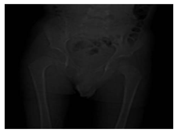
Anteroposterior (AP) radiograph of pelvis revealed a broad left coxa vara (Figure 1). The articular surfaces were smooth with no erosions.
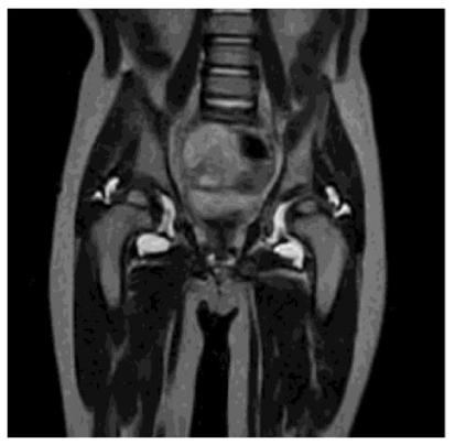
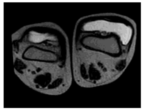
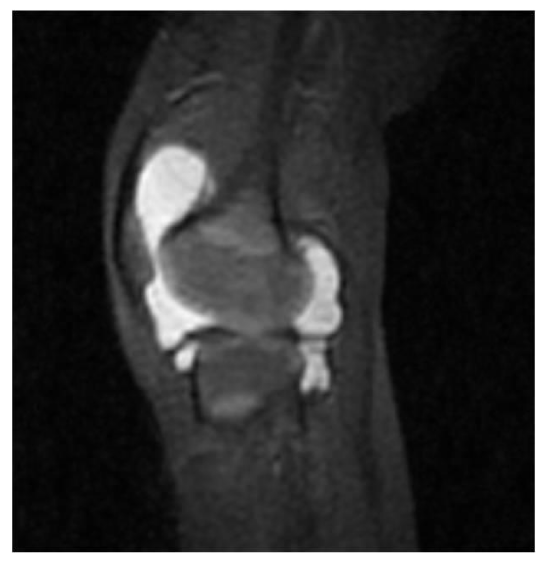
MRI of the hip showed a moderate bilateral hip joint effusion. The synovium was mildly thickened and showed enhancement (Figure 2) conspicuously, there was absence of cartilage destruction. Knee MRI Axial T2WI showed marked bilateral knee joint effusion more at the left side (Figure 3). Also, Sagittal T2WI showed the marked knee joint effusion (Figure 4).
Discussion
Camptodactyly-arthropathy-coxa vara-pericarditissyndrome is a rare genetic condition due to a mutation in the gene proteoglycan 4 (PRG4) - a chondroitin sulfateproteoglycan that acts as a lubricant for the cartilage surfaces. This gene is also known as lubricin. This condition is inherited as an autosomal recessive.
The gene responsible for this condition is located on the long arm of chromosome 1 (1q). The encoded protein is a glycoprotein of ~345 kDa [3], specifically synthesized bychondrocytes located at the surface of articular cartilage, and also by some synovial lining cells. The cDNA encodes a protein of 1,404 amino acids (human a isoform) with asomatomedin B homology domain, heparin-binding domains, multiple mucin-like repeats, a hemopexin domain, and an aggregation domain. There are 3 consensus sequences for N-glycosylation and 1 chondroitin sulfate substitution site.
This condition was first described in 1986 [4] and is a syndrome of camptodactyly, arthropathy, coxa vara and pericarditis [5]. It may also include congenital cataracts [6]. The cause of this syndrome was discovered in 1999 [7].
Children with this syndrome often present with a joint effusion that is cool and resistant to anti-inflammatory therapy. The arthropathy principally involves large joints such as elbows, hips, knees, and ankles [6]. Pericarditis may be a presenting feature or may occur later in the course of the disease but in our case we found Mitral regurge with normal systolic function. Coxa vara occurs in 50–90% of cases and non-inflammatory pericarditis in 30%.The main line of treatment is physiotherapy and supportive analgesics like paracetamol [7].
Conclusion
CACP syndrome should be considered in all patients who present with a non-inflammatory arthropathy or with “atypical juvenile idiopathic arthritis,” particularly if radiographs reveal an absence of erosions. Increasing awareness of this familial condition is required to prevent confusion with other inflammatory musculoskeletal conditions seen during childhood. Differentiation from JIA is clinically important in view of the different management protocol for the two conditions, and in particular because of the possible severe side effects associated with treatment for JIA.
- Jackie N, Combe B, Emery P (2013) Eular text book on rheumatic diseases.Rheumatoid arthritis: Pathogenesis and clinical features .First edition. Link: https://goo.gl/VhkIHq
- Iain B Mclnnes, Elsa Vieira-Sousa, Joao Eurico Fonseca (2013) Eular text book on rheumatic diseases. Rheumatoid Arthritis: Treatment. First edition. Link: https://goo.gl/nU99mB
- Su JL, Schumacher BL, Lindley KM, Soloveychik V, Burkhart W, et al. (2001) "Detection of superficial zone protein in human and animal body fluids by cross-species monoclonal antibodies specific to superficial zone protein". Hybridoma 20: 149–157. Link: https://goo.gl/yGztlF
- Bulutlar G, Yazici H, Ozdogan H, Schreuder I (1986) A familial syndrome of pericarditis, arthritis, camptodactyly and coxa vara. Arthritis Rheum 29: 436–438. Link: https://goo.gl/cWImG2
- Offiah AC, Woo P, Prieur AM, Hasson N, Hall CM (2005) Camptodactyly-arthropathy-coxa vara-pericarditis syndrome versus juvenile idiopathic arthropathy. AJR Am J Roentgenol 185: 522-529. Link: https://goo.gl/GQoDFd
- Akawi NA, Ali BR, Al-Gazali L (2012) A novel mutation in PRG4 gene underlying camptodactyly-arthropathy-coxa vara-pericarditis syndrome with the possible expansion of the phenotype to include congenital cataract. Birth Defects Res A Clin Mol Teratol. Link: https://goo.gl/odY92i
- Marcelino J, Carpten JD, Suwairi WM, Gutierrez OM, Schwartz S, et al. (1999) CACP, encoding a secreted proteoglycan, is mutated in camptodactyly-arthropathy-coxa vara-pericarditis syndrome. Nat Genet 23: 319-322. Link: https://goo.gl/NQSciz
Article Alerts
Subscribe to our articles alerts and stay tuned.
 This work is licensed under a Creative Commons Attribution 4.0 International License.


 Save to Mendeley
