Open Journal of Orthopedics and Rheumatology
Nuances of Microsurgical and Endoscope Assisted Surgical Techniques to the Cranio-Vertebral Junction: Review of the Literature
Visocchi Massimiliano1*, Signorelli Francesco1, Iacopino Gerardo2 and Barbagallo Giuseppe3
2Department of Experimental Biomedicine and Clinical Neurosciences, School of Medicine, Neurosurgical Clinic, University of Palermo, Italy
3Division of Neurosurgery, Department of Neurosciences, Policlinico “G. Rodolico” University Hospital, Catania, Italy
Cite this as
Visocchi M, Signorelli F, Iacopino G, Barbagallo G (2017) Nuances of Microsurgical and Endoscope Assisted Surgical Techniques to the Cranio-Vertebral Junction: Review of the Literature. Open J Orthop Rheumatol 2(1): 001-008. DOI: 10.17352/ojor.000006Purpose: An update of the technical nuances of microsurgical - endoscopic assisted approaches to the craniocervical junction (transnasal, transoral and transcervical) if provided from the literature in order to better contribute to identify the best strategy.
Methods: A non-systematic update of the review and reporting on the anatomical and clinical results of endoscopic assisted and microsurgical approaches to the craniocervical junction (CVJ) is performed.
Results: Pure endonasal and cervical endoscopic approaches still have some disadvantages, including the learning curve and the deeper surgical field. Endoscopically assisted transoral surgery with 30° endoscopes represents an emerging option to standard microsurgical techniques for transoral approaches to the anterior CVJ. This approach should be considered as complementary rather than an alternative to the traditional transoral-transpharyngeal approach.
Conclusions: Transoral (microsurgical or video-assisted) approach with sparing of the soft palate still remains the gold standard compared to the ‘‘pure’’ transnasal and transcervical approaches due to the wider working channel provided by the former technique. Transnasal endoscopic approach alone appears to be superior when the CVJ lesion exceeds the upper limit of the inferior third of the clivus. Of particular interest the evidence that advancement in reduction techniques can avoid ventral approach.
Introduction
Endoscopic endonasal, transoral and transcervical approaches have recently been developed as promising options to the traditional transoral microsurgery to the CVJ, and may become more mainstream as experience with these approaches increases (cons: learning curve, loss of 3-dimensional visualization) [1,2].
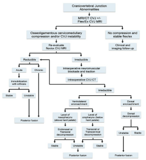
The transoral–transpharyngeal approach historically remains the ‘‘gold standard’’ for anterior approaches to the upper cervical spine when indicated according to the Menezes algorithm (Figure 1) [3]. However, there are still technical difficulties with the operating microscope, such as the need to see and work through a narrow opening in a deep cavity; to improve visualization; soft palate splitting and even hard palate resection along with extended maxillotomy are occasionally required. To overcome such complications, endoscopic assisted procedures for CVJ decompression have been developed. The endoscopic approaches to the CVJ include endoscopic endonasal approach, endoscopic transoral approach, robot- assisted endoscopic transoral approach, combined endoscopic transnasal and transoral approach, and endoscopic transcervical approach [4,5]. The aim of the current review is to give an update on the anatomic fundamentals of endoscopic assisted surgery to the CVJ and to report on the available clinical results.
Anatomic studies on endoscopic craniocervical approaches
The he most commonly used endoscopically assisted approaches to the craniocervical junction include transnasal, transoral and transcervical routes so far (Table 1) [2,6-22].
Endoscopic transoral approach: In 2004, de Divitiis et al., studied an endoscopic transoral– transclival intradural approach on 15 cadavers, without maxillotomy or mandibulotomy, and estimated a safe entry zone through the clivus endoscopically [7].
In 2006, Balasingam et al., conducted a cadaveric anatomical study to assess the area of surgical exposure and the available liberty of action for instrument manipulation by four surgical different approaches to the extracranial periclival region: traditional transoral route, transoral with a palate split, LeFort I osteotomy, and median labioglos- somandibulotomy [8].
In 2009, Pillai et al., performed an odontoidectomy in 9 specimens by a direct transoral approach; endoscope assisted (5 cases) or combined endoscopic––microscopic aid, evaluating the surgical working area and the surgical freedom; the authors conclude that endoscope and image guidance allow to approach the ventral CVJ transorally with minimal tissue dissection, no palatal splitting, and no compromise of surgical freedom [10] (Figure 1).
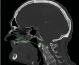
Endoscopic endonasal approach: The main advantages of this endonasal approach to the ventral craniovertebral junction are minimal invasiveness, unlimited surgical access to the rostral midline craniocervical junction, avoidance of palatal split, and overall less operative morbidity compared with the transoral approach. Thanks to a relatively inclined surgical trajectory, in a rostral to caudal direction, the compressive pathology of basilar invagination, including the lower clivus and odontoid tip, may be removable without removing C1 anterior arch, thus maintaining stability of C1–C2 [23]. In 2009, Kassam’s team published the concept of the ‘‘Nasopalatine line’’ (NPL) [23]. The NPL is a reliable predictor of the maximal length of inferior dissection, and odontoid surgery can safely be performed according to the preoperative radiological study of the potential anatomical limitations of the endonasal approach. In 2012 Aldana et al proved that a line in the midsagittal plane, the nasoaxial line (NAxL), connecting the midpoint of the distance from rhinion to the anterior nasal spine of maxillary bone and the C2 vertebra, tangential to the posterior nasal spine of palatine bone, accurately predicted the lowest limit of this approach on the cervical spine [15] (Figure 2).
Endoscopic transcervical approach: In 2011, Russo et al., [18] described the microsurgical anatomy and limits of exposure of the endoscopically assisted high anterior cervical, submandibular, approach to the clivus and foramen magnum; optimal route to access pathologies located ventral to the pontomedullary region. Two extensions of the approach were studied and described: an extended anterior far-lateral clivectomy and an inferior petrosectomy, thus extending the exposure to the anterior foramen magnum and the anterior cerebellopontine.
Comparison studies: In a study on 9 cadaver heads, in 2009 Baird et al., assessed surgical access to the craniocervical junction using three endoscopic approaches: endonasal, transoral and transcervical. Data suggested that the surgical goals of lower clival and odontoid decompression were achieved using the endonasal and transoral approach, and the distance to the target area was shorter in the first one. The transcervical approach was unable to achieve more than 1 cm of lower clival resection, thus not allowing complete odontoid resection [19]. In 2010, Seker et al., Stated that transnasal endoscopic approach provided the shorter route to the CVJ while the transoral exposure gained a wider opening [20].
However, the two approaches should be considered complementary rather than alternative. When removing large lesions that extend from the upper clivus to below C2, the transnasal and transoral route may be successfully used combined. The transcervical approach has the clear clinical advantage of reducing the risk of meningitis, of cerebrospinal fluid leak, to maintain a sterile surgical field, familiar approach, and an optimal surgical trajectory for pathological findings lower than C2.
In 2012, Dallan et al. [11], investigated a new robotic surgical setting, DaVinci system, in two cadavers, comparing the traditional transoral, and the combined transoral transnasal procedures to the CVJ. They concluded that the lower the placement of the robotic arms, the easier the dissection of the rhinopharynx, basisphenoid and upper clivus.
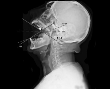
Visocchi et al., compared the surgical exposition angle and the working channel volume of both transnasal and transoral approaches in the cadaver by means of a comparative neuroradiological ‘‘real time’’ study. They concluded that the transnasal approach, as widely discussed, is a viable strategy to reach the CVJ, but has limited angular (nostrils, choanae) and linear (NPL) surgical exposure, which in our view makes it suitable only for certain types of diseases and prevents its systematic applicability in other conditions, such as lateral tumors and pathologies caudal to C2. However, an obvious advantage of this approach is that there is no need to cut the soft palate, which minimizes the potential postoperative morbidity, like swallowing disturbances and hyper-nasal speech, which have a major negative impact on the quality of life (lack palatine veil); the transoral approach provides a better exposure of the CVJ, both on the sagittal plane and on the transverse plane. Finally, the combination of the two approaches must be considered as an option to accomplish a particular surgical goal. From a purely anatomic point of view, the results of Visocchi et al., seem to suggest that in normal conditions, the transnasal approach to the CVJ is an oblique approach, which allows only the piecemeal removal of CVJ pathology and is not recommended for large tumors and low, and far laterally sited CVJ pathologies. The transnasal approach is limited in the caudal direction down to the NAxL; otherwise the transoral approach is limited in the rostral direction with a maximum to the foramen magnum in a normal specimen [24]. In a further study Visocchi has confirmed the NAxL to be a reliable preoperative predictor of the maximal extent of inferior dissection for transnasal approach. Moreover the Author has identified the corresponding palatal line for evaluating the upper limit of the transoral approach (from the inferior dental arch up to the hard palate) which represents the maximal extent of superior dissection and called it the Surgical Palate Inferior Arcade (SPIA); interestingly it can be drawn by a simple lateral head X Ray examination by open mouth. The NAxL appears to vary more than the SPIA. Finally Pros and cons of the each approach have to be taken into account as well as the choice of a combined transoral and transnasal approach. [22] (Figure 3).
Surgical studies (Table 2) [3,25-45]: About associated complications, Valero et al., 2015 [42] in a comprehensive literature search of several databases indexing English-language literature published from 1990 to November 13, 2014, reported CSF leak in 18%. However, it was present in only 3 patients (4.2%) postoperatively. One patient developed meningitis that was complicated by sepsis and death, resulting in a procedure-related mortality of 1.4%. Transient velopharingeal insufficiency was seen in 3 cases (4.2%) and 2 patients had respiratory failure in the perioperative period.
Liu et al., 2015 [46] published the technical nuances used in their institution. In particular they use 2 surgeons (neurosurgeon and otolaryngologist) 3 to 4 hand approach via a binostrils access. They start with a 30° angles HD 4 mm endoscope. A 0° endoscope is preferred in cases of cranial settling in which the odontoid is located very high, above the hard palate. A pedicled naso-settal flap is prepared on both sides. In some cases of platybasia, it may be necessary to perform a sphenoidotomy and extend the midline incision from the floor of the sphenoid sinus down to the inferior clivus, especially if the odontoid process is located in a retroclival position.
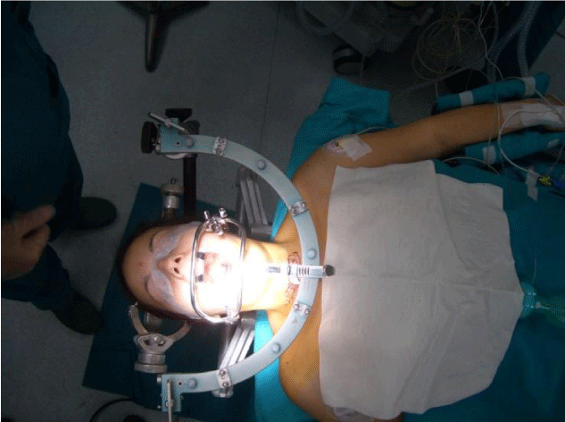
Menezes at al., [3] underlines the importance of the intraoperative reduction strategies. If reduction cannot be achieved, a 360° procedure may be necessary in some cases (depending on the pathology), the patient is positioned supine for a ventral decompression followed by posterior incision and posterior fixation. Moreover all patients undergo neck flexion\extension MRI of the CVJ. The patient is positioned supine with crown halo with traction; an intraoperative 3D CT is obtained in traction. The patient is then placed prone and another 3D CT is obtained. The algorithm is updated.
Conclusions
The progressive worldwide blooming of transoral procedures, thanks to the intensive care and the improvements in intraoperative neurophysiological monitoring techniques (once considered pioneering and very selective), is spreading the expertise in this field of surgery to a new population of surgeons. These techniques are performed alone or in conjunction with posterior procedures [47].
Pure endonasal and cervical endoscopic approach de- serves consideration but still has some disadvantages: (1) the learning curve and (2) the lack of 3-dimensional perception of the surgical field which could be an operationally limiting factor. Image clarity will be diminishevd when endoscopes smaller than 2.7 mm are used. Standard 4-mm endoscopes give a good image quality, but 2.7-mm scopes provide better maneuverability; (3) a limited working channel, according to the variability of the NAxL, which can make it difficult to remove huge tumors.
In our opinion, endoscopically assisted transoral surgery with 30° endoscopes represents an emerging alternative to standard microsurgical techniques for transoral approaches to the anterior CVJ. Used in conjunction with traditional microsurgery and intraoperative fluoroscopy, it provides a safe and improved method for anterior decompression with or without a reduced need for extensive soft palate split- ting, hard palate resection, or extended maxillotomy. Virtually no surgical limitations do exist for endoscopically assisted transoral approach, compared with the pure endonasal and transcervical approaches to the CVJ in normal anatomical conditions.
So far, the endoscope deserves an interesting role as ‘‘support’’ to the standard transoral microsurgical approach since 30° angulated endoscopy strongly improves the visual but not the working channel and volume, even though the soft palate splitting is often still required. In our opinion transoral (microsurgical or video-assisted) approach with sparing of the soft palate still remains the gold standard compared to the ‘‘pure’’ transnasal and transcervical approaches due to the wider working channel provided by the former technique. Transnasal endoscopic approach alone appears to be superior when the CVJ lesion exceeds the upper limit of the inferior third of the clivus. Furthermore, combined transnasal and transoral procedures can be tailored according to the specific pathological and radiological findings. Finally, experience is required with greater numbers of patients and long-term follow-up to further validate all the endoscopic techniques. In our opinion and in agreement with other authors, the endoscopic endonasal approach, rather than an alternative, should be considered a complementary approach to the standard transoral- transpharyngeal route [42].
According to a recent anatomical study, the lower incidence of post- operative dysphagia with the endonasal approach is likely related to the lower density of neuronal elements from the pharyngeal plexus above the palatal plane [21].
However, the time to extubation and oral feeding was significantly shorter in the endonasal group. Similarly Ponce-Gómez and colleagues reported their own series of patients treated using both approaches and found comparable rates of neurological improvement after odontoidectomy, with less time to extubation and oral feeding, as well as shorter hospital stay in the endonasal group [35].
- Visocchi M (2011) Advances in videoassisted anterior surgical approach to the craniovertebral junction. Adv Tech Stand Neurosurg 37: 97–110. Link: https://goo.gl/Q0YMyA
- Visocchi M, Di Martino A, Maugeri R, González Valcárcel I, Grasso V, et al. (2015) Videoassisted anterior surgical approaches to the craniocervical junction: rationale and clinical results. Eur Spine J 24: 2713-2723 Link: https://goo.gl/IddoYU
- Dlouhy BJ, Dahdaleh NS, Menezes AH (2015) Evolution of transoral approaches, endoscopic endonasal approaches, and reduction strategies for treatment of craniovertebral junction pathology: a treatment algorithm update. Neurosurg Focus 38: E8. Link: https://goo.gl/IfQFMM
- Funaki T, Matsushima T, Peris-Celda M, Valentine RJ, Joo W, et al. (2013) Focal transnasal approach to the upper, middle, and lower clivus. Neurosurgery 73: 155–190. Link: https://goo.gl/2whkAi
- Komatsu F, Komatsu M, Di Ieva A, Tschabitscher M (2013) Endoscopic far-lateral approach to the posterolateral craniovertebral junction: an anatomical study. Neurosurg Rev 36: 239–247. Link: https://goo.gl/WX9zUf
- Ammirati M, Bernardo A (1998) Analytical evaluation of complex anterior approaches to the cranial base: an anatomic study. Neurosurgery 43: 1398–1407. Link: https://goo.gl/fByHRS
- de Divitiis O, Conti A, Angileri FF, Cardali S, La Torre D, et al. (2004) Endoscopic transoral-transclival ap- proach to the brainstem and surrounding cisternal space: anatomic study. Neurosurgery 54: 125–130. Link: https://goo.gl/VfkYyo
- Balasingam V, Anderson GJ, Gross ND, Cheng CM, Noguchi A, et al. (2006) Anatomical analysis of transoral surgical approaches to the clivus. J Neurosurg 105: 301–308. Link: https://goo.gl/wbEu2W
- Youssef AS, Guiot B, Black K, Sloan AE (2008) Modifications of the transoral approach to the craniovertebral junction: anatomic study and clinical correlations. Neurosurg 62: 145–154. Link: https://goo.gl/qRl53V
- Pillai P, Baig MN, Karas CS, Ammirati M (2009) Endoscopic image-guided transoral approach to the craniovertebral junction: an anatomic study comparing surgical exposure and surgical freedom obtained with the endoscope and the operating micro- scope. Neurosurgery 64: 437–442. Link: https://goo.gl/cjd5fw
- Dallan I, Castelnuovo P, Montevecchi F, Battaglia P, Cerchiai N, et al. (2012) Combined transoral transnasal robotic-assisted nasopharyngectomy: a cadaveric feasibility study. Eur Arch Otorhinolaryngol 269: 235–239. Link: https://goo.gl/fPzwQb
- Alfieri A, Jho HD, Tschabitscher M (2002) Endoscopic endonasal approach to the ventral cranio-cervical junction: anatomical study. Acta Neurochir (Wien) 144: 219–225. Link: https://goo.gl/gVxPLb
- Messina A, Bruno MC, Decq P, Coste A, Cavallo LM, et al. (2007) Pure endoscopic en- donasal odontoidectomy: anatomical study. Neurosurg Rev 30: 189–194. Link: https://goo.gl/PoAlR6
- Ciporen JN, Moe KS, Ramanathan D, Lopez S, Ledesma E, et al. (2010) Multiportal endoscopic ap- proaches to the central skull base: a cadaveric study. World Neurosurg 73: 705–712. Link: https://goo.gl/aykIxo
- Aldana PR, Naseri I, La Corte E (2012) The naso-axial line: a new method of accurately predicting the inferior limit of the endoscopic endonasal approach to the craniovertebral junction. Neurosurgery 71: 308–314. Link: https://goo.gl/2c8RC1
- Little AS, Perez-Orribo L, Rodriguez-Martinez NG, Reyes PM, Newcomb AG, et al. (2013) Biomechanical evaluation of the craniovertebral junction after inferior- third clivectomy and intradural exposure of the foramen magnum: implications for endoscopic endonasal approaches to the cranial base. J Neurosurg Spine 18: 327–332. Link: https://goo.gl/qqBw7v
- Perez-Orribo L, Little AS, Lefevre RD, Reyes PR, Newcomb AG, et al. (2013) Biomechanical evaluation of the craniovertebral junction after anterior unilateral condylectomy: implications for endo- scopic endonasal approaches to the cranial base. Neurosurgery 72: 1021–1029. Link: https://goo.gl/MhilEL
- Russo VM, Graziano F, Russo A, Albanese E, Ulm AJ (2011) High anterior cervical approach to the clivus and foramen magnum: a microsurgical anatomy study. Neurosurgery 69: 103–114. Link: https://goo.gl/vGCrLg
- Baird CJ, Conway JE, Sciubba DM, Prevedello DM, Quiñones-Hinojosa A, et al. (2009) Radiographic and anatomic basis of endoscopic anterior craniocervical decompression: a comparison of endonasal, transoral, and transcervical approaches. Neurosurgery 65: 158–163. Link: https://goo.gl/f0FDne
- Seker A, Inoue K, Osawa S, Akakin A, Kilic T, et al. (2010) Comparison of endoscopic transnasal and transoral approaches to the craniovertebral junction. World Neurosurg 74: 583–602. Link: https://goo.gl/6boMT2
- Van Abel KM, Mallory GW, Kasperbauer JL, Moore EJ, Price DL, et al. (2014) Transnasal odontoid resection: is there an anatomic explanation for differing swallowing outcomes? Neurosurg Focus 37: E16. Link: https://goo.gl/x1Ibuv
- Visocchi M, Pappalardo G, Pileggi M, Signorelli F, Paludetti G, et al. (2015) Experimental endoscopic angular domains of transasal and transoral routes to the craniovertebral junction: Light and Shade. Spine (Phila Pa 1976) 41: 669-677. Link: https://goo.gl/qM6lls
- de Almeida JR, Zanation AM, Snyderman CH, Carrau RL, Prevedello DM, et al. (2009) Defining the nasopalatine line: the limit for endonasal surgery of the spine. Laryngoscope 119: 239–244. Link: https://goo.gl/HWX7Ag
- Visocchi M, La Rocca G, Della Pepa GM, Stigliano E, Costantini A, et al. (2014) Anterior video-assisted approach to the craniovertebral junction: transnasal or transoral? A cadaver study. Acta Neurochir (Wien) 156: 285–292. Link: https://goo.gl/HNfyvi
- Frempong-Boadu AK, Faunce WA, Fessler RG (2002) Endoscopically assisted transoral-transpharyngeal approach to the craniovertebral junction. Neurosurgery 51: S60–S66. Link: https://goo.gl/ckYwz8
- Liu JK1, Patel J, Goldstein IM, Eloy JA (2015) Endoscopic endonasal transclival transodontoid approach for ventral decompression of the craniovertebral junction: operative technique and nuances. Neurosurg Focus 38: E17. Link: https://goo.gl/J51S9p
- Husain M, Rastogi M, Ojha BK, Chandra A, Jha DK (2006) Endoscopic transoral surgery for craniovertebral junction anomalies. J Neurosurg Spine 5: 367–373. Link: https://goo.gl/FRnRMx
- Wolinsky JP, Sciubba DM, Suk I, Gokaslan ZL (2007) Endoscopic image-guided odontoidectomy for decompression of basilar invagination via a standard anterior cervical approach. Technical note. J Neurosurg Spine 6: 184–191. Link: https://goo.gl/mqL5DC
- Wu JC, Huang WC, Cheng H, Liang ML, Ho CY, et al. (2008) Endoscopic transnasal transclival odon- toidectomy: a new approach to decompression: technical case report. Neurosurgery 63: E92–E94. Link: https://goo.gl/1KLt9N
- Mc Girt MJ, Attenello RJ, Sciubba DM, Gokaslan ZL, Wolinsky JP (2008) Endoscopic transcervical odontoidectomy for pediatric basilar invagination and cranial settling. J Neurosurg Pediatr 1: 337–342. Link: https://goo.gl/PAkhC8
- Menezes AH (2008) Surgical approaches: postoperative care and complications ‘‘transoral–transpalatopharyngeal approach to the Craniocervical Junction’’. Childs Nerv Syst 24: 1187–1193. Link: https://goo.gl/Dne6GQ
- Perrini P, Benedetto N, Guidi E, Di Lorenzo N (2009) Transoral approach and its superior extensions to the craniovertebral junction malformations: surgical strategies and results. Neurosurgery 64: 331–342. Link: https://goo.gl/8GpabV
- El-Sayed IH, Wu JC, Ames CP, Balamurali G, Mummaneni PV (2010) Combined transnasal and transoral endoscopic approaches to the craniovertebral junction. J Craniovertebr Junction Spine 1: 44-48. Link: https://goo.gl/Q993bb
- Lee A, Sommer D, Reddy K, Murty N, Gunnarsson T (2010) Endoscopic transnasal approach to the craniocervical junction. Skull Base 20: 199–205. Link: https://goo.gl/EhFLkN
- Visocchi M, Della Pepa GM, Doglietto F, Esposito G, La Rocca G, et al. (2011) Video-assisted microsurgical transoral approach to the craniovertebral junction: personal experience in childhood. Childs Nerv Syst 27: 825–831. Link: https://goo.gl/gsENWU
- Salunke P, Sharma M, Sodhi HB, Mukherjee KK, Khandelwal NK (2011) Congenital atlantoaxial dislocation: a dynamic process and role of facets in irreducibility. J Neurosurg Spine 15: 678–685. Link: https://goo.gl/qhhNKX
- Dhaliwal PP, Hurlbert RJ, Sutherland GS (2012) Intraoperative magnetic resonance imaging and neuronavigation for transoral approaches to upper cervical pathology. World Neurosurg 78: 164–169. Link: https://goo.gl/mCYTrV
- Gladi M, Iacoangeli M, Specchia N, Re M, Dobran M, et al. (2012) Endoscopic transnasal odontoid resection to decompress the bulbo-medullary junction: a reliable anterior minimally invasive technique without posterior fusion. Eur Spine J 21: S55–S60. Link: https://goo.gl/oUWjO1
- Dasenbrock HH, Clarke MJ, Bydon A, Sciubba DM, Witham TF, et al. (2012) Endoscopic image-guided transcervical odontoidectomy: outcomes of 15 patients with basilar invagination. Neurosurg 70: 351–359. Link: https://goo.gl/hTLS6P
- Choi D, Crockard HA (2013) Evolution of transoral surgery: three decades of change in patients, pathologies, and indications. Neurosurgery 73: 296–303. Link: https://goo.gl/J8G9S3
- Hickman ZL, McDowell MM, Barton SM, Sussman ES, Grunstein E, et al. (2013) Transnasal endoscopic approach to the pediatric craniovertebral junction and rostral cervical spine: case series and literature review. Neurosurg Focus 35: E14. Link: https://goo.gl/Klpy3T
- Morales-Valero SF, Serchi E, Zoli M, Mazzatenta D, Van Gompel JJ (2015) Endoscopic endonasal approach for craniovertebral junction pathology: a review of the literature. Neurosurg Focus 38: E15. Link: https://goo.gl/oUYuNK
- Chaudhry NS, Ozpinar A, Bi WL, Chavakula V, Chi JH, et al. (2015) Basilar Invagination: Case Report and Literature Review. World Neurosurg 83: 1180. Link: https://goo.gl/p9cOU3
- Ponce-Gómez JA, Ortega-Porcayo LA, Soriano-Barón HE, Sotomayor-González A, Arriada-Mendicoa N, et al. (2014) Evolution from microscopic transoral to endoscopic endonasal odontoidectomy. Neurosurg Focus 37: E15. Link: https://goo.gl/LUFIm5
- Burns TC, Mindea SA, Pendharkar AV, Lapustea NB, Irime I, et al. (2015) Endoscopic Transnasal Approach for Urgent Decompression of the Craniocervical Junction in Acute Skull Base Osteomyelitis. J Neurol Surg Rep 76: e37-42. Link: https://goo.gl/F0S2Oa
- Liu JK, Patel J, Goldstein IM, Eloy JA (2015) Endoscopic endonasal transclival transodontoid approach for ventral decompression of the craniovertebral junction: operative technique and nuances. Neurosurg Focus 38: E17. Link: https://goo.gl/IwRaLJ
- Visocchi M, Pietrini D, Tufo T, Fernandez E, Di Rocco C (2009) Pre-operative irreducible C1–C2 dislocations: intra-operative re- duction and posterior fixation. The ‘‘always posterior strategy’’. Acta Neurochir (Wien) 151: 551–559. Link: https://goo.gl/IE9rrv
Article Alerts
Subscribe to our articles alerts and stay tuned.
 This work is licensed under a Creative Commons Attribution 4.0 International License.


 Save to Mendeley
