Open Journal of Orthopedics and Rheumatology
How Challenging is the “Scaling Up” of Orthopaedic Simulation?
Mohamed Mediouni1*, Neil Vaughan2, Sunil H Shetty3, Manit Arora3,4, Alexander Volosnikov5 and Amal Khoury6
2Department of Computing and Informatics Bournemouth University, UK
3Padmashree Dr. D.Y. Patil Hospital, Navi Mumbai, India
4St. George Clinical School, University of New South Wales, Australia
5Federal State Budgetary Institution, Russian Ilizarov Scientific Center, Restorative Traumatology and Orthopaedics of Ministry of Healthcare, Russian Federation
6Department of Orthopaedics, Hadassah Hebrew University Medical Center, Jerusalem, Israel
Cite this as
Mediouni M, Vaughan N, Shetty SH, Arora M, Volosnikov A, et al. (2016) How Challenging is the “Scaling Up’’ of Orthopaedic Simulation?. Open J Orthop Rheumatol 1(1): 012-014. DOI: 10.17352/ojor.0000031. The orthopaedic innovation policy
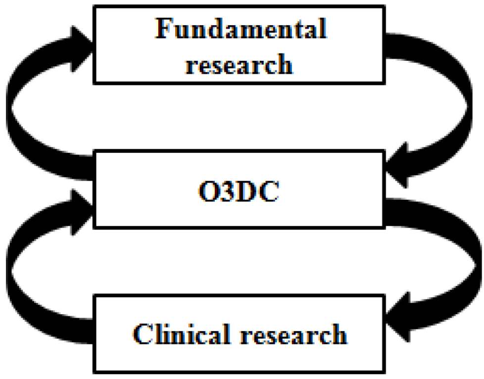
Innovation is a process that brings progress. In public health, there are many definitions of innovation because this concept covers many areas. Michael Porter [1], said that ‘’innovation includes both improvements in technology and better methods or ways of doing things. It can be manifested in product changes, process changes, new approaches to marketing, new forms of distribution, and new concepts of scope.’’ According to Peter Drucker [2], innovation is ‘’change that creates a new dimension of performance’’. Innovation is an idea that needs to be reproducible, to have an economic cost, and must meet a specific need. Innovation must be credible, observable, and relevant. The challenge of innovation is their implementation on the large scale. For that purpose, we must explain what is meant by scaling up, constraints and the strategies to succeed the development of innovation. There have been few attempts to explain what is meant by the scaling up and the term has often been used alongside the terms ‘’going to scale’’ and ‘’at scale’’. In the field of health, Simons et al. [3], mentioned that scaling up is used to increase the coverage of health interventions in order to benefit from the expertise and support political programs on a large scale. It is necessary to identify the constraints to increase the advancement of research and improve clinical outcomes. Several constraints may influence the ability of scaling up include the lack of clear strategies, lacks adequate infrastructure and equipment, lack of human resources, economic factors, skill level and motivation of team work. In orthopaedics, the scaling up requires a systematic planning in which innovations can be tested and implemented in a larger scale to produce a wider impact. (Figure 1)
The success of scaling up depends on the following conditions:
• Orthopaedic community must perceive a need and motivate to implement it. Does the innovation respond to a priority and who are exactly the advocates of this innovation?
• We must make sure that timing and circumstances are right.
• Maximize the opportunities and minimize the constraints for better implementation.
• Build a strong team, network of supporters include individuals and institutions and find an appropriate collaboration. It is important to ensure that managerial expertise and skills in advocacy are well integrated.
• Identify the financial resources, the organization capacities and the limitations. We need to mobilize funding for the resource team.
In the following sections, we will explain the need for simulation for training, in order to explain our platform O3DC as an example to challenging the problem of scaling up.
2. A look to the future: An efficiency simulation
Today, a certain degree of expertise is required to practice orthopaedics in the operating room. Becoming a skilled surgeon requires the ability to perform a high number of surgical procedures in training, which a simulator can enable. With the advancement of technology of virtual reality, residents can develop their motor skills in-vitro. Simulators provide 3D graphics giving a focused and effective way of repeatable practicing technical skills without risks [4,5]. The simulation approach standardized clinical situations that prepare students to consolidate their skills. Katri et al. [6], discuss that video gaming can have a good impact on the acquisition of skills. Furthermore, Thomas Kuivila states that in the operating room, residents do not have the time to look around the knee, for example, and catch some nuances because they need to keep things moving for the sake of the patient [7]. Oliphant et al. [6], explains the concept of Virtual Journal Club (VJC) as an e-learning tool to acquire the skills. It is an environment to facilitate the development of critical evaluation to detail the forces and the limits of new simulators (Figure 2).
Orthopaedic simulators are growing in popularity. Orthopaedic trainees can gain several skills from VR simulator training, including arthroscopy [8], endoscopy, knee, shoulder and hip surgery [9]. As well as being useful for teaching new skills, 16 clinical studies have shown that VR simulators can be an objective method for assessment of orthopaedic skills, to identify experience and skill level of trainees or experts [9]. Future orthopaedic simulators make use of cutting edge technology such as hands free motion tracking, acquired from data using Microsoft KinectTM sensor, or wireless cameras using image processing algorithms. These wireless tracking devices could also be used to track surgical tools during the actual procedure in-vivo to provide alerts and assistance.
Still today, simulators for orthopaedic surgery and not as advanced as simulators for other procedures [9], however trainees are becoming more exposed to computers and laboratory training. This eases the work load and costs due to time constraints on the expert trainers. Compared to cadaver training for orthopaedic procedures such as hip replacement, the virtual simulator training can reduce cost, travel, and improve the availability which speeds up the training process.
Another benefit of VR training is the ability to model patients of all different sizes, shapes and BMI. In-vivo surgical procedures vary between patients due to anatomical variation. The cutting edge simulators are able to model the patient variation using physical modelling of soft tissue biomechanics. By practicing on obese patients or difficult surgical cases, even experts can gain skill from VR simulators and it improves the availability of practice for difficult and complicated cases which in-vivo are rarer.
For the sake of realism, we need a methodology for obtaining a 3D model from 2D data include Magnetic Resonance Imaging (MRI), Computed Tomography, angiography, ultrasound, and Magnetic Resonance Spectroscopy. These techniques give different views of the organs morphology, as well as their functional characteristics. Computer-assisted, model based planning can cover all anatomical structures as well as other phenomena such as fluid dynamics, mechanical loads and diffusion processes [10]. After practices, we need debriefing which constitute a judgement of the simulation feedback [11]. It enhance trainee cognitive and help to explore a behaviors that reveal performance gaps. Debriefing can improve the discussion between professors and students and targeted instructions can enhance future performance.
3. Understanding a common language: O3DC
The idea of O3DC [12,13], is to develop a transition strategy in a large scale, in order to transport the advanced achievements in the technology framework and the pilot projects for benefit in research. Among the tasks of O3DC is the reclassification of human fracture. Using two dimensional (2D) x-ray images is very important to decide the treatment of fractures. The traditional classification methods are not enough for orthopaedist in terms of reliability. Orthopaedic surgeons need access to accurate classification on 3D CT images. Osteoporotic fractures, especially hip fractures, constitute a large problem for the elderly population and, in terms of health care costs [14]. To correct these fractures, surgeons need to use drilling, which constitutes a difficult task even for well-trained and experienced clinicians [15]. Using simulation O3DC will provide the orthopaedic community with an overview of the level of temperature in drilling for each fracture to avoid the problem of necrosis. To perform the simulation process, some parameters must be provided, such as overall length, point angle, diameter, material of drill, speed of rotation, and axial load at the contact point of the drill and bone. Based on this information, the model can predict the variation of bone temperature with drill time for different rotating speed of drill and the variation of bone temperature with drill time for different forces applied. This information will help trainee surgeons to improve their skills when drilling to correct a complex fractures (Figure 3).
Simulating the bone is helpful to predict and measure pathogenesis, progression, diagnosis and treatment outcomes for fragility. Many methods have been used successfully to characterise bone [16]. O3DC will provide a simulation of micro and nano bone. To understand what constitutes bone quality and how it can be measured may lead to better predictions of fracture risk [16]. The information provided by O3DC will have definitely beneficial impacts on the accuracy in operation and positively influence the clinical results. In addition, the residents and junior surgeons can find the update of new simulations around the world and avoid the problems of scaling up. The key of O3DC is the preparation of both of trainees and trainers. We will create courses including distributed simulation that facilitate the engagement of participants around the world.
Conclusion
Today with the challenges in orthopaedics, we need to increase the innovation by creating a bridge between orthopedists and professors in academia to produce a new strategy of education using 3D simulation. For that purpose, the message to the orthopaedic community is clear: ‘’it is time to change’’.
The authors would like to thank World Dental Network for supporting the idea of this article.
- Porter ME (1990) The Competitive Advantage of Nations. New York, Free Press 74-91.
- Marshall BJ (2006) Helicobacter connections. ChemMedChem 1: 783-802.
- Simmons R, Fajans P, Ghiron L (eds) (2007) Scaling Up Health Service Delivery: From Pilot Innovations to Policies and Programmes. Geneva: World Health Organization.
- Mediouni M, Volosnikov A (2015) The trends and challenges in orthopaedic simulation. J Orthop 12: 253-259.
- Mediouni M (2015) The What, Where and Why of Orthopaedic Simulation. Int J Surg Res Pract 2: 3.
- (2014) Self-assessment of Skills on a Virtual-reality Orthopaedic Trauma Simulator - Does Practice Lead to Insight of Performance? Int J Surg12: 13-117.
- (2015) Orthostreams, https://orthostreams.com/2015/01/a-look-to-the-future-teaching-the-next-generation-of-orthopedic-surgeons/.
- Leali A, Rebolledo B, Hamann J, Ranawat A (2016) Arthroscopy skills development with a surgical simulator: a comparative study in orthopaedic surgery residents. Bone Joint J 98: 143-143.
- Vaughan N, Dubey VN, Wainwright TW, Middleton RG (2015) Can virtual-reality simulators assess experience and skill level of orthopaedic surgeons?. IEEE Science and Information Conference (SAI) 105-108.
- Zdravkovic V, Bilic R (1990) Computer-assisted preoperative planning (CAPP) in orthopaedic surgery. Comput Methods Programs Biomed 32: 141-146.
- Rudolph JW, Simon R, Rivard P, Dufresne RL, Raemer DB (2007) Debriefing with good judgment: combining rigorous feedback with genuine inquiry. Anesthesiol Clin 25: 361-376.
- Mediouni M, Khoury A (2016) O3DC: The Curiosity in Orthopaedics. Journal of Clinical & Experimental Orthopaedics 2: 10.
- Mediouni M (2016) Orthopaedic 3D Collection: Mission and Impact. M J Ortho 1: 004.
- Byberg L, Gedeborg R, Cars T, Sundström J, Berglund L, et al. (2012) Prediction of fracture risk in men: a cohort study. J Bone Miner Res 27: 797-807.
- Pandey RK, Panda SS (2013) Drilling of bone: A comprehensive review. J Clin Orthop Trauma 4: 15-30.
- Abel RL, Prime M, Jin A, Cobb JP, Bhattacharya R (2013) 3D Imaging Bone Quality: Bench to Bedside. Hard Tissue 2: 42.
Article Alerts
Subscribe to our articles alerts and stay tuned.
 This work is licensed under a Creative Commons Attribution 4.0 International License.


 Save to Mendeley
