Journal of Surgery and Surgical Research
Maxillary sinus floor augmentation through bone densification
Kelly Zaia Barel* and Paulo Sérgio Zaidan Maluf
Cite this as
Barel KZ, Zaidan Maluf PS (2020) Maxillary sinus floor augmentation through bone densification. J Surg Surgical Res 6(2): 149-151. DOI: 10.17352/2455-2968.000119Objective: Presenting a case of maxillary sinus lifting through alveolar crest by means of bone compacting from bone densification with drills and immediate placement of the dental implant (fixture). Patients who suffer from alveolar bone insufficiency present a common problem in the recovery from osseointegration implants, when a minimum remaining bone extension is necessary for implant stability.
On those patients who present great bone loss in the posterior maxilla it is necessary to perform a sinus lifting surgery. The elevation of the sinus floor can be performed in two different ways, as described in the field literature: accessing the lateral window or accessing the maxillary sinus through alveolar crest by means of bone densification, thus allowing oral rehabilitation through dental implants.
Introduction
The sinus lift technique through alveolar crest is minimally invasive and less traumatic, making use of hydropneumatics counterclockwise rotating instruments, which allows the osseodensification by elevating the maxillary sinus floor without touching on the Schneiderian membrane, allowing it to be lifted at a minimum risk of perforation [2].
Considering the low density of the posterior maxilla and that successful implants in that area are also related to bone density, the technique of sinus crest lifting through osseodensification enhances bone density, thus providing a larger bone extension surface in contact with the implant and, consequently, greater possibility of primary torque and long term implant success in that area [6].
Osseodensification, a technique developed by Huwais in 2013, has as its objective the use of a series of specially designed drills bits, aimed to increase bone density by progressively expanding the osteotomy. The Densah® technique is based on a bone preparation entitled “osseodensification”[3]. Contrary to the traditional dental perforation techniques, osseodensification does not remove bone tissue, which is instead compacted and simultaneously grafted into the external part, expanding from the osteotomy. When a Densah® drill bit is used at high speed counterclockwise, without a cutting edge and with constant external irrigation, a dense and strong bone tissue layer is formed along the walls of the base of the osteotomy [1].
These drill bits combine the advantages of the osteotomous with the controlled expansion during perforation, without the removal of the bone tissue and without fracturing the trabecula. As the drill bit does not possess a cutting edge counterclockwise, this decreases the risk of perforation of the sinus membrane. The drills also allow bone preservation and condensation by means of self-grafting through compacting during the osteotomy, thus accounting for higher initial stability of the implant [1,2].
The stability of the initial implant is extremely important to the osteointegration of the same, and it is related to the extension of bone that surrounds the implant. As such, maintaining a quality bone tissue in sufficient quantity allows it to have greater contact with the implant threads and, consequently, it will provide greater possibility of a successful osteointegration of the implant [4].
The technique which makes use of Densah® is prescribed for cases of sinus maxillary with a minimum height of 4 to 6 millimeters and an alveolar bone with a 4-millimeter width. Planning is done by measuring first the bone height up to the sinus floor and clockwise perforation with a pilot drill bit up to 1 millimeter before the sinus floor (800-1,500 rpm), with abundant irrigation [2,3].
Clinical case report
A female patient, age 58, came to the Associação Paulista do Cirurgião Dentista – APCD (São Paulo State Association for the Dental Surgeon) implants clinic, in São Caetano do Sul (greater São Paulo city area) aiming to rehabilitate the posterior edentulous area to the left with dental implants.
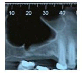
During clinical examination of the oral cavity and tomography image analysis, a healthy maxillary sinus was identified with, nonetheless, bone tissue loss in the section of elements 15 and 16, which thus prevented implants in that area (Figure 1).
After data gathering and complementary laboratory and imaging exams were made, showing no changes that could lead to the unlikelihood of performing the surgery, we made a choice for the sinus lifting technique making use of densifying drill bits Densah®, due to the remaining bone width being compatible with the sinus procedure and concomitantly insertion of an implant (Figure 2).
The patient was prescribed two pills of Clavulin DB 1 gram each (a combination of amoxicillin 875 milligrams, associated to 125 milligrams of Clavulanate Potassium) one hour before the procedure, plus two pills of Dexamethasone of 4 milligrams each, before the surgery, as a prophylactic procedure with antibiotic therapy.
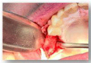
The patient was laid on the chair in a thirty-degree supine position. The local antisepsis intra and extra oral was performed with aqueous Chlorhexidine 2%, and sterile surgical drapes were put in place. Local anesthesia was performed with Articaine 4%. The surgical access was performed with an intra oral incision on the left posterior alveolar ridge area and mucoperiosteal flap detachment and gingival removal, exposing the alveolar crest area (Figure 3).
The initial drilling was performed according to the established Densah® technique protocol, being initiated with a pilot 1.5 mm drill toward the ridge, at a distance of 1 millimeter before the sinus floor, in 1,200 rpm clockwise rotation and being abundantly irrigated with a 0.9% saline solution. After the initial drilling, a sequence for expansion and bone compacting was performed in the lateral apical directions, according to the selected implant. Perforations were made anticlockwise, at 1,200 rpm rotation speed, and abundant irrigation, lifting to 3 millimeters the sinus membrane.
After reaching the sinus membrane, we decreased the rotation to 200 rpm, removed the saline solution input, consequently suspending the irrigation, and we then began the insertion of the chosen material for grafting, thus allowing the material to passively lift the membrane, keeping it in its new position (Figure 4).
We used as graft material biomaterial associated to LPFR (Figure 5) to foster new bone formation in that area. The implants were of a Cone Morse type, with 3.75 mm X 8.5 mm in the tooth 15 area and 3.5 mm X 11.5 mm in the tooth 16 area, having been installed in the oral cavity with a 40N torque (Figure 6).
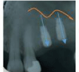
The flap was maintained with intra oral sutures using nylon 4.0. As post-surgery medication, we prescribed the continuation of Clavulin BD 1 gram in 12-hour intervals for 10 days, and analgesics were also prescribed in case of painful sensitivity. Instructions regarding cleaning of the nasal cavity with a saline solution were given, as well as rigorous and assisted oral hygiene actions. The patient also received post-surgery orientation in writing and was asked to return to the clinic for a radiography exam, which revealed gain measurements of 7 millimeters in the 15 tooth area, and of 9 millimeters in the tooth 16 area Figure 7.
Conclusion
Existing new researches and, consequently, the appearance of new surgical techniques for sinus lifting, have made possible to perform sinus augmentation in a less invasive manner and with results that surpass by far the ones derived from techniques previously employed.
The osseodensification with drill bits is a surgical alternative with the least traumatic impact possible for patients with an edge of 4 mm in bone height, allowing in most of the cases lifting the sinus floor within reduced heights and placing an implant concomitantly to the first surgery, due to the increase of bone density and thus presenting greater primary stability, with high torques at the moment of implant placement.
Due to the bone impact in the apical direction, we did not experience perforation of the sinus membrane, which increases the success of the surgery in the long term. Some factors also cooperate with the success of the chosen surgery technique, such as minimum bone width, which favors bone compacting and increases the primary stability of the implant, preserving the integrity of sinus membrane and the overall health of the sinus floor. There was no post-surgery incident and the technique employed is more comfortable and less traumatic than the conventional one based on the lateral window for lifting of the sinus floor. Furthermore, the technique here employed is an effective one in terms of immediate implant placement recovery.
- Huwais S, Mazor Z, Ioannou AL, Gluckman H, Neiva R (2018) A multicenter retrospective clinical study with up-to-5-year follow up utilizing a method that enhance boné density and allows transcrestal sinus augementation through compaction grafting. Int J Oral Maxillofac Implants 33: 1305-1311. Link: https://bit.ly/2J2PUi7
- Trisi P, Berardini M, Falco A, Podaliri Vulpiani M (2016) New Osseodensification implant site preparation method to increase bone density in low-density bone. In vivo evaluation in sheep. Implant Dent 25: 24-31. Link: https://bit.ly/35qQrC8
- Huwais S (2018) Osseodensifation facilited Densah lift protocol.
- Huwais S, Meyer E (2017) A new osseous densification approach in implant osteotomy preparation to increase biomechanical primery stability, bone mineral density, and bone-to-implant contact. Int J Oral Maxillofac Implants.32: 27-36.
- Meredith N (1998) Assessment of implant stability as a prognostic determinant. Int J Prosthodont 11: 491-501. Link: https://bit.ly/3kruvNr
- Trisi P, Perfetti G, Baldoni E, Berardi D, Colagiovanni M, et al. (2009) Implant micromotion is related to peak insertion torque and bone density. Clin Oral Implants Res. 20: 467-471. Link: https://bit.ly/34r1ziM
Article Alerts
Subscribe to our articles alerts and stay tuned.
 This work is licensed under a Creative Commons Attribution 4.0 International License.


 Save to Mendeley
