Journal of Surgery and Surgical Research
Primigravida with diffuse uterine leiomyomatosis and IUGR necessitating a caesarean section and hysterectomy
Layla Hassan, Candace P Beoku-Betts and Oluseye A Oyawoye*
Cite this as
Hassan L, Beoku-Betts CP, Oyawoye OA (2020) Primigravida with diffuse uterine leiomyomatosis and IUGR necessitating a caesarean section and hysterectomy. J Surg Surgical Res 6(1): 84-086. DOI: 10.17352/2455-2968.000103Diffuse uterine leiomyomatosis is uncommon and often associated with infertility. To date, few successful pregnancies have been published within the context of the condition. Of the known cases, all but one have occurred after medical, radiological or surgical management. Here, we report a case of diffuse uterine leiomyomatosis in a 39 year-old diagnosed intrapartum.
Caesarean hysterectomy was performed at 30+5 week gestation due to massive peripartum haemorrhage secondary to diffuse nodular leiomyomatosis. All her antenatal scans reported multiple uterine fibroids and diffuse leiomyomatosis was not suspected until the time of caesarean section. At 6320 gm this is the largest sized uterine tissue so far published.
Introduction
Diffuse Uterine Leiomyomatosis (DUL) is a rare, histologically benign condition in which the myometrium is replaced by multiple poorly defined nodules which almost completely replace the myometrium [1]. This poorly recognised condition typically affects women of reproductive age and presents with abdominal pain, menorrhagia and infertility [2].
To date, few successful pregnancies have been published within the context of DUL and all but one have occurred after medical, radiological or surgical management. Our literature search revealed only 44 cases of DUL with only two speculative papers on its pathophysiology. Conservative treatment options for DUL are limited and hysterectomy remains the gold standard of symptomatic treatment [3].
Here we report a case of DUL in a 39 year-old woman diagnosed peri-partum. At 6320g this is the largest and heaviest uterine tissue published thus far.
Case report
A 39 year old, primigravida of Bangladeshi origin presented at our antenatal clinic at 15 weeks gestation with a history of iron deficiency anaemia, menorrhagia and abdominal pain. The patient had previously reported to her general practitioner 8 years before the index pregnancy due to severe dysmenorrhea and irregular vaginal bleeding and inability to conceive after 5 years of marriage. Ultrasound scan then showed “a grossly enlarged uterus measuring 14×8.8×11cm. There was a 6 cm posterior wall fibroid. The remainder of the uterine body had a coarse echo pattern suggestive of diffuse fibroid changes”. She also had an open myomectomy 4 years before conception, 30 fibroids were removed.
Of note, detail foetal scan at 22 weeks was restricted by poor image quality due to huge fibroids with symphysio-fundal height at 37 cm. Repeat scan at 27+4 weeks gestation showed reduced amniotic (AF) fluid, limited intrauterine space but foetal growth was within normal limits. Further scan at 29+3 weeks gestation reported normal foetal growth and liquor volume with foetal movements restricted by multiple large fibroids. At 30+1 weeks, the patient presented following spontaneous rupture of her membranes and was commenced on erythromycin and two doses of steroid injections. Her final pregnancy scan at 30+4 weeks gestation revealed growth well below 3rd centile, severe oligohydramnios and poor diastolic flow on umbilical artery Doppler study and transverse lie with head to the right. A grade 3 Caesarean was therefore arranged.
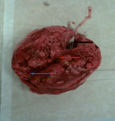
At Caesarean section the lower uterine segment and main body of the uterus was replaced by extensive diffuse nodular growths with no definitely-formed lower segment. A transverse incision in the lower uterus only revealed a narrow uterine cavity with the foetus squashed into a small space transversely across the uterine fundus. Transverse lower uterine incision was extended midline form an ‘inverted -T’ to deliver the baby. A 860g baby was delivered and the placenta was found morbidly adherent to the uterine walls (Figures 1). The whole uterine walls were rigid and not distensible, and the incisional edges bled freely due to lack of uterine retraction. Our Unit’s protocol for massive obstetric haemorrhage was activated with administration of oxytocics. These did not reduce haemorrhage and it was obvious the uterus was unable to retract because the walls had mostly been replaced by nodular non-retractable fibroid tissues. There was little normal uterine tissue at the edges thus making closure impossible.
A sub-total hysterectomy was therefore performed. Estimated blood loss was 5 litres. She was transfused 8 units of packed red blood cells, 4 units of FFP, 1 unit of cryoprecipitate, and 1 unit of platelets. The infant was transferred to a tertiary unit for further care where she recovered satisfactorily. The mother was discharged home five days post operatively without further event and was able to breastfeed her baby successfully.
Pathology
Macroscopically the uterus was enlarged weighing 6320g, with a smooth serosal surface. The myometrium was uniformly expanded with a multinodular appearance (Figures 1).
Microscopically, there were multiple well-defined nodules dissecting the myometrium, composed of spindle cells arranged in fascicles. There was evidence of infarct type necrosis, hyalinisation and haemorrhage, consistent with a non-malignant smooth muscle tumour. Portions of the myometrium were separated from chorionic villi by a layer of fibrinoid material rather than decidua. Placenta increta was present and the placenta showed features of chorangioma.
Immunohistochemistry showed low proliferative neoplastic cells and negative staining for P16 and P53, with no intravascular growth on CD34 staining.
Discussions
This rarity of DUL may in part be due to poor reporting or poor recognition of the condition, which was first described and published under the name of uterine leiomyomatosis in 1979 [4]. The aetiology is incompletely understood. Baschinsky, et al. suggested that DULmay be an exuberant example of multiple uterine leiomyomas budding into each other to the extent that the single nodules could not be readily identified by gross examination. MRI is probably the diagnostic method of choice with uterus diffusely enlarged with multiple innumerable ill-defined leiomyomas without discrete margins [5].
There are several novel therapeutic options available for DUL, however hysterectomy remains the method of choice for symptomatic relief [4]. Of the 44 well documented DUL cases, only in nine was hysterectomy avoided in lieu of more conservative methods for symptomatic relief and the restoration of fertility. Authors such as Yen et al have reported successes in five patients with hysteroscopic myomectomy in terms of uterine preservation [6]. Similarly, Shimizu reported a successful pregnancy post hysteroscopic myomectomy following administration of Nafarelin acetate over a 6-month period. Others have utilised uterine artery embolisation [7-9].
Grignon, et al. reported the first documented case of pregnancy in the presence of DUL, with a pregnancy complicated by spontaneous premature rupture of membranes and intrapartum haemorrhage. Similar to our case, peri-partum massive haemorrhage occurred due to non-contractility of the leiomyomatous nodular uterine walls.
Ideally, DUL should be diagnosed pre-pregnancy so that patients are forewarned of the potential complications associated with the rare cases where pregnancies occur. This will ensure the family is mentally prepared for the possibility of hysterectomy. In our case the patient was first seen during pregnancy and clear diagnosis of DUL was intra-partum. Pre-pregnancy or antenatal diagnosis will also ensure that such women are delivered in obstetric units with facilities to cater for severe peri-partum haemorrhage. Otherwise, mortality risk from severe post partum haemorrhage among women with DUL will be high.
DUL must be considered as a differential in women presenting with symptoms of abdominal pain, abnormal uterine bleeding, dysmenorrhea, menorrhagia and pressure symptoms. This is especially important as uterine and fertility-sparing treatments have better prognosis in early-stage disease [6].
Conclusion
This report further draws attention to a rare and underreported condition with enormous clinical and emotional implications for affected women. Modern imaging techniques, in addition to patients’ symptomatology and demography, will probably come up with criteria for early diagnosis. More research into its pathophysiology may also lead to earlier diagnosis and an understanding of a possible role in the burden of infertility and obstetric haemorrhage.
- Baschinsky DY, Isa A, Niemann TH, Prior TW, Lucas JG, et al. (2000) Diffuse leiomyomatosis of the uterus: a case report with clonality analysis. Hum Pathol 31: 1429-1432. Link: https://bit.ly/2YwxN8i
- Fedele L (1982) Diffuse uterine leiomyomatosis. Acta Eur Fertil 13: 125-131.
- Wallach EE, Vlahos NF (2004) Uterine myomas: an overview of development, clinical features, and management. Obstet Gynecol 104: 393-406. Link: https://bit.ly/37tYXRr
- Fedele L, Bianchi S, Zanconato G, Carinelli S, Berlanda N (2004) Conservative treatment of diffuse uterine leiomyomatosis. Fertil Steril 82: 450-453. Link: https://bit.ly/2N1c2rL
- Ueda H, Togashi K, Konishi I, Kataoka ML, Koyama T, et al. (1999) Unusual appearances of uterine leiomyomas: MR imaging findings and their histopathologic backgrounds. Radiographics 19: S131-S145. Link: https://bit.ly/2ULfVFx
- Yen CF, Lee CL, Wang CJ, Soong YK, Arici A (2007) Successful pregnancies in women with diffuse uterine leiomyomatosis after hysteroscopic management. Fertil Steril 88: 1667-1673. Link: https://bit.ly/3fn1GPa
- Shimizu Y, Yomo H, Kita N, Takahashi K (2009) Successful pregnabasncy after gonadotropin-releasing hormone analogue and hysteroscopic myomectomy in a woman with diffuse uterine leiomyomatosis. Arch Gynecol Obstet 280: 145-147. Link: https://bit.ly/2B94iRU
- Chen L, Xiao X, Wang Q, Wu C, Zou M, et al. (2015) High-intensity focused ultrasound ablation for diffuse uterine leiomyomatosis: A case report. Ultrason Sonochem 27: 717-721. Link: https://bit.ly/2UI7j2o
- Purohit R, Sharma JG, Singh S (2011) A case of diffuse uterine leiomyomatosis who had two successful pregnancies after medical management. Fertil Steril 95: 2434.e5-2434e6. Link: https://bit.ly/2AtpYZf
Article Alerts
Subscribe to our articles alerts and stay tuned.
 This work is licensed under a Creative Commons Attribution 4.0 International License.


 Save to Mendeley
