Journal of Surgery and Surgical Research
Rare primary sternal tumours – Reports of two cases
Sunil Kumar Rout1*, Chandrabhanu Parija2 and Devidutta Mohanty3
2Consultant Cardiothoracic & Vascular Surgeon, Kalinga Hospital Ltd, Bhubanesewar, Odisha, India
3Consultant Plastic Surgeon, Sunshine Hospital, Bhubanesewar, Odisha, India
Cite this as
Rout SK, Parija C, Mohanty D (2019) Rare primary sternal tumours – Reports of two cases. J Surg Surgical Res 5(1): 043-044. DOI: 10.17352/2455-2968.000068Primary tumours arising from sternum are rare. We came across two patients with primary tumours arising from sternum, one malignant (Ewings sarcoma) and the other benign in nature (giant cell tumour) which are very uncommon. After wide local excision the reconstruction was done by using autologous free rib graft in one and titanium mesh in other. In both the cases the coverage was provided with pectoralis major muscles. After six months both were disease free with chest wall integrity well maintained. We believe the reconstructive options adopted by our team for these cases are simple, bio-compatible and reproducible.
Introduction
Primary tumours arising in sternum are extremely rare. We came across two patients having primary tumours arising from sternum, evaluated them for clinical diagnosis and treated them surgically by performing wide local excision and reconstruction. Histopathological examination of the excised specimens revealed one of them to be Ewing’s sarcoma and the other giant cell tumour. These tumors are very uncommon as evidenced from published English literature. We adopted two different techniques of reconstruction in both the cases based on the need and feasibility. We consider both the reconstructive techniques to be novel and ideal for such situation. After a six month follow up both the patients were doing well in terms of function and aesthesis of the operated region.
Case Reports
Case 1
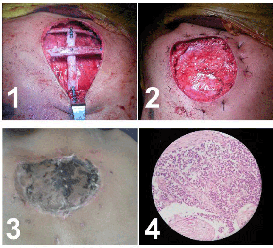
A 16 years old boy presented with a swelling over the sternal region noticed over a period of six months. The lesion was subjected to incisional biopsy 1 month before the patient attended the outpatient department of our hospital. The patient was evaluated by our team of thoracic surgeons and the plastic surgeons and treatment planned. The previous incisional biopsy showed none of the features of malignancy. Hence the treatment was planned considering it a benign tumour. MRI depicted the lesion expanding into the mediastinum and externally as well. The tumour was found to involve medial ends of both the clavicles and sternal end of the upper two ribs. Wide local excision was performed including the tumour with 2 centimeters of healthy margin from sternum, clavicles and the ribs. The tumour was free from adhesion with the mediastinal tissues and could be taken out with ease and it was removed with its overlying skin. Hence the excision left a defect of significant size with the mediastinum and the pericardium exposed to the exterior without any tissue overlying them (Figure 1).
From the reconstructive point of view the bony wall of the thorax along with overlying soft tissue and skin were deficient. The part was planned to be reconstructed with rib grafts with a cover of pectoralis major muscle flap and a split thickness skin graft over it. An inframamary approach was used to harvest the muscle flap as well as the ribs. The ribs were fixed with titanium miniplates (2 mm) and cross head titanium screws (2x10 mm). Reconstructed bony wall was covered with pectoralis major muscle and split thickness skin graft was put over that. The recovery was uneventful. Post excisional histopathology study established the diagnosis of Ewing’s sarcoma. The patient was referred to the department of medical oncology for adjuvant chemotherapy. He was observed doing well after 6 months of surgery and completion of chemotherapy.
Case 2
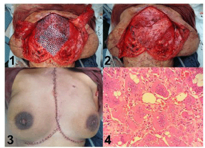
A 32 years old female presented with a swelling over sternal region observed by the patient over a period of 8 months. It was growing slowly. She was operated for giant cell tumour of lower end of left radius 2 years prior to this observation. Radiographic evaluation suggested it to be a giant cell tumour of sternum. The tumour was expanding into the mediastinum with very minimal growth towards the surface. The patient was planned for surgery by thoracic and plastic surgery team. The tumour was excised with palpable healthy margin of 2.5 centimeters with a small area of scarred adherent skin at the center. The defect was very large with deficiency of entire sternum except xiphisternum, medial ends of clavicles and upper five ribs. There was apparently no skin deficiency. Hence reconstructive need was replacement of bony as well as muscular tissue covering it.
Considering the size of the bony defect we preferred to proceed for allo-implant. Titanium mesh was used for reconstruction of bony defect, covered with pectoralis major muscle flaps from both the sides. Skin wound could be closed primarily without any difficulty. The patient was followed up for six months and not found to develop any complication. She probably had the necrosis or atrophy of medial part of pectoralis muscle flap which was evident from the contour of the reconstructed area during follow up (Figure 2).
Discussion
Primary tumours of sternum are very rare [1]. The bone is mostly affected secondarily due to metastasis from other malignant tumours like breast, ovary, kidney etc [2]. Ewings sarcoma is the second most common primary malignancy of sternum [3]. Very few cases of primary Ewings sarcoma and primary giant cell tumour arising in sternum have been reported. The sternal reconstruction is necessary in order to achieve protection to the exposed thoracic viscera and aesthetic appearance of the chest wall. Available materials and techniques are plenty to choose [4]. It includes metallic/alloplastic prostheses, autogenous bone graft, cryo-preserved sternal allograft or homograft as skeletal substitutes which need to be covered with soft tissue like pectoralis major or latissimus dorsi, rectus abdominis muscle or myocutaneous flap or omental flap [5,6]. Rib grafts and pectoralis major muscles we used in first case for reconstruction, are in close proximity of these defects and can easily be obtained by any reconstructive surgeon. Bone banks are available only in select healthcare delivery centers. Titanium mesh is well tolerated without much of complications and suitable for large sternal defects [7]. Titanium mesh we used in the second case was essential due to size of the defect. The techniques we used for reconstruction of post excisional defects are technically reproducible, cost effective and bio-compatible. Both the options used for reconstruction do not pose any threat in terms of adjuvant radiation or chemo therapy. The negativity of incisional biopsy in the case of Ewings sarcoma of course was an issue considering retrospectively since neo-adjuvant chemotherapy is recommended for such cases before contemplating to surgical excision. We presume it to be a procedural inadequacy in part of the surgeon performed this.
Disclosure
We disclose no conflict of interest in these cases and no financial support is received from any source for these cases.
- Eng J, Sabanathan S, Pradhan GN (1989) Primary sternal tumours. Scand J Thorac Cardiovasc Surg 23: 289-292. Link: https://tinyurl.com/y6erq9g7
- Bandi V, Lunn W, Ernst A, Eberhardt R, Hoffmann H, et al. (2008) Ultra- sound vs. CT in detecting chest wall invasion by tumor: a prospective study. Chest 133: 881-886. Link: https://tinyurl.com/y4dwyy9d
- Delattre O, Zucman J, Melot T (1994) The Ewing family of tumors – a subgroup of small-round-cell tumors defined by specific chimeric tran-scripts. N Engl J Med331: 294-299. Link: https://tinyurl.com/y4la9san
- Sanna S, Brandolini J, Pardolesi A, Argnani D, Mengozzi M, et al. (2017) Materials and techniques in chest wall reconstruction: a review. J Vis Surg 3: 95. Link: https://tinyurl.com/y4k7yz8f
- Nosotti (2012) Sternal reconstruction for unusual chondrosarcoma: Innovative technique. J Cardiothorac Surg 7: 40. Link: https://tinyurl.com/y3ar8mzh
- Seder CW, Rocco G (2016) Chest wall reconstruction after extended resection. J Thorac Dis 8: S863-S871. Link: https://tinyurl.com/y6fupflp
- Zhang Y, Li JZ, Hao YJ (2015) Sternal tumor resection and reconstruction with titanium mesh: a preliminary study. Orthop Surg 7: 155-160. Link: https://tinyurl.com/y48tfpj5
Article Alerts
Subscribe to our articles alerts and stay tuned.
 This work is licensed under a Creative Commons Attribution 4.0 International License.


 Save to Mendeley
