Journal of Surgery and Surgical Research
Bacterial isolation from environment and nosocomial pathogens in burned patient, with their susceptibility pattern in burn and plastic surgery department, Aljalla Hospital Benghazi
Mohammed Idres El Hassy1, Suad El Senussi2 and Khaled Elgazwi3*Samah M Elakeili4
2Head of infection control office, Infection Control Department, Aljalla Hospital Trauma, Benghazi Libya, Libya
3Head of surgical departments, Infection Control Department, Aljalla Hospital Trauma, Benghazi Libya, Libya
Cite this as
El Hassy MI, El Senussi S, Elgazwi K, Elakeili SM (2018) Bacterial isolation from environment and nosocomial pathogens in burned patient, with their susceptibility pattern in burn and plastic surgery department, Aljalla Hospital Benghazi. J Surg Surgical Res 4(2): 023-028. DOI: 10.17352/2455-2968.000056Background: 1) Hospital environment is a potential reservoir of bacterial pathogens, therefore Burn patients are at high risk of developing nosocomial infection because of their destroyed skin barrier and suppressed immune system, compounded by prolonged hospitalization and invasive therapeutic procedures.
Aim: The aim of this study was to detect rate of nosocomial pathogens in burned patients with their susceptibility pattern, and to determine the environmental contamination in Burn and plastic surgery department, aljalla hospital, Benghazi.
Method: In a prospective study and statistical analysis was performed for 120 samples were collected from different wound sites in burn and plastic surgery department, aljalla hospital during year 2016, additional to 217 samples were collected from patient zones, floor areas, walls, bed-frames, door handles, light switches, sinks, besides corridors, tables, autoclave, cupboards, medical waste transport cart, dressing room, nursing station, drums, trolley, nasal and hand swabs for nursing staff, then cultured on blood agar and MacConkey agar, Isolation and identification of microorganisms was done according to standard procedure.
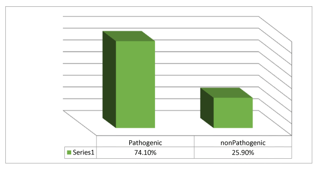
Results: A total of 195 samples were collected from patient zones and the environment, the culture positive was seen in 143 (73%), 106 (74.1%) of them were pathogenic. The most predominant bacterial isolate was Staph.albus (33.6%), followed by klebsialla ( 29.5%), N.L.F (14.7%), Pseudomonas(11.47%).E.coli (5%),N.H.S(4.1%), Staph.aureus (1.6%). of the 11 nasal swabs were obtained from nursing staff in the department,(81.8%) of them were pathogenic, predominant bacterial isolate was Staph.albus (30.7%), N.H.S(30.7%),N.L.F(23%), Klebsialla (15.3%), while11 hand swabs were obtained, (54.5%) were pathogenic, Staph.albus (50%), N.H.S(25%), N.L.F(15%), Klebsialla (10%). A total of 31 burned patient, 21 (67.7%) were females and 10 (32.2%) males, 120 burn swabs were collected from them, the predominant bacterial isolate was Pseudomonas (50.8%), Staph.aureus (16.7), Klebsialla (13.3%) ,Acinetobacter (10%), Enterobacter (2.5%), E.coli (2.5%), N.H.S (2.5%), Proteus (0.8%), Staph.Albus(0.8%), Among these isolates P. aeruginosa was found washighly resistant for most the antibiotic tested, while it had less resistance to ciprofloxacin and sensitive for colisten also present study showed that staph aureus was highly resistant for Augmantine, ceftriaxone, ciprofloxacin, ceftazidim, Acinetobacter and Klebsiella where highly resistance for Augmantine, Ceftrixone, Ciprofloxacin, Ceftazidim, Gentamicin, Amikacin and highly sensitive to colisten.
Introduction
Burns are a serious global public health problem. There are over300 000deathsper/ year from fires alone with more deaths from other types of burn, including scalds, and electrical and chemical burns. In addition, millions more are left with lifelong disabilities and disfigurements, often with resulting stigma and rejection. All of these result in further personal difficulties and economic losses for victims and their families.
The vast majority (over 95%) of these burns occur in low- and middle income countries. Rates of fire-related burn deaths in low- and middle income countries are 5.5 deaths/100 000 people per year. This is nearly six times higher than the 0.9 deaths/100 000 people per year in high-income countries [1]. Nepal has about 1700 burn deaths a year for a population of 20 million, giving a death rate 17 times that of Britain [2].
A burn is defined as damage to the skin caused by excessive heat or caustic chemicals. The most common burn injuries result from exposure to heat and chemicals. Full-thickness burns usually develop, causing immediate cell death and matrix destruction, with the most severe damage on the wound surface, Additional heat and inflammation induce tissue injury beneath the nonviable surface, which can either progress over time to healing or can deteriorate to further necrosis, depending on the approach to treatment [3].
Mortality and morbidity have been markedly reduced due to overall major improvements in critical care, metabolic support, infection control, and wound management [3].
The major challenge for a burn team is nosocomial infection in burn patients, which is known to cause over 50% of burn deaths, because of their destroyed skin barrier and suppressed immune system, compounded by prolonged hospitalization and invasive therapeutic and diagnostic procedures [4].
Literature Review
Many studies on nosocomial infection in burn patients focused on burn wound infection, one of them was during the second part of 2010, 164 burn patients were admitted to the Motahari Hospital, Tehran, Iran included in this study. Of the 164 patients, 717 samples were taken and 812 bacteria were identified, the bacterial isolate was 325 (40%) Pseudomonas, 140 (17%) Acinetobacter, 132 (16%) S. aureus, and 215 (27%) other bacteria [5].
In the Taleghani Burn Hospital which is an academic educational hospital affiliated to Ahvaz Jundi- Shapour University of Medical Sciences, A total of 182 patients were admitted to the hospital during the one year period (from May 2003 to April 2004), 173 bacterial isolates were obtained. The most predominant bacterial isolate was Pseudomonas aeruginosa 65 (37.5%) followed by Staphylococcus aureus 35 (20.2%), Acinetobacter baummanni 18(10.4%), Escherichia coli 10 (5.7%), Proteus mirabilis 9 (5.2%), co-agulase negative Staphylococus 9 (5.2%), Enterobacteraerogenes 9 (5.2%), Klebsiellapneumoniae 6 (3.4%), Enterococcus spp. 4 (2.3%), Citrobacterfreundii 3(1.7%), Serratiamarcescens 2 (1.1%), Alcaligenes spp. 2(1.1), haemolytic group A Streptococcus1 (0.5%) [4].
This study included 220 patients who were admitted in the burn unit in Al-Hada Military Hospital Taif, Saudi, during the 6 months period from March 2015 to August 2015, Gram-positive bacteria Staphylococcus epidermis was the predominant organisms with rate of 50 (22.2%), followed by Staphylococcus aureus 44 (20%), Enterococcus facium 10 (4.5%).Gram-negative rod E. coli was the predominant organisms with high rate of 89 (40%), followed by Pseudomonas aeruginosa 87 (39.5%), klebsiellapneumoniae 62 (28.1%), Acinetobacterbaumannii 43 (19.5%), Proteus mirabilis 35 (16%), and Morganellamorganii 25 (11.3%) [6].
Insulaimania burn hospital,reconstructive and plastic surgery hospital, in Iraq from (2008 to April 2009),A total 1126 samples ,bacterial isolated were found in 935 ,total organisms isolated 1402 organisms. Staphylococcus aureus (33%) was the most commonly isolated bacteria among burn patients with burn wound infectionfollowed by P. aeruginosa(18%),klebsiallapneumoniae (15%),other non-fermenting (9%), coagulase negative staphylococcus spp.(8%), Enterobacter cloacae(4%), E.coli (2%) [7].
Another study done in University of Gondar Teaching Hospital from November 2010 to February 2011, 220 specimens of pus, nasal, hand and surfaces were isolates. A total of 268 bacterial pathogens were recovered from all specimens processed in the study. Most of the isolates, 142 (52.9%) were from the hospital environments such as medical devices, inanimate objects and air, The most commonly isolated Gram-positive bacteria from the surgical units were co-agulase negative staphylococci 69 (68.3%) followed by S. aureus 31 (30.7%) and Enterococcus species 1(1%).Similarly, Klebsiella species 11 (26.8%), E. coli 10 (24.3%),P. aeruginosa 8 (19.5%), Proteus species 5 (12.2%) and Enterobacter 4 (9.8%) were common among Gram-negative isolates, Citrobacter 2 (4.9%), Serratia and Enterococcus species each 1 (2.4%) were the least isolated [8].
Distribution of probable nosocomial pathogens in a government hospital in Nigeria was investigated. A total of 56 samples, Gram positive (45) and Gram negative (11) bacteria were isolated. Gram positive cocci were the highest number of isolates of which Staphylococcus epidermidis (22; 39.2%) occurred the most especially from the skin in all the wards. This was followed by Staphylococcus aureus (16; 28.5%) and the least being Streptococcus spp. (5; 8.9%). Among the Gram negative bacilli, Escherichia coli was the highest (4; 7.1%). Others were Klebsiella pneumonia (3; 5.3%), Proteus spp. (2; 3.5%) and Enterobacteraerogenes (2; 3.5%) [9].
From April 2006 to January 2007, 1208 samples (1156 wet swabs, eight water dialysis and 44 hand washing samples) were taken from surface and medical instruments in different hospitals' wards. The present study 57% of samples were positive and more than 10 species were isolated. Co-agulase negative staphylococci (36.1%) and Klebsiellapneumoniae (8.9%) were the predominant isolates among Gram-positive and negative bacteria, respectively. Hands (79.5%), kitchen (71.4%), staffs' room (61.1%) and equipments (57.8%) were the most infected sites. Gram-negative enteric bacilli (50%) in food service personnel and Gram-positive cocci (46.6%) in medical personnel were predominant isolates from hand specimens, 60% of Staphylococcsaureus yielded methicillin resistant (MRSA) [10].
Objectives
The aim of this study was to detect rate of nosocomial pathogens in burned patients with their susceptibility pattern, and to determine the environmental contamination in Burn and plastic surgery department, aljallahospital, Benghazi.
Materials and Methods
In a prospective study and statistical analysis was performed for 120 samples were collected from different wound sitesin burn and plastic surgery department, aljalla hospital during one year 2016,additionl to 217 samples were collected from patient zones, floor areas, walls, bed-frames, door handles, light switches, sinks, besides corridors, tables, autoclave, cupboards, dressing room, nursing station, drums, trolley, nasal and hand swabs for nursing staff ,then cultured on blood agar and MacConkey agar, Isolation and identification of microorganisms was done according to standard procedure.
Disk diffusion test were performed for 120 samples were collected by recommended method by Clinical and Laboratory Standard Institute (CLST). A suspension of each isolate was made, so that the turbidity was equal to 0.5 McFarland standard then plated onto nutrient agar plate, Antibiotic disk (Oxoid) was applied to each plate , After incubation at 35cº for 24hr, zone size was measured.
Results
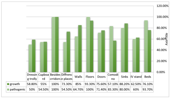
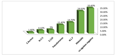
A total of 195 samples were collected from patient zones, dressing room, floor areas, walls, bed-frames, door handles, light switches, sinks, besides corridors, tables, autoclave, cupboards, trolley, drip stand in Burn and plastic surgery department ,aljalla hospital,the culture positive was seen in 143(73%),106(74.1%) of them were pathogenic. The most predominant bacterial isolate was Staph.albus (33.6%), followed by klebsialla (29.5%), N.L.F (14.7%), Pseudomonas (11.47%). E.coli (5%), N.H.S (4.1%), Staph.aureus (1.6%) (Figures 1-4).
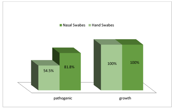
A total of 11 nasal swabs were obtained from nursing staff in the department,(81.8%) of them were pathogenic, predominant bacterial isolate was Staph.albus (30.7%),N.H.S(30.7%),N.L.F(23%), Klebsialla (15.3%),while11 hand swabs were obtained, (54.5%) were pathogenic, Staph.albus (50%), N.H.S (25%), N.L.F (15%), Klebsialla (10%) (Figures 5-6).
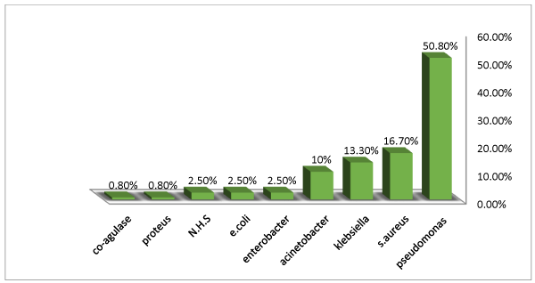
Of the 31 burn cases, 21 (67.7%) were females and 10 (32.2%) males, 120 burn swabs were collected from them, the predominant bacterial isolate was Pseudomonas (50.8%), Staph. Aureus (16.7), Klebsialla (13.3%), Acinetobacter(10%), Enterobacter (2.5%), E.coli (2.5%), N.H.S (2.5%), Proteus(0.8%), Staph. Albus (0.8%) (Figure 7).
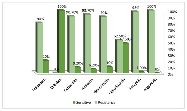
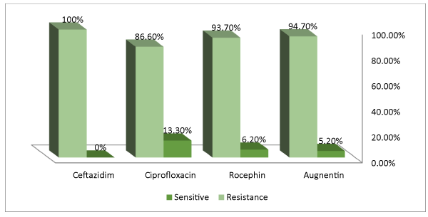
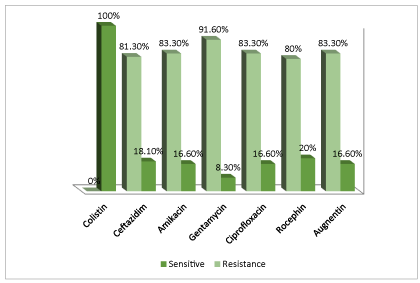
Antibiotic sensitivity tests were undertaken for Augmentin, Ceftrixone, Ciprofloxacin, Imipenem, Ceftazidime, Amikacin, Gentamicin and Colisen. The Figures show percentage of cultures resistant and sensitive to each of these antibiotics (Figures 8-11).
Discussion
Hospital environment is a potential reservoir of bacterial pathogens since it houses both patients with diverse pathogenic microorganisms and a large number of susceptible individuals. The increased frequency of bacterial pathogens in hospital environment is associated with a background rise in various types of nosocomial infections.
In this study 195 samples were collected from various environmental sources in Burn and plastic surgery department included inanimate objects (such as floor areas, walls, bed-frames, door handles, light switches, sinks, besides corridors, tables, autoclave, cupboards), animate objects (stands for infusion apparatus, trolleys, drums, Medical Waste Transport Cart).
The most prevalent bacteria isolated in this study was co-agulase negative (33.6% ), this result is consistent with a study in northwest Ethiopia ,which were co-agulase negative the most frequently isolated from all the samples collected from the wards and operating room, Other studies in Taiwan and Nigeria also demonstrate similar finding on patient’s medical chart and X-ray machine contamination with co-agulase negative staphylococci (15).The reason of their high prevalence may because of the fact that staphylococci are members of the body flora of health and sick individuals that can be spread by their hands, expelled from respiratory tract to the immediate environment. In our study the second isolated pathogen was klebsiella (29.5%), This finding is consistent with Iran which were klebsiella pneumonia (8.9%),but it is higher as compared to their study. Thus, effective cleaning is important condition in the prevention and control of microbial spread, as well as the type of disinfectants used to diminish risks of cross infections during healthcare assistance.
Results of the nasal swabs culture of this study indicated that nursing staff carried coagulase negative staphylococcus(30.7%)This finding is in line with a study in Northwest Ethiopia where the prevalence of coagulase negative staphylococci(58.3%).
This study also showed that (54.5%) hands of nursing staff had bacterial contaminations mostly with coagulase negative staphylococcus, this result was lower than that observed by a study in Northwest Ethiopia, which was (88.4%) [8, 10].
Colonization of pathogens in burn wounds and their systemic invasion may cause severe complications and death. The two most common pathogens responsible for burn wound infections are Staphylococcus aureus and Pseudomonas aeruginosa, Burn wards have been reported to harbor multi-drug resistant strains of P. aeruginosa which can colonize burn wounds and lead to infection. This pathogen has been reported as the most common source of burn wound infection in the United States [11], According to the CDC protocol, Pseudomonas and Acinetobacter are members of nosocomial microorganism.in some countries such as Iraq, S.aureus can be considered as a major cause of nosocomial infection in burn wounds [7]. In this present study the predominant bacterial isolates was pseudomonas (50.85), staphylococcusaureus (16.7%) This finding is in line with CDC protocol and Iran which were the predominant bacterial isolate was pseudomonas aeruginosa (37.5%) followed by staphylococcus aureus (20.2%), and contravened with Iraq which were staphylococcusaureus (33%) the most commonly isolated bacteria among burn patients with burn wound infection followed by pseudomonas aeruginosa (18%).
The change in the pattern of bacterial resistance in the burn unit is important both for clinical settings and epidemiological purposes. The results of antimicrobial sensitivity showed that pseudomonas aeruginosa was highly resistant for most the antibiotic tested, while it had less resistance to ciprofloxacin and sensitive for colistin also present study showed that staph aureus was highly resistant for Augmantine, ceftriaxone, ciprofloxacin, ceftazidim.
Acinetobacter and Klebsiella where highly resistance for Augmantine, Ceftrixone, Ciprofloxacin, Ceftazidim, Gentamicin, Amikacinand highly sensitive to colisten (Table 1).
Conclusion
Burn patients are highly susceptible to wound infection, because of the loss of the barrier function of skin, the immune dysregulation that accompanies severe burn injury.
The most predominant bacterial isolated from patient zone were Staph.albus, followed by klebsialla, Pseudomonas, E.coli, N.H.S and Staph.aureus.
Burn patients were most commonly infected with P. aeruginosa, S. aureus and they wereresistant of most of the antibiotics tested.
A total of 195 samples were collected from patient zones and environment, 106 of them were pathogenic, this indicates about lack of effectiveness of cleaning and disinfection in the ward.
Hands and surfaces (e.g., beds, side rails, tables, and equipment) are often heavily contaminated with organisms from the patient and may be transmitted to other patients if strict barriers are not maintained and appropriate decontamination carried out.
Prevention of infection in burn patient is an important issue that should be considered in burns unit. Isolation of these patients, health policy such as control of staff and nurses, cleaning and disinfection of the animate or inanimate objectives and other equipment related to these patients, and preparation of optimum care conditions of burn patients can be helpful to treat of them.
Use of some antimicrobial agents must be restricted, due to the high resistance to them.
A nosocomial infection surveillance system must be introduced to reduce the rate of nosocomial infections among burn patients, and for better therapeutic options.
Routine surveillance wound cultures should be obtained when the patient is admitted and at least weekly until the wound is closed. Many burn centers recommend obtaining wound cultures two or three time a week for patients with large burn injuries [12].
Based on these finding it is recommended that need to additional attention for hand hygiene applying and disinfection of inanimate objects in the hospital environment to limit the transfer of pathogens from patient to patient or to another departments.
- W.H.O (2011) Burn prevention: Success stories and lessons learned. link: https://tinyurl.com/ybkg83lc
- Hettiaratchy S, Dziewulski P (2004) ABC of burns. BMJ 328: 1366-1368. link: https://tinyurl.com/y98ecqbb
- Desanti L (2005) Extra pathophysiology and current management of burn injury. Wound Care Journal. link: https://tinyurl.com/y7zl28bo
- Ekrami A, Kalantar E (2007) Bacterial infections in burn patients at a burn hospital in iran. Indian J Med 126: 541-544. link: https://tinyurl.com/ydy4gls8
- Azimi l, Motevallian A, Ebrahimzadeh Namvar A, Asghari B, Lari AR (2011) Nosocomial infections in burned patients in motahari hospital, Tehran, Iran. Dermatology research and practice 2011:436952 link: https://tinyurl.com/y9h4xwtw
- Al-Aali K (2016) Microbial profile of burn wound infection in burn patients, Taif, Saudi Arabia. I Med pub Journal 7: 2-15. link: https://tinyurl.com/ybqbcnmz
- Ruder A, Muhhamad JA (2010) Nosocomial infection in sulaimani burn hospital, Iraq. Annals of burns and fire disasters 23:177-181. link: https://tinyurl.com/yavenv68
- Gelaw A, Gebre-Selassie S, Tiruneh M, Mathios E, Yifru S (2014) Isolation of bacterial pathogens from patient with post-operative surgical site infections and possible sources of infections at the university of Gondar hospital, northwest Ethiopia. Journal of environmental and occupational science 3: 103-108. link: https://tinyurl.com/y9frucp5
- Chikere CB, Omoni VT, Chikere BO (2008) Distribution of potential nosocomial pathogens in a hospital environment. African Journal of biotechnology 7:3535-3539. link: https://tinyurl.com/ydhb5b5t
- Alireza. Ekrami, Kayedan, Abbas, Jahangir, Et al. (2011) Isolation of common aerobic bacterial pathogens from the environment of the environment of seven hospitals, Ahvaz, Iran, Jundishapur. Journal of microbiology 4:75-82. link: https://tinyurl.com/y7fcrg3b
- Othman N, Babakir-Mina M, Noori CK, Rashid PY (2014) Pseudomonas aeruginosa infection in burn patients in sulaimaniyah, Iraq: risk factors and antibiotic resistance rates. JInfectDevCtries 8: 1498-1502. link: https://tinyurl.com/y9asjqjc
- Sharma B, Singh VP, Bangar S, Gupta N (2005) septicemia: the principal killer of burns patients. American journal of infectious diseases1:132-138. link: https://tinyurl.com/yczt8ns5
Article Alerts
Subscribe to our articles alerts and stay tuned.
 This work is licensed under a Creative Commons Attribution 4.0 International License.




 Save to Mendeley
