Journal of Surgery and Surgical Research
Treatment of varicose veins – Surgery/ Laser
Goran Batrićević*, Perica Maraš, Davor Musić, Milosav Smiljanić, Aleksandar Filipović and Aleksandar Vulić
Cite this as
Batrićević G, Maraš P, Musić D, Smiljanić M, Filipović A, et al. (2017) Treatment of varicose veins – Surgery/ Laser. J Surg Surgical Res 3(2): 025-029. DOI: 10.17352/2455-2968.000040Introduction: According to statistics damage venous circulation of the lower extremities are among the most common diseases in the world. Although the treatment varicose veins (VV) modernized using non-invasive-endovenous procedures, forecast on short term is not quite satisfactory.
Aim of this study was to examine the results of treatment vv lower extremities to the appearance of improving the quality of life and the emergence of recurrence 1 year after operating (OP) and endovenous laser ablation (EVLA) treatment.
Methods: VV patients who were operated classic surgical method (OP) were the OP group (n-36) and the patients who were treated EVLA method were the EVLA group (n-36). We studied quality of life the patients after 1 year of treatment by Aberdeen’s questionnaire and the occurrence of recurrence.
Results: Between the two groups there was no difference in terms of risk factors before OP / EVLA treatment, as well as in terms of disease severity assessed CEAP classification. Quality of life after 1 year showed that there were no differences between the two groups (5.288 / 7.444, p <0.076). However, symptoms of patients in both groups improved after 1 year of intervention, the pain is to reduce the OP group to 61%, in EVLA group by 87% (p <0.017) Also, after 1 year there was no significant difference in recurrence occurs between OP and EVLA group. (10/36 to 27.8% in the group of OP 6/36 to 16.7 %% in EVLA group). Of the risk factors in multivariate analysis on the phenomenon of recurrence a previous deep vein thrombosis was significantly influenced (p = 0.011).
Conclusion: Patients after 1 year of treatment OP or EVLA of varicose veins had significantly fewer symptoms, particularly patients who were EVLA group Differences between groups were not significant in terms of the recurrence and the quality of life.
Introduction
Varicose veins (vv) are a disease that is mentioned centuries ago. So, there are written records in the Ebers Papyrus-in even in ancient Egypt (1580 BC) [1].
Of the many epidemiological studies on vv point out that in the adult population of Western countries the prevalence of varicose veins was 20% (range 21.8 to 29.4%), 5% have skin changes, venous edema and venous ulcers [2].
Dysfunction of the venous system can occur due to degeneration of the walls of veins, post-thrombotic damage valvular apparatus, chronic venous obstruction or dysfunction of muscle pump [3]. Venous disease complicated by Venous Stasis Syndrome (VSS), which manifests clinically as chronic edema of the legs with painful skin, hyperpigmentation and thickening (induration) of tissue, and in the worst case as venous ulcers [4]. In order to standardize the classification of chronic venous disease (CVD) with various events it was introduced in 1994 yrs. classification system (CEAP) for comparison of the various manifestations of the disease in patients with varicose veins (vv).
This classification is based on certain criteria such as: clinical (C), etiologic (E), anatomical (A) and pathophysiological (P) [5,6].
Etiological varicose veins
1. Primary, congenital valvular incompetence or agenesis apparatus and weakness of the venous wall construction material (weakness of the connective tissue) and 2.Secondary as a result various factors of which is deep vein thrombosis (DVT) is the most [5]. Of risk factors (RF) the most important are: family history of venous disease, older age, prolonged standing, obesity (increased body mass index), conditions that require prolonged sitting (sedentary lifestyle), a condition in which the elevated estrogen, and pregnancy [7-11].
Early event in chronic venous insufficiency (CVI) is the increase in endothelial permeability leading to the extravasation of water and nutrients as a basis for the edema formation and extravasation of red blood cells leading to hyperpigmentation of skin and all these combined lipodermatosclerosis [12].
The importance of preventing the development of CVI and timely start treating patients, especially in stage C0s, when patients have symptoms despite the absence of visible signs of CVI is that it can normalize venous physiology and postpone further development of the disease [11].
Early treatment, for the prevention of venous hypertension, inflammation and reflux, can alleviate symptoms CVI and decrease the risk of ulcers in the terminal stages of CVI, which considerably reduces the quality of life and increases the price of the treatment several times [4,11].
CVI of lower limbs has the effect on both physical to psychological status of the patient that substantially reduces the quality of life, pain and progresses to reduce physical activity [13]. To measure the quality of life in patients with varicose veins, among other, in many countries is used Aberdeen Varicose Vein Questionnaire (AVVQ) introduced 1993 years by Andrew Garratt (which was translated into many languages) [14]. The questionnaire consists of diagrams for plotting varicose veins, estimates of the size pain, swelling ankles, the use of compression stockings, interference with activities and cosmetic changes.
Treatment of superficial veins insufficiency has great medical and socio-economic importance, quite complex and is based on the principles of pathogenic and CEAP classification venous system [15]. There are many treatment options in the treatment of varicose veins and all improve the quality of life (QoL), but none of them led to a complete and lasting effect [16,17].
In addition to conventional surgery for the treatment of varicose veins, which is still the gold standard, thermal ablation of vein saphenous magna (VSM) and vein saphenous parva (VSP) are increasingly in use. These methods are by many authors at least as safe and effective as conventional surgery but with a much faster recovery and better cosmetic effect [18,19]. In addition to the less invasive, the advantage is that these procedure takes less time and can be performed under local (tumescent) anesthesia [20].
The aim was to determine if patients with symptomatic varicose vein of lower extremities after treatment or conventional surgery or endovenous laser therapy have recurrence after 1 year and that of the used methods has better results in relation to the quality of life.
Methodology
VV patients who have been operated classic surgical method (OP) were the OP group (n-36) and that have been treated with EVLA have been EVLA group (n-36). We studied patients clinically after 1 year of treatment on the occurrence of recurrence, while we examined their quality of life after 1 year by Aberdeen’s questionnaire.
Criteria for inclusion were
Diagnosis of varicose veins.
Criteria for exclusion were
Cardiac patients, patients suffering from cancer or other diseases in advanced degree. The diagnosis was set before surgery by clinical inspection and by duplex scans. Severity of the disease is estimated at the reception CEAP classification C0-C6.
In all 36 patients of the OP group were applied a common surgical technique. After preparation of the saphenous-femoral junction, saphenous vein ligation (VSM) at the junction ,simultaneously with the ligation of side branches (crossectomia), then cut in the level of the medial malleolus After identifying where venotomy knife is placed flexible endoluminal catheter that after crosscutting veins in the distal ligation fixed to a blood vessel in the proximal part .Distal part is ligated at the end, after fixation of the catheter in the groin ligation approach to draw a complete venous stable (stripping).
For EVLA procedure we used Biolitek The Ceralas TM E laser according to the protocol provided (Model 980nm / 15w / 200uM (photo number). Patients were treated with energy (depending on the diameter of diseased veins) 70-100 J / cm and power of 14W continuous 200 um fiber diameter throughout the length of diseased veins.
We used local (tumescent) anesthesia. .Puncture a vein was at the predetermined position with the aid of ultrasound, J placing the flexible wire is made hardly visible incision and placing a dilator (sheath). Placing the laser probe (pre-connection to a apparat) through a catheter in which the previously extracted in the dilator position below the saphenous-femoral junction
Statistical methods
The statistical analysis were used methods of descriptive statistics. For calculating differences between continuous variables both groups used the hi2-test and t-test. Multivariate analysis was used for calculating the factors that have influenced the appearance of recurrence.
Results
On the table 1, were registered that the female gender is present in more than 80%, middle age in both groups were <50 years. Patients of both groups were obese, mean weight of patients was> 25 kg / cm2. About 50% of patients had bilateral varicose veins, Of these 9 patients were done bilateral EVLA., Clinical characteristics estimated CEAP classification and were very similar in both groups, the majority of patients belonged to the C3 group.
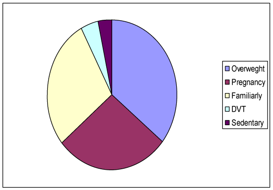
Risk factors shown in figure 1 were: overweight is prevailed (46/72 to 63.8%), pregnancy about 50% of patients, familiarly presence had 36 patients (50%) sedentary occupation / permanent standing was not significantly represented in both groups (5 in the IOP group and 6 in the EVLA group). Also, previous deep thrombosis had a small number of patients in both groups 6 patients (8%).
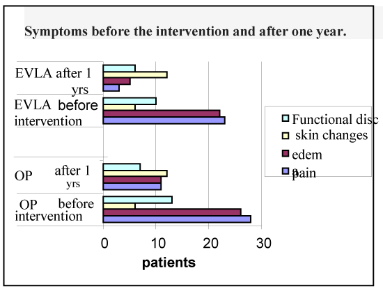
Figure 2 shows that the number of patients with varicose veins that had symptoms is not significantly different in both groups before the intervention. However, after 1 year significantly fewer patients had pain as the OP group, pain was reduced to 61%, but the difference was not significant, while the group EVLA pains were reduced by 87%, which was statistically significant (p <0.017).
The difference between the patients with the edema before and after 1 year. in both groups was also expressed, the edema are reduced in the OP group to 58%, in the EVLA group edema decreased up to 77%, the difference was not significant (p <0.91).
Skin changes in both groups were worsened in the OP by 50% in the EVLA group by 50%, also.
Disturbances in everyday activity were decreased in both groups in the OP group to 46% in EVLA group 40%.
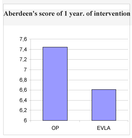
Aberdeen’s score after 1 year showed that is lower in patients who were in the EVLA group than in patients in OP group (Figure 3).
Recurrence was lower in patients who had EVLA compared to OP patients, (6/36 to 16.7 %% in EVLA group / 10/36 to 27.8% in the OP group). Correction of the number of infected leg did not give significant differences between groups (6/45 to 13.3% in the group EVLA / 10/36 to 27.8% in the OP group, p = 0.221) (Figure 4).
Of the many risk factors for recurrence previous DVT was significantly affected (p <0.011)) (Table 2).
Discussion
Many studies have examined the relationship between the surgical and endovenous treatment options to get the answer, which is more effective method, in short and long term, to be recommended as a method superior [21]. The patients in our study were in most cases women (about 80%), the average age was about 49 years and were the most in C3 clinical group to CEAP classification (55%), which is in agreement with the available randomized trials [22].
In comparison, patients who were operated varicose veins of the lower extremities (IP) with patients who had intervention endovenous techniques such as laser (EVLA) after interventions have less absence from work in our study. This, faster recovery after interventions in patients who have had EVLA in relation to the OP patients. Have registered and other studies [22-24]. Many authors emphasize the advantages of outpatient treatment and local anesthesia [24]. So, we can say that minimally invasive procedures have a favorable socio-economic effect.
Symptomatology after one year of intervention
In order to assess the effectiveness of both methods for the short term, ie. For one year of intervention, we examined patients in terms of their pain, the edema of legs, skin changes (dispigmentation) or possibly other serious changes and the difficulties in performing everyday activities. Results showed that, looking at total, all patients had a significant improvement in terms of pain and swelling of legs. Only changes in the skin worsened, in terms of the existence of dark skin, most likely due to insufficient absorption hematoma after intervention. Also to improve the daily activity in both studies groups. Rasmussen et al. study found that pain reduced after the intervention [23]. In another randomized study de Medeiros was found that there was no difference in postoperative pain, but it was smaller edema in EVLA group [25].
Quality of life after one year of intervention
We used a Aberdeen’s questionnaire in testing the quality of life in patients after 1 year of intervention. In our study, after one year of intervention Aberdeen’s middle score was lower in EVLA patients, which means better quality of life in relation to the OP patients, but the difference was not significant, even when adjusted to the number of diseased leg. Studies using simultaneously Aberdeen’s questionnaire and SF-36 questionnaire for testing the efficacy EVLA and the OP showed a significant improvement in quality of life [23]. In other randomized studies which have compared the quality of life at EVLA and OP patients with Aberdeen’s scoring system there was no a significant difference between the term, but was improved in both groups in study Darwood and the authors [27].
In any of the non-randomized studies not found advantage in key parameters, but has found significant superiority in measuring quality of life in the short-term and long-term monitoring in EVLA group of patients compared to surgical stripping [26].
Recurrence
In our study we found that for one year follow-up recurrence was more often in the OP group compared to EVLA groups. Examining which of the risk factors influenced the emergence of recurrence, we found that the previous deep thrombosis (DVT) significantly affected the appearance of recurrence. Recurrence in our patients was minimal in the thigh, which we explain as neovascularization. Other authors also believe that the neovascularization is one of the causes of recurrence especially in the operated patients [21].
Causes of recurrence according to Rasmussen and all are-various - technical errors, wrong evaluation reflux (which can be minimized by using Ultrasound preoperatively), neo-vascularization, and the disease progression [21,28].
Conclusion
Patients after 1 year of treatment of varicose veins of the lower extremity with EVLA or OP had fewer symptoms, particularly patients who were in EVLA group had less pain and swelling. The difference between groups was not significant in terms of recurrence and quality of life.
- Ido Weinberg (2011) History of varicose veins. Vascular Medicine. Link: https://goo.gl/Z6QQgM
- Kaplan RM, Criqui MH, Denenberg JO, Bergan J, Fronek A (2003) Quality of life in patients with chronic venous disease: San Diego population study. J Vasc Surg 37: 1047-1053. Link: https://goo.gl/J8nsmR
- Gloviczki P, Mozes G (2009) Development and anatomy of the venous system. In: Gloviczki P, editor. Handbook of venous disorders: guidelines of the American Venous Forum. 3rd ed. London: Hodder Arnold 12-24. Link: https://goo.gl/U2UdWV
- Bergan JJ, Geert WSS, DC Smith, Andrew NN, Michel RB et al. (2006) et al. Chronic venous disease. N Engl J Med 355: 488-498. Link: https://goo.gl/KP5hKM
- Kistner RL, Eklof B (2009) Classification and etiology of chronic venous disease. In: Gloviczki P, editor. Handbook of venous disorders: guidelines of the American Venous Forum. 3rd ed. London: Hodder Arnold 37-46. Link: https://goo.gl/U2UdWV
- Peter Gloviczki, Comerota AJ, Dalsing MC, Eklof BG, Gillespie DL, et al. (2011) The care of patients with varicose veins and associated chronic venous diseases: Clinical practice guidelines of the Society for Vascular Surgery and the American Venous Forum. J Vasc Surg 53: 2S-48S. Link: https://goo.gl/TvawBt
- Cornu-Thenard A, Boivin P, Baud JM, De Vincenzi I, Carpentier PH (1994) Importance of the familial factor in varicose disease. J Dermatol Surg Oncol 20: 318-326 Link: https://goo.gl/SfTQKZ
- Criqui MH, Jamosmos M, Fronek A, Denenberg JO, Langer RD, et al. (2003) Chronic venous disease in an ethnically diverse population: The San Diego Population Study. Am J Epidemiol 158: 448-456. Link: https://goo.gl/YrvGMh
- Tuchsen F, Hannerz H, Burr H, Krause N (2005) Prolonged standing at work and hospitalisation due to varicose veins: a 12 year prospective study of the Danish population. Occup. Environ Med 62: 847-850. Link: https://goo.gl/naRfsP
- Aneel AA, Silverstein MD, Lahr BD, Petterson TM, Bailey KR, et al. (2009) Risk factors and underlying mechanisms for venous stasis syndrome: a population-based case–control study. Vasc Med 14: 339-349. Link: https://goo.gl/dnXb1U
- Ramelet AA, Perrin M, Kern P, Bounameaux H (2008) Phlebology. 5th ed. Issy les Moulineaux, France: Elsevier Masson.
- Bergan JJ, Schmid-Schönbein G, Coleridge-Smith P, Nicolaides A, Boisseau M, et al. (2006) Chronic venous disease. N Engl J Med 355:488-498. Link: https://goo.gl/XB5oCu
- Nicolaides AN, Hussein MK, Szendro G, Christopoulos D,Vasdekis S, et al.(1993) The relation of venous ulceration with ambulatory venous pressure measurements. J Vasc Surg 17: 414–419. Link: https://goo.gl/vmwBCv
- Rabe E, Pannier-Fischer F, Bromen K, Schuldt K, Stang A, et al. (2003) Bonner Venenstudie der Deutschen Gesellschaft für Phlebologie Epidemiologische Untersuchung zur Frage der Häufigkeit und Ausprägung von chronischen Venenkrankheiten in der städtischen und ländlichen Wohnbevölkerung. Phlebologie 32: 1-28. Link: https://goo.gl/Epo6Vv
- D Kelleher, Lane TRA, Franklin IJ, Davies AH (2012) Treatment options, clinical outcome (quality of life) and cost benefit (quality-adjusted life year) in varicose vein treatment. Phlebology 27: 16-22. Link: https://goo.gl/9eKsYC
- Labropoulos N, Tiongson J, Pryor L, Tassiopoulos AK, Kang SS, et al. (2003) Definition of venous reflux in lower-extremity veins. J Vasc Surg 38: 793-798. Link: https://goo.gl/nBwywK
- Radak DJ, Vlajinac HD, Marinković JM, Maksimović MŽ, Maksimović ZV (2013) Quality of life in chronic venous disease patients measured by short Chronic Venous Disease Quality of Life Questionnaire (CIVIQ–14) in Serbia. Journal of Vascular Surgery 58: 1006–1013. Link: https://goo.gl/FFA23e
- Müller-Bühl U, Leutgeb R, Engeser P, Achankeng EN, Szecsenyi J, et al. (2012) Varicose veins are a risk factor for deep venous thrombosis in general practice patients. Vasa. 41: 360–365. Link: https://goo.gl/LAzqLU
- Weiss RA (2002) Comparison of endovenus radiofrquenci versus 810nm diode laser occlusion of large veins in an animal model.Dermatol Surg 28: 56-61. Link: https://goo.gl/HWRMtp
- Goldman MP, Mauricio M, Rao J (2004) J.Intravascular 1320-nm laser closure of the great saphenous vein: a 6-to12 –month follow-up study. Dermatol Surg 30: 1380-1385. Link: https://goo.gl/GGXA5t
- Min RI, Zimmet SE Isaacs MN, Forrestal MD (2001) Endovenous laser treatment of the incompetent geater saphenous vein.J Vascu Interv Radiol 12: 1167-1171. Link: https://goo.gl/yctZX5
- Rasmussen LH, Bjoern L, Lawaetz M, Lawaetz B, Blemings A, et al. (2010) Randomized trial comparing endovenous laser ablation of the great saphenous vein with with stripping of the great saphenous vein: clinical outcome and recurrence after 2 years. Eur J Vasc Endovasc Surg 39: 630–5. Link: https://goo.gl/jhXifm
- Christenson JT, Gueddi S, Gemayel G, Bounameaux H (2010) Prospective randomized trial comparing endovenous laser ablation and surgery for treatment of primary great saphenous varicose veins with a 2-year follow-up. J Vasc Surg 52: 1234-1241. Link: https://goo.gl/cbxDeu
- Rasmussen LH, Lawaetz M, Bjoern L, Vennits B, Blemings A, et al. (2011) Randomized clinical trial comparing endovenous laser ablation, radiofrequency ablation, foam sclerotherapy and surgical stripping for great saphenous varicose veins. Br J Surg. 98: 1079–1087. Link: https://goo.gl/kGP7aU
- D Carradice, AI Mekako, FA K.Mazari, et al. (2011) Endovenous laser ablation compared with conventional surgery for great saphenous varicose veins. British Journal of Sugery.
- de Medeiros CA, Luccas GC (2005) Comparison of endovenous treatment with an 810 nm laser versus conventional stripping of the great saphenous vein in patients with primary varicose veins. Dermatol Surg 31: 1685-1694. Link: https://goo.gl/zkPJpb
- Mekako AI, Hatfield J, Bryce J, Lee D, McCollum PT, et al. (2006) A nonrandomised controlled trial ofendovenous laser therapy and surgery in the treatment of varicose veins. Ann Vasc Surg 20: 451-457. Link: https://goo.gl/s7jPDM
- Darwood RJ, Theivacumar N, Dellagrammaticas D, Mavor AI, Gough MJ (2008) Randomized Clinical trial comparing endovenous laser ablation with surgery for the treatment of primary great saphenous veins. Br J Sug 95: 294-301. Link: https://goo.gl/vcdyKZ
- Perrin MR, Guex JJ, Ruckley CV, dePalma RG, Royle JP, et al.(2000) Recurrent varices after surgery (REVAS), a consensus document. REVAS group. Cardiovasc Surg 8: 233-245. Link: https://goo.gl/QSEDgR
Article Alerts
Subscribe to our articles alerts and stay tuned.
 This work is licensed under a Creative Commons Attribution 4.0 International License.


 Save to Mendeley
