Journal of Surgery and Surgical Research
Anterior and Posterior Capsule-Periosteal sleeve avulsion as an unusual cause of Shoulder Instability
Eduvigis Aranda-Izquierdo, Olga Pérez-Moro, Marcos Fernández-Cuadros*, Ana Maria Valverde-Villar, Alejandro Ortíz-Espada and Rafael Llopis-Miró
Cite this as
Aranda-Izquierdo E, Pérez-Moro O, Fernández-Cuadros M, Valverde-Villar AM, Ortíz-Espada A, et al. (2017) Anterior and Posterior Capsule-Periosteal sleeve avulsion as an unusual cause of Shoulder Instability. J Surg Surgical Res 3(1): 020-024. DOI: 10.17352/2455-2968.000039Traumatic anterior shoulder instability is a typical condition in young people and athletes. There are many different injuries that may be present, producing or coexisting with shoulder instability, in addition to classic Bankart lesion.
Post-traumatic shoulder dislocation is much less common, between 2% to 5% of all shoulder dislocation, ascending to 10% on contact sports people. The most common causes are seizures, high-energy trauma and electrocution. Traumatic multidirectional instability without hypermobility is extremely rare. We have found no reports in literature about a similar case.
The purpose of this case report is to describe an unusual case of a patient who suffered an anterior and posterior shoulder dislocation simultaneously during self-defense training. Besides the complete description of a new and unusual type of injury, difficulty in diagnosis, surgical treatment and the used rehabilitation protocol are reviewed.
Introduction
Traumatic anterior shoulder instability is a typical condition in young people and athletes. There are many different injuries that may be present, producing or coexisting with shoulder instability, in addition to classic Bankart lesion, and including the glenoid bone deficiency (bony Bankart), bone loss of the humeral head (Hill-Sachs lesion), rotator cuff tears, capsular injury with avulsion of the glenohumeral ligaments (HAGL lesions), SLAP lesions, circumferential lesions of the labrum and anterior labrum-ligamentous periosteal sleeve avulsion (ALPSA lesions). Neviaser [1], in 1993, was the first to adopt the term “ALPSA injury” to describe an arthroscopic finding consisting of an anterior periosteal labrum-ligamentous detachment, with a clear separation between the labrum and capsule.
Post-traumatic shoulder dislocation is much less common, between 2% to 5% of all shoulder dislocation, ascending to 10% on contact sports people [2]. The most common causes are seizures, high-energy trauma and electrocution. Athletes who play contact sports such as wrestling, hockey, rugby and soccer have an increased risk of traumatic posterior shoulder dislocation. Other athletes who perform repetitive movements or releases such as volleyball, basketball, baseball, tennis or swimming can develop shoulder instability because of micro trauma or overuse. Posterior dislocations of the shoulder are rarely isolated, and often other injuries are associated, such as fractures of the humeral neck and reverse greater or lesser tuberosity Hill-Sachs lesions. Rotator cuff tears are present in up to 13% of cases. Besides, second posterior glenohumeral dislocation palsy is rare, less than 1% of cases, and the axillary nerve is the one most commonly injured.
Traumatic multidirectional instability without hypermobility is extremely rare. We have found no reports in literature about a similar case.
The purpose of this case report is to describe an unusual case of a patient who suffered an anterior and posterior shoulder dislocation simultaneously during self-defense training. Arthroscopic findings are shown with peel periosteal lesions in both the anterior and the rear face of the glenoid, and the subsequent partial nerve injury from suprascapular nerve elongation. Besides the complete description of a new and unusual type of injury, difficulty in diagnosis, surgical treatment and the used rehabilitation protocol are reviewed.
Case Report
A case of a 35 years old male, bodyguard profession, who practices different sports, wrestling and self-defense among others, is presented. In April 2013, during a self-defense training, he reported blunt trauma with entrapment to his right arm, in which the opponent turned his arm into external rotation, and then inwards and backwards, being out of balance and falling down, and finally his arm staying behind his head.
He underwent conventional MRI one month later, without any significant findings. However, the patient continued to feel unstable, preventing him from doing his daily life activities, for example, “drawing his weapon.” One month later a CT study was performed and no bone lesions were found, so subsequently an arthro-MRI was done. This was informed as well positioned glenohumeral joint without displacement of the inferior glenohumeral labrum-ligamentous complex. Chondral defect in the anterior part of the glenoid, close to the labrum, was found. No bone lesions in the anterior glenoid rim, or Hill-Sachs lesions were observed.
5 months later the patient came to our clinic. Atrophy of the suprascapular area was seen on inspection. Sulcus maneuver was negative and the apprehension maneuvers were difficult to perform because the patient did not let to explore the arm in any direction (Figure 1).
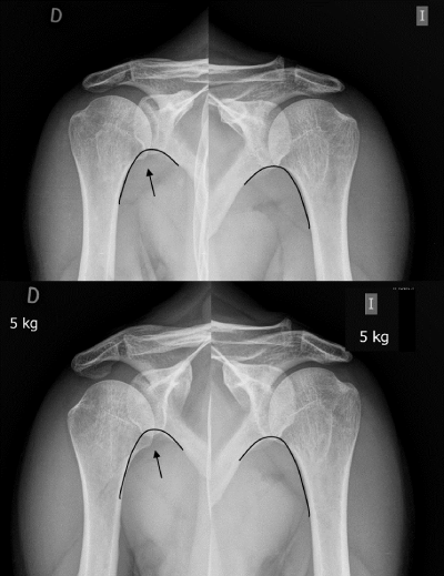
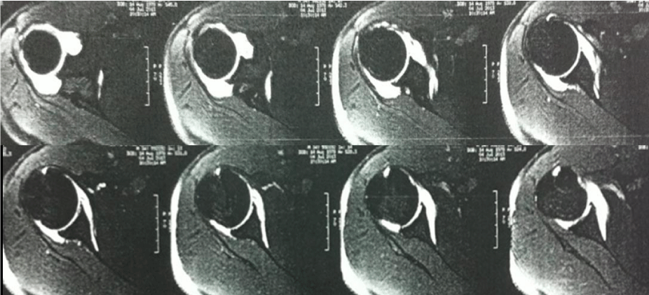
Imaging studies were reviewed and a comparative simple X-ray study of both shoulders was performed. Right shoulder inferior subluxation was found, as shown by the brake up of the normal “gothic arch” formed by the lower border of the scapula to the lower border of the humerus. This subluxation brake up increased with 5 kg extra weight (Figure 2). Reviewing arthro-MRI, capsule-periosteal detachment both on the anterior and posterior neck of the glenoid could be seen (Figure 3). EMG was requested, reporting very severe traumatic injury of the right suprascapular nerve (branches to supra and infraspinatus muscles), which is characteristic in incomplete axonotmesis with greater intensity in the infraspinatus branch. Mild axonal injury of the right axillary nerve was reported.
Arthroscopic surgery was decided with the diagnosis of shoulder instability caused by anterior and posterior capsule-periosteal detachment (ALPSA) and the diagnosis of suprascapular nerve injury. Before surgery, he was evaluated by the Rehabilitation department and started treating neurological injury and muscle toning.
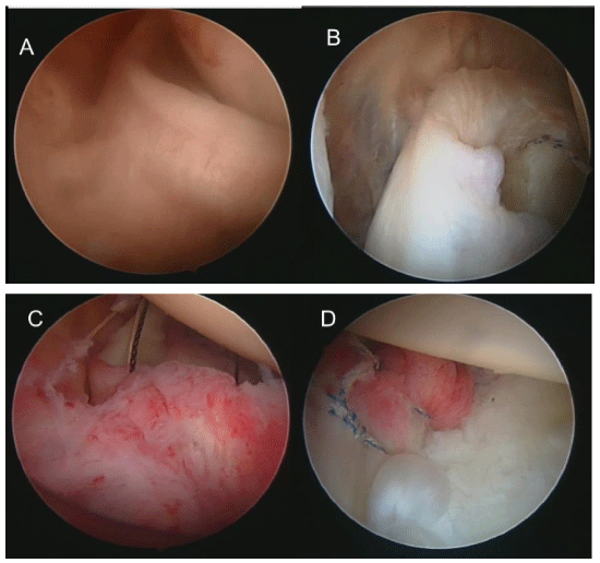
Arthroscopy was performed seven months after trauma. It was found a congruent glenohumeral joint, well located on the rim of the glenoid labrum and a capsular detachment exposing the entire neck of the glenoid both in the front, and the back faces (Figure 4). Treatment consisted of anterior capsule reinsertion “Bankart-type” and the subsequent reintegration of the capsule to the glenoid rim. The patient went to early rehabilitation program, which initially involved gentle active/assisted movements as tolerated, avoiding inadequate movements to protect performed fixations, but without forgetting the rigidity caused by lack of mobility, and treatment of neurological injury.
Rehabilitation treatment was multidisciplinary and designed to improve and fasten functionality, called “Fast Track” or protocol for rapid action to return the patient to his earlier range of motion, and a rapid return to functional activity levels without increasing risk of injury.
This rehabilitation protocol is started within 24 hours after surgery. Active mobilization in assisted range of motion (forward flexion, rear flexion, abduction), flexion and extension exercises of wrist and elbow, postural control and mobilization exercises of the cervical spine and shoulder exercises fighting circular muscle tension and excessive protection of surgery were performed. The patient should perform a home-based exercise program as it was stated in the informative discharge card, as well as a day and night recommendations protocol in order to diminish pain and to protect the surgery.
At one year follow up, the patient had recovered complete mobility and had come back to its usual sport practice (Figure 5).
Discussion
Lereux et al. [3], studied the prevalence of anterior shoulder dislocation in the Ontario city population and stated that it appeared in 23.1 cases per 100.000 people a year, although the incidence is higher in young men. In contact sports, such as rugby, the prevalence may be up to 14.8% [4].
According to Robinson et al. [5], the prevalence of posterior shoulder dislocation is low, and the percentage of recurrences in the first year is 17.7%. The risk is higher in younger than forty years people and after seizures.
Multidirectional shoulder instability without hypermobility was classified by Gerber and Nyffeler [6], by including them within the dynamic instabilities (class B (B4)). They are extremely rare and it requires at least two lesion traumas with two episodes of instability, one anterior and one posterior, as it was in our case report. Physical examination reveals an anterior and posterior positive apprehension test, with a negative Sulcus maneuver.
The mechanism to dislocate anteriorly a shoulder typically requires to force abduction and external rotation. On contact sports, such causes usually involve the collision with another athlete or an inanimate object, including the ground. To dislocate posteriorly a shoulder, a force in flexion, adduction and internal rotation [7], is required. In our case report, both maneuvers were performed simultaneously by a sports opponent.
In the diagnosis of shoulder instability is very important to make a correct clinical interview. Inspection should be comparative to the contralateral side and it should be able to show whether there are bone prominences alterations (shoulder epaulet), atrophy of the deltoid, supra or infraspinatus muscles, or if there is an internal or external rotation attitude of the affected arm. Physical examination may reveal painful spots, blockages in mobility, and whether there is associated laxity (sulcus sign). Sometimes examination is difficult because of patient defense.
Image studies should include standard X-rays with a minimum of two projections, AP (anterior to posterior) and Velpeau [8]. In our case report, the comparative study between the two shoulders shows the alteration of the Gothic arch that should form the lower border of the scapula and the lower border of the humerus (Figure 1). The CT should indicate whether there is evidence or suspicion of fracture or bone injury in the glenoid or humeral head, in order to identify and to quantify bone loss. MRI is essential not only to identify labral lesions, but also to allow the diagnosis of other conditions that may coexist or cause instability, involving the rotator cuff, glenohumeral ligaments, cartilage and capsule. Kim et al. [9], indicated that the incidence of SLAP lesions associated with anterior shoulder dislocation amounts up to 42.8% in acute anterior dislocations and 32.0% in recurrent dislocation.
Arthro-MRI is more effective [10], in the diagnosis of labrum-ligamentous lesions, such as ALPSA type lesions, which are sometimes unnoticed with conventional MRI, and according to Forsythe [11], it should be preferred in young athletes. Schreinemachers et al. [12], discussed whether to perform arthro-RM in different positions, to detect ALPSA type injuries and they conclude that the results of conventional arthro-MRI in neutral position is comparable to that performed in ABER position (abduction and external rotation). However, Tian et al. [13], refer the arthro-MRI in ABER position to be more effective to detect this type of injury.
Capsule-labrum-ligamentous lesions reported in these studies are associated with anterior shoulder instability, as in the case of [14]:
- Classic Bankart Injury: the most frequent labral lesion. In this lesion, there is a rupture of the anterior-inferior area of the labrum associated with rupture of the periosteum. If the injury is associated by a bone lesion of the anterior margin of the Glena, it is called bony Bankart injury.
- Perthes Injury: This is a lesion of the labrum with the periosteum intact. The labrum is well located on MRI.
- ALPSA lesion (Anterior Labrum-ligamentous Periosteal Sleeve Avulsion): defined as an avulsion or detachment and medial displacement of the labrum-ligamentous complex by the scapular neck.
- GLAD Injury (Glenoid Labral Articular Defect): An injury of the anterior-inferior part of the labrum associated with injury of the anterior-inferior zone of the cartilage.
- SLAP lesion (Superior Labrum Anterior to Posterior) type 5: lesion affecting the anterior-superior area of the labrum including the biceps.
- HAGL lesion (Humeral avulsion of Anterior Glenohumeral Ligament): disinsertion at the humerus level of the inferior glenohumeral ligament (anterior portion). It is very rare. In the arthro-MRI is characteristic to see in the coronal sections a J-shaped capsular distension instead of a U-shape, which is the norm. Also, there is a bony HAGL lesion, in which the ligaments are pulled together with a fragment of humeral bone, the latter being less frequent.
- GAGL Injury (Glenoid Avulsion of the Glenohumeral Ligaments): It is very uncommon. This lesion involves the avulsion of the inferior glenohumeral ligament of the glenoid without associated injury of the lower labrum.
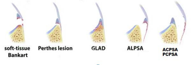
This case report is difficult to fit into one of these injuries. It could be a GAGL lesion that continues to detach the periosteum, but it does not have an intact labrum so it would be partly a GLAD lesion since it has an anterior-inferior chondral lesion with an injured labrum, or an ALPSA lesion but with the Labrum well located. It could be defined as an ACPSA (Anterior Capsule-Periosteal Sleeve Avulsion) lesion (Figure 6).
As for the injuries associated with subsequent instability, we can date:
- Inverse Bankart Injury: same as Classic Bankart Lesion but in the back area. We could also have an inverted bony Bankart.
- Kim’s injury: occluded and incomplete avulsion of the posterior-inferior zone of the labrum.
If we try to locate the posterior lesion of our case report, we are not able neither to define it nor to classify it. It would be a capsule-periosteal pullout injury with intact labrum. This lesion could be called PCPSA (Posterior Capsule-Periosteal Sleeve Avulsion) (Figure 6).
Neurological involvement following a shoulder dislocation varies in literature between 12.5% and 21% of cases and the axillary nerve is the most commonly affected. Sensitive deficits may persist after reduction of the dislocation in 25% of cases [15]. The suprascapular nerve is very rare to be injured after direct trauma, fractures or dislocations of the shoulder. It has been described in relation to massive rotator cuff tears, although most of these tears have no suprascapular nerve lesion [16]. It is also been described suprascapular nerve injury in the context of shoulder instabilities by overuse in some sports such as tennis or volleyball [17], or as recently published by Arriaza et al. [18], the association of this nerve injury in swimmers. In our case it is a lesion caused by trauma, and maintained over time by the traction exerted by slight sagging of the glenohumeral joint, because of the large anterior and posterior capsular lesion presented.
Recurrence after arthroscopic repair of ALPSA injury, according to the study by Ozbaydar et al. [19], is twice the recurrence after repair of Bankart lesions. Lee et al. [20], concluded that the group of ALPSA lesions are more likely to have recurrent dislocations after arthroscopic repair, and they also have a higher number of preoperative dislocations, synovitis and larger Hill-Sachs lesions.
Accelerated rehabilitation decreases postoperative pain, improves joint range of motion, promotes fast return of patients to their activities of daily living including sports, and improves patient satisfaction, all of this without increasing morbidity.
Conclusion
The multidirectional instability is described and requires double traumatism on the shoulder, as was observed in our case report. For its diagnosis, as for other types of instabilities, the arthro-MRI is very important since with normal MRI we could underdiagnose capsule-ligamentous lesions.
In this case, a new variant of anterior and posterior capsule-periosteal lesion with the labrum well located, as an unusual cause of shoulder instability is described.
- Neviaser TJ (1993) The anterior labroligamentous periosteal sleeve avulsion lesion: a cause of anterior instability of the shoulder. Arthroscopy 9: 17-21. Link: https://goo.gl/t5NFp7
- Tannenbaum EP, Sekiya JK (2013) Posterior shoulder instability in the contact athlete. Clin Sports Med 32: 781-796. Link: https://goo.gl/OJjTSS
- Leroux T, Wasserstein D, Veillette C, Khoshbin A, Henry P, et al. (2014) Epidemiology of primary anterior shoulder dislocation requering closed reduction in Ontario, Canada. Am J Sport Med 42: 442-450. Link: https://goo.gl/EjR1I7
- Kawasaki T, Ota C, Urayama S, Maki N, Nagayama M, et al. (2014) Incidence of and risk factors for traumatic anterior shoulder dislocation: an epidemiologic study in high-school rugby players. J Shoulder Elbow Surg 23: 1624-1630. Link: https://goo.gl/PFeCn3
- Robinson CM, Seah M, Akhtar MA (2011) The epidemiology, risk of recurrence, and functional outcome after an acute traumatic posterior dislocation of the shoulder. J Bone Joint Surg Am 93: 1605-1613. Link: https://goo.gl/FPzdDB
- Gerber C, Nyffeler RW (2002) Classification of glenohumeral joint instability. Clin Orthop Relat Res: 65-76. Link: https://goo.gl/Hcz3Qm
- Abrams JS, Bradley JP, Angelo RL, Burks R (2010) Arthroscopic management of shoulder instabilities: Anterior, posterior and multidirectional. AAOS Intr Course Lect 59: 141-155. Link: https://goo.gl/ZsZVLc
- Rouleau DM, Herbert-Davies J, Robinson CM (2014) Acute traumatic posterior shoulder dislocation. J Am Acad Orthop Surg 22: 145-152. Link: https://goo.gl/Srw9T3
- Kim DS, Yi CH, Yoon YS (2011) Arthroscopic repair for combinated Bankart and superior labral anterior posterior lesions: a comparative study between primary and recurrent anterior dislocation in the shoulder. International Orthop 35: 1187-1195. Link: https://goo.gl/X5mVB0
- Mutlu S, Mahirogullari M, Güler O, Uçar BY, Mutlu H, et al. (2013) Anterior glenohumeral Instability: Classification af pathologies of anteroinferior labroligamentous structures using MR arthrography. Adv Orthop 2013: 473194. Link: https://goo.gl/lx9lxD
- Forsythe B, Frank RM, Ahmed M, Verma NN, Cole BJ, et al. (2015) Identification and treatment of existing copathology in anterior shoulder instability repair. Arthroscopy 31: 154-166. Link: https://goo.gl/i6V6TE
- Schreinemachers DA, Van der Hulsr VP, Jaap WW, Bipat S, Van der Woude HJ (2009) Is a single MR arthrography series in ABER position as accurate in detecting anteroinferior labroligamentous lesions as conventional MR artrhography? Skeletal Radiol 38: 675-683. Link: https://goo.gl/38Pb5s
- Tian CY, Cui GQ, Zheng ZZ, Ren AH (2013) The added value of ABER position for the detection and classification of anteroinferior labroligamentous lesions in MR arthrography of the shoulder. Eur J Radiol 82: 651-657. Link: https://goo.gl/leKmUV
- Te Slaa RL, Wijffels MP, Brand R, Marti RK (2004) The prognosis following acute primary glenohumeral dislocation. J Bone Joint Surg Br 86: 58-64. Link: https://goo.gl/nlwc7x
- Perron AD, Ingerski MS, Brady WJ, Erling BF, Ullman EA (2003) Acute complications associated whit shoulder dislocation at an academic Emergency Department. J Emerg Med 24: 141-145. Link: https://goo.gl/Lgr7JJ
- Collin P, Treseder T, Lädermann A, Benkalfate T, Mourtada R, et al. (2014) Neuropathy of the supraescapular nerveand massive rotator cuff tears: a prospective electromiografic study. J Shoulder Elbow Surg 23: 28-34. Link: https://goo.gl/u6jcDs
- Corò L, Azuelos A, Alexandre A (2005) Supraescapular nerve entrapment. Acta Neurochir Suppl 92: 33-34. Link: https://goo.gl/8L3Fsq
- Arriaza R, Ballesteros J, López-Vidriero E (2013) Supraescapular neuropathy as e cause of swimmer´s shoulder: results after arthoscopic treatment in 4 patients. Am J Sports Med 41: 887-893. Link: https://goo.gl/tyYuCQ
- Ozbaydar M, Elhassan B, Diller D, Massimini D, Higgins LD, et al. (2008) Results of arthroscopic capsulolabral repair: Bankart lesión versus anterior labroligamentous periosteal sleeve avulsion lesion. Arthoscopy 24: 1277-1283. Link: https://goo.gl/akJY1p
- Lee BG, Cho NS, Rhee YG (2011) Anterior labroligamentous periosteal sleeve avulsion lesion in arthroscopic capsulolabral repair for anterior shoulder instability. Knee Surg Sport Traumatol Arthrosc 19: 1563-1569. Link: https://goo.gl/M1dpMj
Article Alerts
Subscribe to our articles alerts and stay tuned.
 This work is licensed under a Creative Commons Attribution 4.0 International License.


 Save to Mendeley
