Journal of Novel Physiotherapy and Physical Rehabilitation
Exercise Tolerance in Children with Simple Congenitally Corrected Transposition of the Great Arteries: A Comparative Study
Tony Reybrouck*, Marc Gewillig, Werner Budts and Roselien Buys
Cite this as
Reybrouck T, Gewillig M, Budts W, Buys R (2017) Exercise Tolerance in Children with Simple Congenitally Corrected Transposition of the Great Arteries: A Comparative Study. J Nov Physiother Phys Rehabil 4(3): 066-070. DOI: 10.17352/2455-5487.000050Background: The aim of our study was to investigate the exercise capacity of children with congenitally corrected transposition of the great arteries without significant associated heart defects (l-TGA) in comparison with children with the classical type of TGA (d-TGA) and a healthy control group.
Methods: Seven children with isolated l-TGA (11.2 ± 3,2 years), 17 children after a Senning operation (13.4 ± 1,6 years ), 26 children with an arterial switch operation (11.0 ± 2,3 years) and 34 healthy controls (12.0 ± 2,9 years) performed a maximal graded cardiopulmonary exercise test on a treadmill, during which oxygen uptake (VO2) and heart rate (HR) were registered.
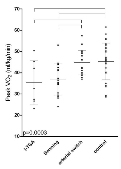
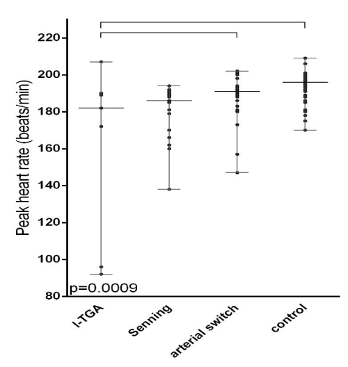
Results: Significant differences were present between groups for peak VO2 (p<0,001) and peak HR (p<0,001). Compared to the control group, l-TGA patients had a significantly lower peak VO2 (35.4 ± 10.5 mL.min-1.kg-1 vs 45.3 ± 8.65 mL.min-1.kg-1) and peak HR (161 ± 47 beats. min-1 vs 193 ± 9 beats. min-1). Between the arterial switch group and the control group, no significant differences were found, nor between the l-TGA group and the Senning group.
Conclusion: Children with simple l-TGA and children who underwent a Senning procedure for d-TGA have similar exercise capacity, which is significantly lower when compared to arterial switch patients and healthy controls. The underlying mechanism for the impaired exercise capacity seems to be rather HR-related in children with l-TGA, whereas in children with Senning operation reduced right ventricular function and therefore reduced increase in stroke volume with exercise is more present
Introduction
Congenitally corrected transposition of the great arteries (l-TGA), also called levi-transposition of the great arteries is a very uncommon type of transposition of the great arteries (TGA) which is characterized by the presence of atrioventricular discordance with ventriculoarterial discordance [1]. It is especially rare when no concomitant heart defects are present (simple l-TGA). In this condition, the blood is normally oxygenated, so no immediately identifiable symptoms are detectable and the anomaly is often not diagnosed or recognised in early life. However, in the patients’ third or fourth decade, problems such as arrhythmia’s and tricuspid valve regurgitation may arise [1]. In addition, the right ventricle may eventually become hypertrophic and dysfunctional due to increased pressure and further contribute to the development of symptoms such as dyspnoea or fatigue [1]. These symptoms typically arise at first during exercise.
In this light, cardiopulmonary exercise testing might be a sensitive tool to detect the first signs of a reduced cardiac function as objectivised by an impaired exercise capacity.
Only few studies, in adults or a mixed age group, were performed in patients with l-TGA and reported reduced exercise capacities [2-6]. Based on these studies, it seems that the reduction in exercise capacity is present, even when the patients are asymptomatic. The role of right ventricular dysfunction was put forward as an important cause for the reduced exercise tolerance [2-5].
Indeed, in adults with a systemic right ventricle such as patients with Mustard or Senning operation for TGA, reduced exercise capacities are present and a relation with right ventricular function exists [7,8]. Winter et al. documented the exercise tolerance of a group of adult patients with a systemic right ventricle and also reported reduced exercise capacities [9]. They however did not investigate patient groups separately even though they admit the fact that patients with l-TGA are different from those with an atrial switch operation. Giardini et al. reported similar exercise capacities in adults with l-TGA when compared to adults who underwent the Senning operation for d-TGA [5].
Patients after an atrial switch operation are known to have diminished exercise capacities already during childhood [10,11], whereas patients after an arterial switch operation [12,13], show mildly diminished to normal exercise capacities. In patients with l-TGA, complications such as right ventricular dysfunction usually only arise during adulthood. Whether the impaired exercise capacity is already present during childhood in this population, is not known. To the best of our knowledge, the exercise capacity in children with l-TGA without other heart defects has never been investigated and compared to children with d-TGA and an atrial or arterial switch operation.
The aim of this study was therefore to investigate the exercise performance and cardiovascular response during graded exercise of children with simple l-TGA in comparison with children operated for d-TGA by the atrial or arterial switch procedures and with a control group of healthy children.
Methodology
Study design and patient selection
This study was a single-centre retrospective investigation of the exercise capacity of children with different types of TGA in comparison with each other and with healthy controls.
All patients with simple l-TGA were selected from a database of exercise tests performed at the department of paediatric cardiology at the University Hospital of Leuven, Belgium. Furthermore, all patients with Senning operation and with arterial switch operation for d-TGA were selected. Only patients who were asymptomatic at the moment of testing were included in this study. The control group consisted of a random sample of healthy children of comparable age who were referred to the paediatric cardiology unit for exercise testing but in whom a diagnosis of a morphologically and functionally normal heart was made. Children mentally or physically unable to perform an exercise test until exhaustion were excluded. Other exclusion criteria were obesity (>95% CI), additional congenital heart defects or other medical conditions such as muscular or endocrine disease, and age below 7 and above 16 years old.
All patients and parents gave informed consent prior to participation. The study was approved by the local medical ethics committee.
Cardiopulmonary exercise testing (CPET)
Prior to exercise testing, height (cm) and weight (kg) were assessed using a wall-mounted stadiometer and a digital scale with children barefoot and wearing light clothes. Both patients and controls performed a graded maximal CPET on a treadmill, of which the speed was set at 5.6 km/h. Inclination of the treadmill was increased by 2% every minute until exhaustion (as defined by shortness of breath and/or fatigue in the legs) or until symptoms arose. The children were verbally encouraged to perform a true maximal effort. Support from the handrails in order to maintain balance was not allowed. During the test, the heart rhythm was continuously monitored by the ECG monitor (Max Personal Exercise Testing, Marquette, WI) and a 12-lead electrocardiogram was recorded at one-minute intervals. Oxygen uptake (VO2) and carbon dioxide output (VCO2) were measured on a breath-by-breath basis by a computerized system with fast-responding electronic gas analysers (MedGraphics Ultima CPX, Medical Graphics Corporation, St Paul, MN). Inspiratory and expiratory flow was measured with a Pitot flow meter (VMM-110; Alpha Technologies, Akron, OH). The system was calibrated before each exercise test with a test gas of known composition.
Peak exercise performance was assessed by means of the peak oxygen uptake (peak VO2), defined as the highest value for VO2 calculated over a 60 second time window. After the exercise test, the ventilatory anaerobic threshold (VAT) was determined by using the V-slope method and expressed as the VO2 at that point of graded exercise [14]. Peak oxygen pulse was calculated by dividing peak VO2 by peak heart rate. Peak exercise values were compared with reference values for healthy children of the same age and gender and expressed as a percentage of predicted [14].
Echocardiography
Routine transthoracic echocardiography was performed in all patients by experienced echocardiographers using standardized views either on an Accuson 128 or Interspec apparatus. Quantitative grading as normal (grade 1), mild (grade 2), moderate (grade 3) and severe dysfunction (grade 4) was performed to asses systemic ventricular function.
Similar quantitative evaluation was used to assess systemic ventricular dilatation as no, mild, moderate or severe dilatation on a scale from 1 to 4. Systemic ventricular hypertrophy was scored as 1 if present and as 0 when no hypertrophy was present.
Statistical analysis
SAS® statistical software, version 9.3 for windows (SAS Institute Inc, Cary, NC, USA), was used for the analyses. QQ-plots and histograms supported the assumption of normally distributed data of all parameters, except for peak heart rate. Data were reported as numbers and percentage, mean and standard deviation or as median and range.
Analysis of covariance and post hoc Scheffé’s comparison of means between more than two groups were performed with age as covariate. Kruskal–Wallis analysis of variance and Wilcoxon signed rank tests with correction for multiple testing were used for non-normally distributed data. All statistical tests were 2-sided at a significance level of ≤0.05.
Results
Patients
The first study group consisted of 7 patients (2 male, 5 female), who were born with a simple l-TGA. The second study group consisted of 17 (14 male, 3 female) patients who were born with d-TGA and underwent an atrial switch operation by the technique of Senning. The third study group consisted of 26 patients (16 male, 10 female) who underwent an arterial switch operation for d-TGA. Thirty four (20 male and 14 female) healthy children of comparable age were included in the control group.
Demographic characteristics of the study groups are reported in table 1. No significant differences were found between sex, weight and height between the four groups. Children with Senning operation were significantly older compared to the three other groups (p<0.05).
Exercise measurements
Exercise parameters of the four groups are summarized in table 2. There were significant differences between groups for peak VO2 (p<0,0001) and peak heart rate (p<0,001) (Figures 1,2). Peak VO2 was significantly lower when compared to the control group for children with l-TGA and for children after a Senning procedure. The two former groups showed also a significant difference with the children who underwent an arterial switch operation (see Figure 1). Differences in peak heart rate between groups are shown in figure 2.
Heart rate could only be increased to a median value of 182 (average 161, range 92 – 207) beats per minute in the patients with l-TGA, which was significantly lower than normal. This result was driven by clear chronotropic limitation in 2 patients. Those two patients presented respectively with a 2nd and 3rd degree AV block at rest.
No significant differences between groups could be found for peak oxygen pulse and oxygen uptake at VAT.
Systemic ventricular function and morphology
Information regarding function and morphology of the systemic ventricle is provided in table 3. Only one of the l-TGA patients showed normal right ventricular morphology, in all others, right ventricular hypertrophy and moderate (or severe in one patient) right ventricular dilatation was present. Only one patient with l-TGA had mild right ventricular dysfunction. Mild to moderate right ventricular dysfunction, right ventricular hypertrophy and mild to moderate right ventricular dilatation was present in all patients with Senning operation. Patients with arterial switch operation and subjects from the control group all had normal left ventricular contractility and showed normal left ventricular morphology.
Discussion
Our study demonstrates that children with a simple l-TGA have a reduced exercise capacity. Peak VO2 from this patient group is similar to that from children with a Senning operation and significantly lower when compared to patients who underwent the arterial switch operation and to healthy controls.
A subnormal peak VO2 in a small group of children with simple l-TGA was revealed, despite the fact that they had no associated heart defects, that all but one child had a normal right ventricular function and that all children were asymptomatic. Moreover we could not document a difference with the exercise capacity in children with Senning operation for d-TGA. These results are in agreement with results from previous studies about this patient group. Previously, these significant lower values were supposed to be due to the presence of systemic right ventricular dysfunction [2,3,15]. Our data can however not support this assumption.
In two of the included patients with simple l-TGA, the heart rate could not be increased appropriately during exercise due to conduction abnormalities. Low exercise capacity in this group is therefore driven by the very small increase in heart rate of these two subjects. Conduction abnormalities are common in l-TGA and the presence of clear chronotropic incompetence in 2 out of 7 patients is therefore not surprising [16]. Underlying reasons for chronotropic incompetence might furthermore consist of dilatation and hypertrophy of the systemic ventricle since in normal hearts [17].
The underlying mechanism for the impaired exercise capacity thus seems to be rather related to chronotropic incompetence in children with l-TGA, whereas children with Senning operation show reduced right ventricular function and consequently reduced increase in stroke volume with exercise while heart rate increase is only mildly subnormal.
The exercise tolerance in the studied group of patients with D-TGA and arterial switch operation was normal and significantly higher than the exercise tolerance of the group with a Senning operation, which was as expected and in line with former studies [12,13]. The non-significant difference between the control group and the children in the arterial switch group is in line with previous reports and can be interpreted as an evidence of the efficiency of the arterial switch procedure [11-13].
Our findings demonstrate that additional attention is necessary regarding follow-up of children with a simple l-TGA especially when it comes to sport participation. Because exercise testing was safe, these children should be encouraged to be fully active during daily life. Exercise or sport activities can be adopted by avoiding isometric or long lasting exercise. The ESC guidelines state that these children may participate in physical activities including low to moderate dynamic or static sports [18]. Nevertheless, physical activity uptake in children with CHD is generally reduced, which has partly been attributed to overprotective parents [19]. Rehabilitation programs might be useful for increasing uptake of physical activity aimed at improving exercise tolerance and motor function in children with CHD, which might at the same time increase the childrens’ confidence in their physical capacity and decrease anxiety and concerns of both the patients and their parents.
Limitations
Simple l-TGA is rare and hardly diagnosed before symptoms occur. We could only include 7 children with this condition in our study, due to which statistical power was low. More studies with a larger group of children with simple l-TGA are necessary to allow for generalizations to be made as well as to allow for the investigation of differences between boys and girls.
Conclusion
Children with simple l-TGA and children who underwent a Senning procedure for d-TGA have similar exercise capacity which is significantly lower when compared to arterial switch patients and healthy controls. The underlying mechanism for the impaired exercise capacity seems to be rather heart rate related in children with l-TGA, whereas in children with Senning operation reduced right ventricular function and consequently reduced increase in stroke volume with exercise seems to mainly determine the subnormal exercise capacity.
- Presbitero P, Somerville J, Rabajoli F, Stone S, Conte MR (1995) Corrected transposition of the great arteries without associated defects in adult patients: clinical profile and follow up. Br Heart J 74: 57-59. Link: https://goo.gl/bgrZ4u
- Grewal J, Crean A, Garceau P, Wald R, Woo A, et al. (2012) Subaortic right ventricular characteristics and relationship to exercise capacity in congenitally corrected transposition of the great arteries. J Am Soc Echocardiogr 25: 1215-1221. Link: https://goo.gl/ZJ9pXg
- Tay EL, Frogoudaki A, Inuzuka R, Giannakoulas G, Prapa M, et al. (2011) Exercise intolerance in patients with congenitally corrected transposition of the great arteries relates to right ventricular filling pressures. Int J Cardiol 147: 219-223. Link: https://goo.gl/ohSpXT
- Fredriksen PM, Chen A, Veldtman G, Hechter S, Therrien J, et al. (2001) Exercise capacity in adult patients with congenitally corrected transposition of the great arteries. Heart 85: 191-195. Link: https://goo.gl/csxE8m
- Giardini A, Lovato L, Donti A, Formigari R, Oppido G, et al. (2006) Relation between right ventricular structural alterations and markers of adverse clinical outcome in adults with systemic right ventricle and either congenital complete (after Senning operation) or congenitally corrected transposition of the great arteries. Am J Cardiol 98: 1277-1282. Link: https://goo.gl/U89jR9
- Ohuchi H, Hiraumi Y, Tasato H, Kuwahara A, Chado H, et al. (1999) Comparison of the right and left ventricle as a systemic ventricle during exercise in patients with congenital heart disease. Am Heart J 137: 1185-1194. Link: https://goo.gl/mSx7Qp
- Buys R, Cornelissen V, Van De Bruaene A, Stevens A, Coeckelberghs E, et al. (2011) Measures of exercise capacity in adults with congenital heart disease. Int J Cardiol 153: 26-30. Link: https://goo.gl/N8C8Bo
- Buys R, Van De Bruaene A, Budts W, Delecluse C, Vanhees L (2012) In adults with atrial switch operation for transposition of the great arteries low physical activity relates to reduced exercise capacity and decreased perceived physical functioning. Acta Cardiol 67: 49-57 Link: https://goo.gl/yF8bP6
- Winter MM, Bouma BJ, van Dijk AP, Groenink M, Nieuwkerk PT, et al. (2008) Relation of physical activity, cardiac function, exercise capacity, and quality of life in patients with a systemic right ventricle. Am J Cardiol 102: 1258-1262. Link: https://goo.gl/ziFztu
- Buys R, Budts W, Reybrouck T, Gewillig M, Vanhees L (2012) Serial exercise testing in children, adolescents and young adults with Senning repair for transposition of the great arteries. BMC Cardiovasc Disord 12: 88 Link: https://goo.gl/UEULdF
- Reybrouck T, Vangesselen S, Gewillig M (2009) Impaired chronotropic response to exercise in children with repaired cyanotic congenital heart disease. Acta Cardiol 64: 723-727. Link: https://goo.gl/hGn7Av
- Muller J, Hess J, Horer J, Hager A (2013) Persistent superior exercise performance and quality of life long-term after arterial switch operation compared to that after atrial redirection. Int J Cardiol 166: 381-384. Link: https://goo.gl/7dtJnW
- Sterrett LE, Ebenroth ES, Montgomery GS, Schamberger MS, Hurwitz RA (2011) Pulmonary limitation to exercise after repair of D-transposition of the great vessels: atrial baffle versus arterial switch. Pediatr Cardiol 32: 910-916. Link: https://goo.gl/8BVihV
- Reybrouck T, Weymans M, Stijns H, Knops J, Van der Hauwaert L (1985) Ventilatory anaerobic threshold in healthy children. Age and sex differences. Eur J Appl Physiol Occup Physiol 54: 278-284. Link: https://goo.gl/Nq2Va8
- Fratz S, Hager A, Busch R, Kaemmerer H, Schwaiger M, et al. (2008) Patients after atrial switch operation for transposition of the great arteries can not increase stroke volume under dobutamine stress as opposed to patients with congenitally corrected transposition. Circ J 72: 1130-1135. Link: https://goo.gl/DJhNMe
- Dyer K, Graham TP (2003) Congenitally Corrected Transposition of the Great Arteries: Current Treatment Options. Curr Treat Options. Cardiovasc Med 5: 399-407. Link: https://goo.gl/m8JeNH
- Lauer MS, Larson MG, Evans JC, Levy D (1999) Association of left ventricular dilatation and hypertrophy with chronotropic incompetence in the Framingham Heart Study. Am Heart J 137: 903-909. Link: https://goo.gl/NgeAPz
- Takken T, Giardini A, Reybrouck T, Gewillig M, Hovels-Gurich HH, et al.(2012) Recommendations for physical activity, recreation sport, and exercise training in paediatric patients with congenital heart disease: a report from the Exercise, Basic & Translational Research Section of the European Association of Cardiovascular Prevention and Rehabilitation, the European Congenital Heart and Lung Exercise Group, and the Association for European Paediatric Cardiology. Eur J Prev Cardiol 19: 1034-1065. Link: https://goo.gl/7FxKum
- Carey LK, Nicholson BC, Fox RA (2002) Maternal factors related to parenting young children with congenital heart disease. J Pediatr Nurs17: 174–183. Link: https://goo.gl/unFUK6
Article Alerts
Subscribe to our articles alerts and stay tuned.
 This work is licensed under a Creative Commons Attribution 4.0 International License.


 Save to Mendeley
