Journal of Novel Physiotherapy and Physical Rehabilitation
Effect of Cervicothoracic Mobilization in Distal Radius Fractures after Plaster Removal
PP Mohanty*, Jaya Arora and Monalisa Pattnaik
Cite this as
Mohanty PP, Arora J, Pattnaik M (2016) Effect of Cervicothoracic Mobilization in Distal Radius Fractures after Plaster Removal. J Nov Physiother Phys Rehabil 3(1): 046-052. DOI: 10.17352/2455-5487.000035Introduction: Distal Radius Fracture is one of the most common fractures in forearm. Chronic pain after these fractures could affect as much as 30% of patients. 22 to 39% incidence of Complex Regional pain Syndrome (CRPS) has been reported in patients with distal radius fractures. Spinal PA mobilization has generalized sympathoexcitatory effects, stimulate pain inhibitory descending pathways from Periaqueductal grey of brain and produce immediate hypoalgesia.
Purpose: To see the effect of cervicothoracic mobilisation on pain, swelling and function over conventional therapy or control in these patients after plaster removal.
Design: Experimental randomized controlled study
Methodology: A total of 30 subjects (males-16,females-14) between the age group of 35 to 60 years with conservatively managed distal radius fractures meeting the inclusion and exclusion criteria were randomly divided into 3 groups.
• GROUP A (experimental) - 10 subjects received central PA mobilization of C7 to T3 spinous processes and conventional therapy including contrast bath and active and passive exercise.
• GROUP B (conventional) - 10 subjects received conventional therapy only.
• GROUP C (control) - 10 subjects received no treatment in first week followed by home exercise (auto assisted active exercises) in the second week.
Treatment was given daily 5 days a week for 2 weeks.
Data collection: Measurements grip strength using dynamometer, swelling using volumeter, patient rated wrist evaluation scale, wrist ROM using goniometer and Heart rate were taken prior to the beginning of treatment (Pretest) and were repeated after completion of first week (Post 1). Final measurements were taken on completion of second week (Post 2)
Data analysis: The dependent variables were analysed using 3 X 3 ANOVA with repeated measures of the second factor. All pair wise post hoc comparisons were done using a 0.05 level of significance.
Results: At the end of 1st week as well as 2nd week both experimental and conventional groups showed significantly better improvements in all the variables than control group. Experimental group was better than conventional group in all variables.
Conclusion: This study showed that conventional physiotherapy has a role in the rehabilitation of patients with distal radius fracture after plaster removal in reducing swelling, pain and improving range of motion, strength and earlier return of function. However cervicothoracic mobilization has additional effects in all above mentioned variables.
Introduction
Distal Radius Fracture is one of the most common fractures of radius in forearm [1]. It is a common consequence of fall on outstretched hand, most often occurs in elderly due to osteoporosis and osteopenia.
An annual incidence of 36.8/10000 person years in women and 9/10000 person years in men has been estimated [2]. Following an injury certain level of pain can be anticipated. Chronic pain however can be described as a pain which persists beyond the point at which tissues are expected to heal [3]. It has been estimated that chronic pain after fracture of distal radius could affect as much as 30% of patients with 11% of patients reporting moderate to severe pain after one year [4]. According to a Canadian estimates,16% of patients following fracture distal radius will experience some form of disability like inability to perform activities of daily living such as dressing, cooking etc.
Considering these, goals of rehabilitation after distal radius fracture is to achieve complete and rapid recovery of ROM, strength, and function of wrist and hand. Typical treatment given for minimally or undisplaced fractures is immobilization in POP cast for 5 to 6weeks followed by rehabilitation period. It has been observed that stable fractures with few or no complications reach maximum recovery at about 6 months after injury [5]. Thus it becomes a challenge for the therapist and patients to complete rehabilitation within a time frame that is not compatible with healing and functional recovery. Reality is probable discharge from therapy before maximum functional improvement. The rationale for physiotherapy is that it addresses the most important principle of fracture management which is movement. Usual treatment after POP cast removal is ice, contrast bath, elevation of hand, active exercises, passive exercises, wax bath, strengthening exercises and functional activities.
Literature surrounding conservative management reveals conflicting results. It has been found that any inflammation enhances excitability of primary afferent nociceptors (peripheral sensitization) which in turn increases the sensitivity of dorsal horn neurons (central sensitization). Decrease inhibitory input to spinal cord projecting neurons from higher centers like raphe nucleus, peri aquiductal grey area, also contributes to central sensitization. This causes secondary hyperalgesia [6].
Furthermore, experimental data suggests that sympathetic nervous system might control peripheral inflammation and nociception activation, even subtle changes in pathophysiology can dramatically change the effect of SNS on pain that is descending inhibitory pathways change to facilitatory [7]. Also immobilization can result in sympathetic dysfunction [8].
An increasing number of studies hypothesize activation of central nervous system resulting in non-segmental hypoalgesia with activation of other neural pathways as potential mechanism of action [9]. In spite of frequent use of spinal manipulative techniques, their application has been largely based on clinical observation and hypothetical models rather than knowledge of physiological processes involved. Cardinal feature of manual therapy technique applied to spine is that they supply very rapid onset analgesic affect. There is inhibitory effect of mobilization on spinal dorsal horn neurons [10]. Also mobilization has sympatho-excitatory effects, stimulate pain inhibitory descending pathways from PAG region of brain and produce immediate hypoalgesia in asymptomatic and symptomatic subjects that are specific to nociception stimulation [11].
Preganglionic fibres supplying upper limb stem from upper thoracic segments T2 –T6or 7 ascending via sympathetic trunk to synapse mainly in cervicothoracic ganglion hence post ganglionic fibres pass to brachial plexus, specifically to hand is from T1 to T3 [12]. Hence mobilization of vertebra corresponding to these segments will help in normalizing sympathetic dysfunction in hand of distal radius fracture patients. Earlier studies have shown that application of manipulation at cervical spine produced immediate bilateral increase in pressure pain threshold on elbow and significant increase in pain free grip. It has been found oscillatory aspect of spinal treatment technique produced the greatest physiological and SNS change. Several studies have provided objective evidence that application of mobilization techniques’ produce significant increase in heart rate, respiratory rate, BP, skin conductance. But paucity of studies are there to implement the neurophysiologic effect of spinal mobilization in improving function in the peripheral joints. Considering these facts and incidence of Distal radius fracture and of RSD, so purpose of our study is to see the effect of cervicothoracic mobilisation on pain ,swelling and function over conventional and control in distal radius fracture patients after plaster removal.
Methodology
Research design: Experimental randomized control study
Sample size: 30 subjects (16 males and 14 females with mean age 46.16)
Sampling: Criteria based random sampling
Inclusion criteria
Distal radius fracture without comminution, managed conservatively with plaster cast, within 10 days of plaster removal, Patients presenting with swelling (Difference of equal to or more than 20 ml from contra lateral side) with pain and stiffness of wrist and hand, Age: 35 to 60 years, Gender: Both Male & Female.
Exclusion criteria
Surgically managed distal radius fracture, previous cervical thoracic surgery and spinal injuries, Diabetes, neurological diseases, frozen shoulder, malignancy, Rheumatoid arthritis, Peripheral nerve entrapment, cardio-vascular problems, Neck pain, any other contraindications of spinal mobilization.
Procedure: After fulfilling the inclusion and exclusion criteria, all subjects were asked to fill the consent form, and then subjects were randomly allocated to:
Before initiating treatment, subjects were assessed for baseline values of all the dependent variables such as grip strength by JAMAR dynamometer is a standardized instrument for grip testing [13], swelling by Volumeter, which is a valid instrument for measuring changes in hand size resulting from localized swelling [14], patient rated wrist evaluation is a 15 item questionnaire designed to measure wrist pain and disability in activities of daily living [15]. This allows patients to rate their levels of wrist pain and disability from 0 to 10 and consists of 2 subscales- Pain (0- no pain, 10 –worse ever) and Function- specific activities and usual activities. PULSE OXIMETER is a medical device that indirectly measures the heart rate and oxygen saturation of a patient’ blood [16] and wrist ROM by goniometer, which is a valid instrument to measure wrist ROM [17]
Therapy was started the day after the measurements were taken
Group A: 10 subjects received conventional along with cervicothoracic mobilization. On each treatment session of cervicothoracic mobilization heart rate was measured before, during and immediately after mobilization to see the sympathetic changes. The baseline value of heart rate was taken after a stabilization period of 10 minutes.
• All patients were asked to lie down calmly for a period of 10 minutes in prone lying position required for mobilization. All patients were instructed not to take tobacco, alcohol or any other products of caffeine four hours before mobilization. After stabilization period pulse oximeter was attached to index finger of uninvolved extremity and baseline measures of heart rate were taken for 1 minute, then central PA mobilization was given over C7 spinous process for 2 minutes at a frequency of 2 Hz with amplitude as tolerated by patient with little discomfort and no pain. Same procedure was repeated for T1, T2 and T3 spinous processes.
• Heart rate was continuously monitored during and immediately after intervention to see the sympathetic changes.
Group B: 10 subjects received only conventional treatment [18] such as contrast bath - firstly hand was immersed in warm water for 4 minutes then in cold water for 1 min. This sequence was repeated 5 times to provide a total treatment of 25 minutes and end with immersion in warm water.
Elevation of hand above elbow, elbow above heart level, active exercises-wrist flexion and extension, radial and ulnar deviations, forearm rotations with elbow at about 90 degrees and tucked inside. Each exercise sustained for 10 second, repeat 5 to 10 times and perform 3 times a day.
Passive stretching of intrinsic, thumb web space, passive ROM exercises for individual joints, resisted exercises for strengthening, Thumb and digit opposition, repeated squeezing of rubber ball.
Group C: 10 subjects received no treatment, only auto assisted home exercises were advised to perform
Total duration of treatment was 5 days per week for 2 weeks
Data collection
Measurements were taken prior to the beginning of treatment (Pretest) and were repeated after completion of first week (Post 1). Final measurements were taken on completion of second week (Post 2).
Data analysis
The dependent variables were analyzed using 3 X 3 ANOVA with repeated measures of the second factor. There was one between factor (Group) with three levels (Conventional along with cervico-thoracic mobilization, conventional alone and control group) and one within factor (time) with three levels (pre, post1, post2). All pair-wise post hoc comparisons were done using a 0.05 level of significance.
Results
Swelling
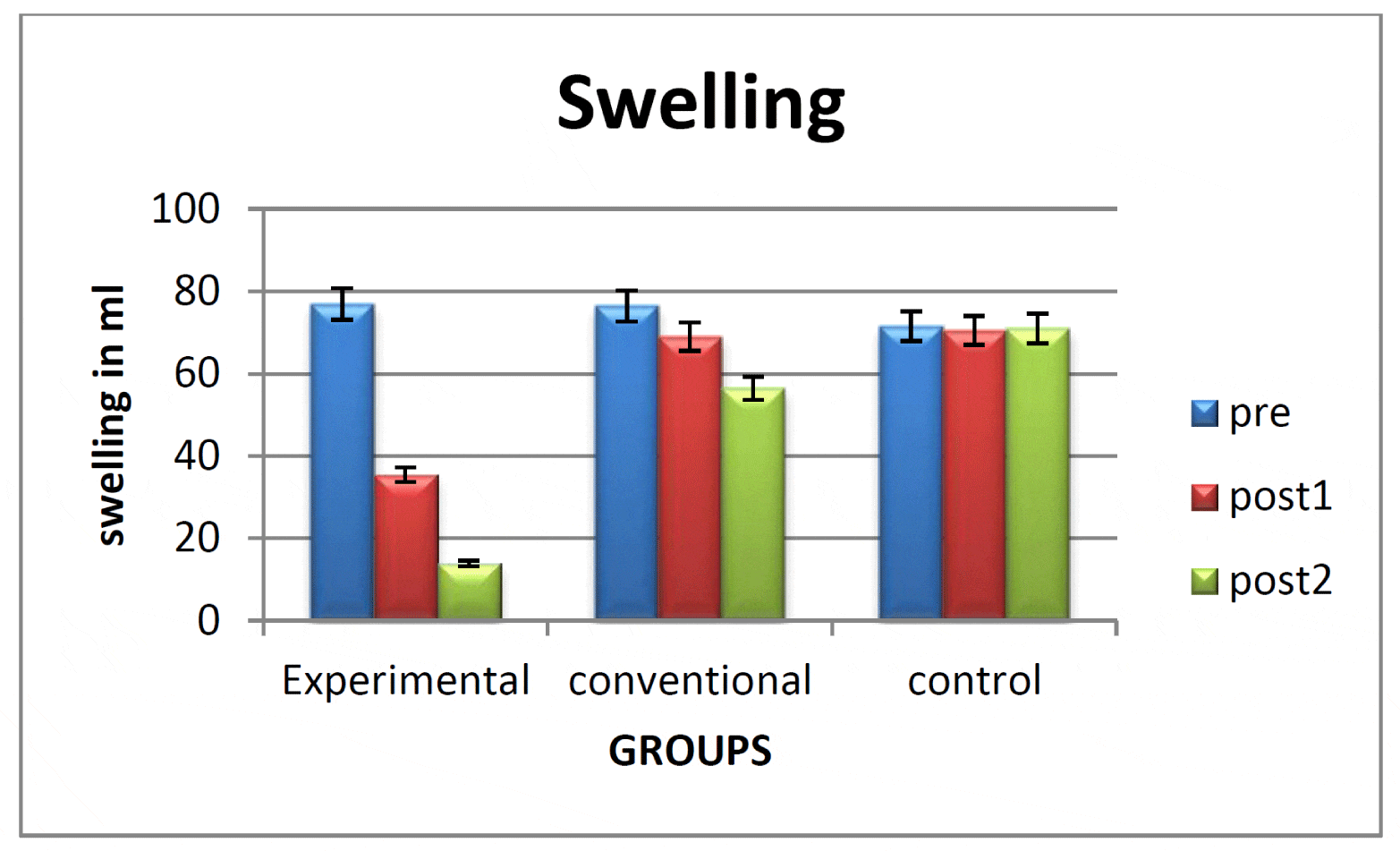
Figure 1 illustrates that there is reduction of swelling over time to a greater extent in experimental group patients than patients in conventional group, while there was no change in control group.
There was a main effect for time F 2,54,0.05 = 60.253, p=0.00
There was also a main effect for group F 2,27,0.05 = 4.196, p=0.026
There was also a main effect for time X group interaction F 4,54,0.05 = 27.606, p=0.00
Tukeys HSD analysis showed that in experimental group there was significant reduction in swelling from baseline to the end of first week as well as second week. While conventional and control group showed no change. However at the end of first week as well as second week experimental group was significantly better than conventional and control group. Similarly conventional group was better than control group only at the end of second week.
Strength
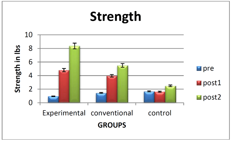
Figure 2 illustrates improvement in strength over time to a greater extent in experimental group patients than patients in conventional group and patients in control group.
There was a main effect for time F 2,54,0.05 = 66.204, p=0.00
There was also a main effect for group F 2,27,0.05 = 5.216, p=0.012
However, there was main effect for time X group interaction F 4,54,0.05 = 14.688, p=0.00
Turkeys HSD analysis showed that experimental group showed significant improvement in strength from baseline to the end of first week as well as second week. Conventional group showed significant improvement in strength only from baseline to the end of first week, while control group showed no significant improvement in strength with time. At the end of first week as well as second week both experimental and conventional groups showed significant greater improvement of strength than control group. However at the end of second week experimental group was significantly better than conventional group.
Prwe
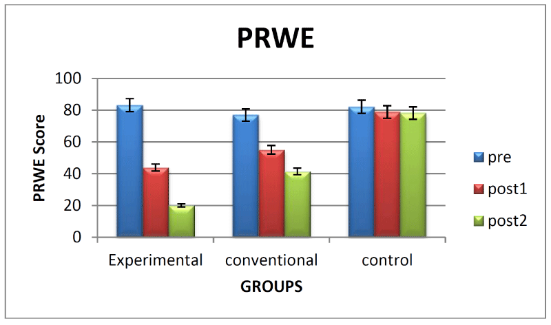
Figure 3 illustrates improvement in scores of Patient Rated Wrist Evaluation Questionnaire over time to a greater extent in experimental group patients than patients in conventional group, while there was no change in control group.
There was a main effect for time F 2,54,0.05 = 164.120, p= 0.00
There was also a main effect for group F 2,27,0.05 = 20.402, p= 0.00
There was also a main effect for time X group interaction F 4,54,0.05 = 40.816, p=0.00
Tukeys HSD analysis showed that both experimental group and conventional group improved significantly in PRWE scores from baseline to the end of first week as well as second week. Control group did not improve significantly with time. However at the end of first week as well as second week experimental group showed significantly greater improvement than other two groups. Similarly conventional group showed significantly greater improvement than control group.
Flexion
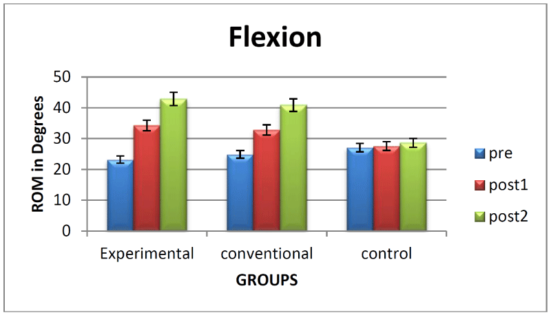
Figure 4 illustrates improvement in flexion range over time to a greater extent in experimental group patients as well as patients in conventional group than the patients in control group.
There was a main effect for time F 2,54,0.05 = 92.273, p= 0.00
However, there was no main effect for group F 2,27,0.05 = 1.075, p= 0.355
There was also a main effect for time X group interaction F 4,54,0.05 = 18.677, p=0.00
Tukeys HSD analysis showed that both experimental and conventional group improved significantly in flexion range of motion from baseline to the end of first week as well as second week. Patients in control group showed no significant improvement with time. At the end of first week as well as second week experimental group and conventional group were significantly better than control group, while there was no significant difference between experimental and conventional group.
Extension
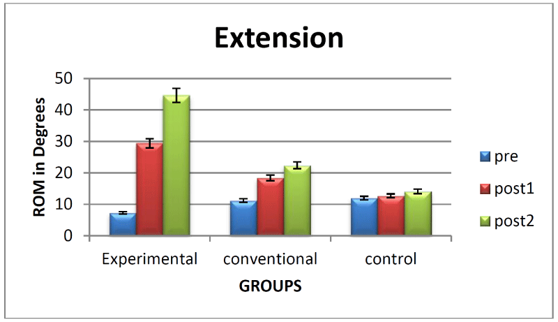
Figure 5 illustrates that there was improvement in extension range over time to a greater extent in experimental group than patients in conventional group, while there was no change in control group.
There was a main effect for time F 2,54,0.05 = 159.559, p= 0.00
There was also a main effect for group F 2,27,0.05 = 16.506, p= 0.00
There was also a main effect for time X group interaction F 4,54,0.05 = 62.594, p=0.00
Tukeys HSD analysis showed that experimental group improved significantly in extension range of motion from baseline to the end of first week as well as second week. Conventional group showed significant improvement in extension only from baseline to the end of first week, while control group showed no significant improvement with time. At the end of first week as well as second week experimental group showed significant improvement than conventional and control group. Similarly conventional group is significantly better than control group.
Discussion
The overall results showed that experimental group improved significantly with time in all the variables at the end of 1st week as well as 2nd week. Conventional group also improved significantly with time in PRWE and flexion range of motion from baseline to the end of 1st week as well as 2nd week. However strength and extension range in this group improved only at the end of 1st week, while control group showed no change.
At the end of 1st week as well as 2nd week both experimental and conventional groups showed significantly better improvements in all the variables than control group. Experimental group was better than conventional group in all variables. While flexion improved to similar extent in both the groups.
Swelling
The findings of the study showed significantly greater reduction in swelling of hand in experimental group as compared to conventional group and control group at end of 1st week as well as 2nd week. Also conventional group showed reduction in swelling with time which differs significantly from control group at the end of 2nd week. This reduction in swelling can be attributed to the fact that in addition to local effects of conventional therapy subjects also got additional benefits of spinal manual therapy. Due to inherent biochemical, histological and mechanical changes during immobilization all patients had swelling and associated pain after plaster removal. Edema is a normal response to injury. However any acute inflammatory process initiated at the the time of injury should be resolved during or shortly after immobilization. Clin Auton et al., in 2005 concluded that post traumatic sympathetic dysfunction can be seen within 46 to 72 days post injury and this plays a decisive role in the genesis of chronic regional pain syndrome I [19]. Chir Nazadov in 1996 has discussed about algodystrophy being a severe complication of distal radius fractures and in 2007 has said that early recognition and immediate commencement of effective therapy gives a real chance of recovery, thereby prevention of the condition is a reasonable approach [20,21]. Preliminary findings in healthy subjects have suggested that limb immobilization may give rise to pain, change in skin temperature and sensitivity [8]. Significant reduction in swelling. In our experimental group suggests the underlying neurophysiological effects of cervicothoracic mobilization which was given only to these patients. As the sympathetic supply to upper limb stem from upper thoracic spinal segments T2-T5, hence the corresponding spinous processes C7,T1,T2,T3 were mobilized [12]. Mobilization of cervicothoracic spine resulted in increased sympathetic activity as shown by 15% increase in heart rate which was measured continuously on pulse oximeter on day to day basis. Activation of SNS results in stimulation of alpha 1 receptors present on veins and arterioles. This results in arteriolar constriction and simultaneous increase the permeability of veins thus causing absorption of fluid from the extracellular spaces into the veins, aiding venous drainage [22]. Hence the blood is redistributed from skin and subcutaneous tissues to the active working muscles. Thereby concluding that SNS activation results in decrease in skin and subcutaneous blood flow (as proven by decrease in skin temperature) and increases in muscle blood flow, thus there is decrease in swelling. Increase in peripheral blood flow which in turn also increases oxygen supply to the peripheral tissues, hence pH increases and proton concentration decreases. Finally the peripheral sensitization decreases and hence the central sensitization also decreases. Thus chronic tissue hypoxia and decrease in ph that occurs in inflammatory states is also improved with sympatho-excitation. Thus there is red of swelling. Ultimately the vicious cycle of pain immobility swelling pain is broken. Our study is consistent with the findings of C J wolf in 1991 according to which optimal treatment of acute pain states should be directed at abolishing peripheral sensitization and to prevent the development of central sensitization [23]. Apart from biomechanical effects of spinal manual therapy many studies have emphasized mainly on neurophysiological effects of spinal manual therapy which triggers hypoalgesia and change in the range of measures which suggests activation of sympathetic nervous system as seen by changes in heart rate, respiratory rate, skin conductance and skin temperature. This is associated with mobilization of descending pain control mechanisms in particular with noradrenergic system. Results of our is consistent with the findings of Mcguiness and B. Vincenzino et al who found 10.5 % increase in heart rate during spinal mobilization [24]. Decrease in skin blood flow is in accordance with the study of Sterling et al who found decrease in skin temperature occurs with cervical spinal mobilization [25]. Daniel et al., in 2004 has shown that generalized exercise induced more hypoalgesia than isometric grip exercise in normal subjects. The results of the study suggested that hypoalgesia was more with generalized exercise group who demonstrated more sympathoexcitation as compared with local exercise group [26]. Decrease in swelling can also be attributed to the fact that manual therapy results in transient increase in EMG, decrease in muscle inhibition and stimulation of muscle spindle evoking a monosynaptic excitatory potential in all the alpha motor neurons of the same muscle [27]. Hence there is facilitation of muscle activity resulting in decrease in swelling. Thirdly there is also direct mechanical effect of mobilization stimulating mechanoreceptors [28]. There is constant sensory input in a rhythmic pattern which goes to the dorsal horn and then to brainstem finally to cortex. This results in reduction of sensitization as well as pain perception. Decrease in pain reduces muscle inhibition and facilitates activity, thus reducing swelling. Conventional group showed significantly better reduction in swelling than control but showed improvement to a lesser extent than experimental group. This can be explained by the fact that though active movements are important to aid venous drainage thereby reduction of swelling but strong muscular contractions and full joint ROM is necessary to accelerate drainage [29] and also severe pain inhibits the patient’s ability to exercise the hand, so natural pumping action of hand is sacrificed permitting increased edema and fibrosis [30]. There was no change in control group because no treatment was given.
Strength
Findings of the study showed significant improvement in strength in experimental group at the end of 1st week as well as at the end of 2nd week while conventional group showed significant improvement only at the end of 1st week while control group showed no improvement. At the end of 1st week and at the end of 2nd week both experimental and conventional groups are significantly better than control group, experimental group is better than conventional only at the end of second week. Improvement in strength at 1st week seen in both experimental and conventional groups can be attributed to reduction of pain inhibition caused due to immobilization. Both groups performed active and passive movements which result in activation of mechanoreceptors thus blocking the sensation of pain through the pain gate theory. At the end of 2nd week improvement was only carried over to experimental group due to sufficient reduction of swelling as confirmed by Young in 1987 that small volume of infusion fluid produces substantial inhibition. Just 20 to 30 ml of infusion produces 50- 60 % reduction in maximum voluntary activation of muscles [31]. Hence insufficient reduction of swelling might have caused reflex inhibition of muscles in conventional group patients.
Spinal manual therapy has demonstrated increase in muscle recruitment in many previous studies. Hence the improvement in strength in the experimental group can also be supported by the study of Brenner et al who showed immediate improvement in contraction of transverses abdominis and multifidus in patients with low back pain after receiving lumbopelvic mobilization [32,33].
In 2006 Bolton et al showed that both thrust and non-thrust manual therapy stimulates different axial sensory beds. Mobilization directed at cervical spine results in reduction of reflex inhibition allowing the muscle to produce a greater force and hence greater strength [34]0.
Fernandes et al showed significant bilateral improvement in Pressure pain threshold over both elbows in asymptomatic subjects after receiving cervical spine mobilization [35].
Patient rated wrist evaluation questionnaire
Findings of the study showed significant reduction in PRWE scores in both Experimental and conventional group at the end of 1st week as well as at the end of 2nd week than control group. However experimental group showed significantly better reduction in PRWE than conventional group both at end of 1st and 2nd week. Improvement in both experimental and conventional group with time can be attributed to the fact that conventional protocol was given to both groups under supervision which included contrast bath, active and passive exercises, strengthening exercises that simulated functional activities of wrist and hand. Patients in both experimental and conventional group were encouraged regularly to increase the hand usage for all ADL from the day of commencement of treatment. Early wrist motions provide functional improvement in wrist and hand functions according to a study by Roumen et al. 1991 [36]. The improvement of scores of PRWE can also be attributed to the improvement in Strength and Range of motion in both experimental and conventional groups. As demonstrated by Taylor et al., in august 2001 that in distal radius fracture patients there were strong and significant correlation between grip strength and functional tasks. Significantly greater improvement in experimental group can be attributed to greater reduction in swelling and greater improvement in extension range of motion and strength [37]. While in control group since no treatment was given there was no change in PRWE due to over cautious behavior of patients there may be functional neglect present that is refusal either conscious or unconscious to use the affected hand. Development of functional neglect in patients can be attributed to fear of pain caused by active movements or a changed body image as the patient regards the hand as sick therefore not useful.
Range of motion
There was significant improvement in flexion Range of motion in experimental group at the end of 1st week and 2nd week than control group. however there is no significant difference in flexion ROM between conventional and experimental group .as the plaster cast is applied with wrist in 20 degrees of flexion and ulnar deviation [38], so the restriction in flexion range was less in both groups at the baseline, hence the residual deficit in range was gained easily with the effect of common conventional protocol given to both groups.
There was statistically significant improvement in extension range of motion in experimental group at the end of 1st week as well as 2nd week than conventional and control group, similarly conventional group is better than control group. In all patients pain was more during extension than flexion, this is due to the fact that as we move towards extension, wrist is moving towards closed pack position which causes joint approximation and pain [39]. Due to significant better reduction of pain and swelling and improvement of strength in experimental group, extension improved more in this group.
Conventional group is better than control group as regarding flexion and extension range of motion as the exercise protocol was given only to the former.
Conclusion
This study showed that conventional physiotherapy has a role in the rehabilitation of patients with distal radius fracture after plaster removal in reducing swelling, pain and improving range of motion, strength and earlier return of function. However cervicothoracic mobilisation has additional effects in all above mentioned variables.
Limitations
Sample size was small, Lack of long term follow up, Sympathetic response could have been measured by galvanic skin conductance.
- Alffram PA, Bauer GCH (1962) Epidemiology of Fractures of the Forearm: A Biomechanical Investigation of Bone Strength. J Bone Joint Surg Am 44: 105-114. Link: https://goo.gl/tt8TtZ
- O'Neill T, Cooper C, Lunt M, Purdie D, Reid D, et al. (2001) Incidence of Distal forearm fracture in British men and Women. Osteoporosis International 12: 555–558. Link: https://goo.gl/tZ5uVM
- Shamley D (2005) An essential text for the allied health professions. Pathophysiology; 160–163. Butterworth Heinemann, England.
- MacDermid J, Roth J, Richards R (2003) Pain and disability reported in the year following a distal radius fracture: A cohort study. BMC Musculoskelet Disord 4:24. Link: https://goo.gl/rb3bzz
- Weber ER (1987) A rational approach for recognition and treatment of colles’ fracture. Hand clin 3: 13-21. Link: https://goo.gl/L5hcXf
- Rowbothan DJ, Pamela E. et al. Clinical pain management.
- Schlereth T, Birklien F (2008) The Sympathetic Nervous System and Pain. Neuromolecular Medicine 10: 141-147. Link: https://goo.gl/4DIcHf
- Terkelsen AJ, Bach FW, Jensen TS (2008) Experimental forearm immobilization in humans induces cold and mechanical hyperalgesia. Anesthesiology 109: 297-307 Link: https://goo.gl/vI71VT
- Schmid A, Brunner F, Wright A, Bachmann LM (2008) Paradigm shift in manual therapy - Evidence for a central nervous system component in response to passive cervical spine joint mobilization. Ma Ther13: 387-396. Link: https://goo.gl/C2gzyy
- Mohammadian P, Gonsalves A, Tsai C, Hummel T, Carpenter T (2004) Areas of Capsaicin-Induced Secondary Hyperalgesia and Allodynia are reduced by a Single Chiropractic Adjustment: A Preliminary Study. Journal of manipulative and physiological therapeutics 27: 381-387. Link: https://goo.gl/im6FJF
- Grant R (2002) Physical therapy for cervical and thoracic spine. 3rd Edition. Link: https://goo.gl/hbrdL8
- Grey H. Greys anatomy.
- Bohannon RW (1999) Inter-tester reliability of hand-held dynamometry: a concise summary of published research. Percept Mot Skills 88: 899-902. Link: https://goo.gl/jY8Pxc
- Hunter JM. Rehabilitation of hand. 156-157.
- McDermid JC (2007) PRWE user manual. School of rehabilitation science, Mcmaster university, Hamilton, Ontario Canada, Clinical Research Lab.
- Kamat V (2002) Pulse Oxymetry. Indian Journal of Anesthesia 46: 251-268. Link: https://goo.gl/EkjCIy
- Norkin CC. Measurement of joint range of motion- A guide to goniometry 84-85.
- Balsky S, Goldford RJ (2000) Rehabilitation protocol for undisplaced colles fractures following cast removal. Journal of Canadian Chiropractice association. 44: 29-33. Link: https://goo.gl/ajmnOl
- Gradl G, Schurmann M (2005) Clinical Autonomic Research.15: 13-14.
- Zyluk A (1996) [Algodystrophy after distal radius fractures]. Chir Narzadow Ruchu ortop Pol 61: 349-355. Link: https://goo.gl/U2Uy59
- Zyluk A (2007) [Prevention of algodystrophy of the upper limb]. Chir Narzadow Ruchu Ortop Pol 72: 424-428. Link: https://goo.gl/3BQh1z
- Seeley−Stephens−Tate (2004) Anatomy and Physiology, 6th edition; 16. Autonomic Nervous System. The McGraw−Hill Companies. Link: https://goo.gl/wZCd3M
- Woolf CJ (1991) Generation of acute pain: central mechanism. Br Med Bull 47: 523-533. Link: https://goo.gl/eLLc5p
- Vincenzino B, Cartwright T (1998) Cardiovascular and respiratory changes produced by lateral glide mobilization of the cervical spine. Manual therapy 3: 67-71. Link: https://goo.gl/pfXFpJ
- Sterling M, Jull G (2001) The effect of musculoskeletal pain on motor activity and control. J Pain. Link: https://goo.gl/tLtQuv
- Drury DG, Stuempfle KJ, Shannon RJ (2004) An investigation of exercise induced hypoalgesia after isometric and cardiovascular exercise. Journal of exercise physiology 7: 1-5. Link: https://goo.gl/XmtMFE
- Boyling JD. Grieves Modern manual therapy- The vertebral column, 3rd edition. Chapter 25: 372.
- Maitland GD, Vertebral Manipulation, 5th edition.
- Sorenson MK (1989) The edematous hand. Phys Ther 69: 1059-1064. Link: https://goo.gl/Mlkuzb
- Morey KR, Watson AH (1986) Team Approach to treatment of the Post traumatic stiff hand. Phys Ther 66: 225-228 Link: https://goo.gl/TI5PyG
- Young A (1993) Current issues in arthrogenous inhibition. Annals of the Rheumatic Diseases 52: 829-834. Link: https://goo.gl/iZW0Qs
- Brenner AK1, Gill NW, Buscema CJ, Kiesel K (2007) Improved activation of lumbar multifidus following spinal manipulation: a case report applying rehabilitative ultrasound imaging. J Orthop Sports Phys Ther 37: 613-619. Link: https://goo.gl/JrvrV7
- Gill NW, Teyhen DS, Lee IE (2007) Improved contraction of the transverse abdominis immediately following spinal manipulation: a case study using real time ultrasound imaging. Man Ther 12: 280-285. Link: https://goo.gl/k5K9Ya
- Bolton P, Budgell B (2006) Spinal manipulation and mobilisation influence different axial sensory beds. Med Hypotheses 66: 258-262. Link: https://goo.gl/qsn16y
- Fernández-Carnero J, Fernández-de-las-Peñas C, Cleland JA (2006) Immediate Hypoalgesic and motor effects after a single cervical spine manipulation in subjects with lateral epicondylalgia. Journal of Manipulative and Physiological Therapuetics 31: 675-681. Link: https://goo.gl/hpJLxc
- Roumen RMM, Hesp W, Bruglish ED (1991) Unstable Colles' and Smith' Fracture in elderly patients. Journal of Bone Joint Surgery. 73B: 307. Link: https://goo.gl/UI8yfy
- Tremayne A, Taylor N, Mcburney H, Baskus K (2002) Correlation of impairment and activity limitation after wrist fracture. Physiotherapy Res Int 7: 90-99. Link: https://goo.gl/C1e4hA
- Aydog S, Keskin D, Ogut B (1994) Rehabilitation of Colles Fracture. Rehabilitation Journal of Islamic academy of Sciences 7: 247-250. Link: https://goo.gl/RJlgHW
- Shahan K, Sarrafian NB. Kinesiology and functional characteristics of upper limb. Atlas of limb prosthetics surgical prosthetic and rehabilitation principles.. Link: https://goo.gl/YFUkYN
Article Alerts
Subscribe to our articles alerts and stay tuned.
 This work is licensed under a Creative Commons Attribution 4.0 International License.

 Save to Mendeley
