Journal of Gynecological Research and Obstetrics
The mysterious extravillous trophoblast
Olaya C Mercedes1* and Franco Z Jorge A2
2Department of Morphology, the Medical School, Pontificia Universidad Javeriana Kra 7a 40-62, Bogota, Colombia
Cite this as
Mercedes OC, Jorge AFZ (2022) The mysterious extravillous trophoblast. J Gynecol Res Obstet 8(3): 025-026. DOI: 10.17352/jgro.000111Copyright License
© 2022 Mercedes OC, et al. This is an open-access article distributed under the terms of the Creative Commons Attribution License, which permits unrestricted use, distribution, and reproduction in any medium, provided the original author and source are credited.To the editor
Extravilluos Trophoblast (EVT) is a truly special cell. It has a temporary character and tumor-like behavior and tumor-like appearance, all of which seem to belong to the malignancy field [1,2]. It is also known as intermediate trophoblast, derived from cytotrophoblast [3-5], having formed between the latter and the syncytiotrophoblast. EVT is capable of reaching the decidua through trophoblast cell columns that connect the anchoring villi to the basal plate in early gestation.
The invasive trophoblast penetrates the decidua, thereby bringing about the remodeling of the maternal arterioles. Two kinds of EVT combine to carry out this function: the endovascular ET and the interstitial ET.
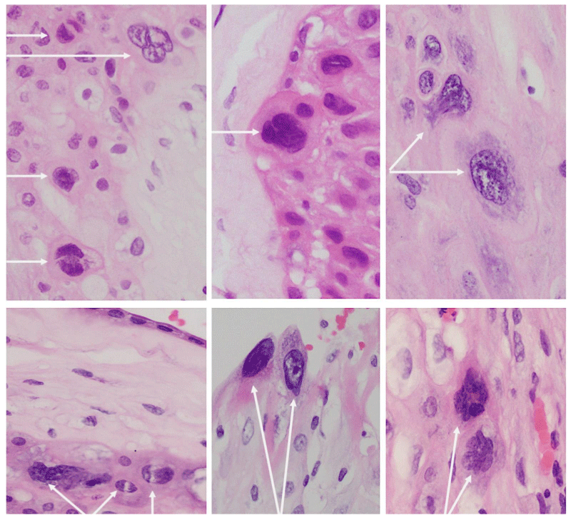
EVT possesses an invasive capacity similar to that of tumoral cells and its appearance is characterized by large polyhedral to a spindled cell whose cytoplasm is usually purple in color, accompanied by anisocytosis. Its cellular makeup could be mono or bi-nucleate, simultaneously exhibiting anisonucleosis, nucleomegaly, nuclear hyperchromasia, and pleomorphic nuclei (Figure 1). In the image included herein, we notice the aberrant, highly conspicuous, and, frankly, monstrous features of EVT in photomicrographs taken of term placentas in routine cases. Despite anticipating the grotesque appearance of EVT, these additional fields draw the pathologist´s attention and always cause the said observer to rule out other diagnoses possibly associated with cells of similar appearance, including those of viral diseases. In these cases, it has therefore been possible to rule out such etiologies.
The features described above make EVT a different and interesting cell, the knowledge of which could aid in understanding not only the bases of alterations in placentation and its consequences, especially preeclampsia but also in recognizing the behavior of tumor cells in their transformation and invasion processes.
Contribution
Olaya-C Mercedes: I declare that I participated in the concept design, acquisition of images, literature review, and drafting of this manuscript; and that I have seen and approved the final version.
Franco Jorge A: I declare that I participated in the acquisition of images, literature review, and drafting of this manuscript; and that I have seen and approved the final version.
- Wagner GP, Kshitiz, Dighe A, Levchenko A. The Coevolution of Placentation and Cancer. Annu Rev Anim Biosci. 2022 Feb 15;10:259-279. doi: 10.1146/annurev-animal-020420-031544. Epub 2021 Nov 15. PMID: 34780249.
- Deng Q, Liu X, Yang Z, Xie L. Expression of N-Acetylglucosaminyltransferase III Promotes Trophoblast Invasion and Migration in Early Human Placenta. Reprod Sci. 2019 Oct;26(10):1373-1381. doi: 10.1177/1933719118765967. Epub 2018 Apr 11. PMID: 29642803.
- Xiao Z, Yan L, Liang X, Wang H. Progress in deciphering trophoblast cell differentiation during human placentation. Curr Opin Cell Biol. 2020 Dec;67:86-91. doi: 10.1016/j.ceb.2020.08.010. Epub 2020 Sep 18. PMID: 32957014.
- Sheridan MA, Zhao X, Fernando RC, Gardner L, Perez-Garcia V, Li Q, Marsh SGE, Hamilton R, Moffett A, Turco MY. Characterization of primary models of human trophoblast. Development. 2021 Nov 1;148(21):dev199749. doi: 10.1242/dev.199749. Epub 2021 Nov 5. PMID: 34651188; PMCID: PMC8602945.
- Castellucci M, Kaufmann P. Basic Structure of the Villous Trees. Pathol Hum Placenta. 2006; 50–120.
Article Alerts
Subscribe to our articles alerts and stay tuned.
 This work is licensed under a Creative Commons Attribution 4.0 International License.


 Save to Mendeley
