Journal of Gynecological Research and Obstetrics
Artificial uterus -research background to improve survival and outcome of extremely low birth weight newborns
Maria Laura Solerte*
Cite this as
Solerte ML (2020) Artificial uterus -research background to improve survival and outcome of extremely low birth weight newborns. J Gynecol Res Obstet 6(3): 067-071. DOI: 10.17352/jgro.000090In clinical research, of worldwide interest, some medical devices are being studied as a new reproductive technology, which can both guarantee the implantation of the embryo or support the premature newborn outside the maternal uterus (human and animal); in focusing attention on the second aspect , exclusively, together with the ethical aspects widely debated by experts in the field, it is necessary to examine how much some researchers , belonging to many world groups, have created postnatal support mechanisms for animals extremely premature. On this ground, a group of experienced scientists are working now, using different technologies, to create an artificial womb, within the next decade, resembles the conditions in a human uterus more closely. Based on the fundamental concept, that every week of extra-uterine suitable care allows to get significant results for preterm newborn outcome and life, the “ medical unit” will have to be studied and then implemented in details, to be used on human premature infants, even born at 22 weeks of gestation, who need specific assistance to ensure the development of severe immature fundamental organs and, therefore, normal cardiorespiratory functions, that are the cause of morbidity and mortality in these frail patients; the scientists will be develop a model destined for clinical use. Unlike current incubators, the prototypes will envelope the preterm babies in liquid and will provide them with oxygen and nutrition via an artificial placenta connected to newborns ‘s umbilical cord.
Introduction
An estimated 15 million babies a year, are born preterm, before 37 completed weeks of gestation; one million die from complications and have significant contributor to childhood morbidity, both related to this condition; unfortunately, this data are bound to increase. Preterm birth is the most common cause of death among infants worldwide, is defined, by the World Health Organization (WHO) as delivery before 37 weeks of pregnancy are completed and is the second leading cause of death globally for children under five years, after pneumonia [1].
There are three sub-categories of preterm babies, based on gestational age: extremely preterm (less than 28 weeks) (Figure 1) , very preterm (28 to 32 weeks), moderate to late preterm (32 to 37 weeks); this is the most used definition of preterm birth [1]. The evolution of care in neonatal intensive care units, aimed at getting better the management of high-risk pregnancy, fetal / perinatal medicine, has significantly improved the outcome of the premature and extremely premature newborn, including new approaches to the old nemesis of bronchopulmonary dysplasia [2], which still affects up to 50% of infants born before 28 weeks’ gestation; moreover prematurity has profound consequences for the course of cardiovascular, metabolic, neurological, and pulmonary diseases throughout life [3]. The studies of an artificial medical technology will be focused to emulate the conditions inside the maternal uterus, which is able to incubate preterm borned human fetuses, and prevent the serious morbidity that occurs in extremely premature babies, by recreating an artificial womb. The last years have seen significant advances in making an artificial uterus that could have facilitated the survival and growth of prematurely born animal fetuses starting around 23-24 weeks of gestation; also according to ethical considerations [4,5], this gestational age is the current goal of “fetal viability” , the point at which a fetus has a chance of survival outside the womb, although morbidity and mortality for premature babies born before approximately 28 weeks of gestation remain high. Elizabeth Chloe Romanis, a lawyer at the University of Manchester, who has explored the bioethics of artificial wombs, warned that the technology would raise questions, including about which babies it should be tested on, as well as the long-term implications of being gestated in an artificial womb ( the law would approach foetuses and babies differently ), adding that there are also questions about how such a gestation might be viewed by society, particularly if it becomes an alternative to a “natural” pregnancy. It is clear that the legal and ethical issues emerging from the technology must be talked about now, in advance of the artificial womb becoming a reality [4,5]. The artificial uterus, as a medical assistive device, which should be ready for human trials within the next five years, according to current scientists’s target in the Netherlands, is an innovative project in that, instead of being an emergency life support, it treats the preterm infant as an unborn fetus [6]. This incredible plan, science fiction for someone, is aimed at building an environment similar to the uterus’s physiology, surrounding the extremely premature baby in artificial amniotic fluid allowing the immature organs to develop as if they remained inside the mother’s body. If successful, this technology could significantly improve health outcomes for infants and perhaps ultimately be committed to supporting the health of pregnant people who have major complications from previous medical conditions or associated with pregnancy itself. This technology could then also be integrated by further ultrasound evaluations, with future customized protocols based on the new vascular resistences, already known and applied for decades [7,8], that could help clinicians in controlling infants inside the artificial womb, both in growth and in hemodynamics, in the various districts that are usually monitored, during the stay of the fetus, who requiring intensive surveillance, in the mother’s uterus.
Study of the foetus in uterus by Leonardo da Vinci
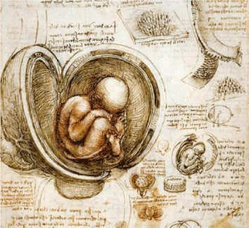
From “Anatomy notebooks” , created thanks to autopsy studies, in Milano from 1509 to 1512, Leonardo da Vinci began to examine (since 1507, with the collaboration of Marcantonio della Torre ) “the fetus in uterus” giving a fundamental innovative contribution, unchanched for over two centuries : in particular, in 18 sheet (Figure 2), the correct position of the fetus in uterus, uterine artery and vascular system of cervix–vagina, was draw with incredible precision, for the first time in the history of medicine and the fetal physiology ( the fetus immersed in the amniotic fluid that does not breath because he would drown ), are touched upon the notes . For the first time, uterus had only one cavity, contrary to its seven chambers as Guido da Vigevano illustrated and contrary to Hippocrates theory that predicted its two cavities; instead, the placenta was from a cow [9,10,11]. Furthermore, Leonardo hypothesized that umbilical cord carried the urine of the fetus out of the uterus, and carried into the fetus the maternal blood.
Ectogenesis
Scientists believe that ectogenesis ( creating life ouside the body ) is not far off and many of them argue that it is an inevitable technology. In 1924, John Burdon Sanderson Haldane, a british scientist known for his works in physiology, biology and genetics, conied ectogenesis ( from the greek ecto, “outer”, and genesis, “generation” ), thinking the growth of an organism outside the body through an artificial womb. The evolutionary scientist imagined that artificial womb might become popular by 2074 ( that only a small minority-fewer than 30% of newborn-would be born of woman) [12]. The ectogenesis’s idea had already started in 1880, by the french obstetrician Etienne Stephane Tarnier, who built a wooden box, with a compartment for a hot-water box, to reduce the mortality of premature babies [13,14]; his design did not become much more technologically until the 1950. By the 1960, was opened the first Amenican newborn intensive care unit, designed by Louis Gluck [15]; the experiments began on incubators ( which provide warmth and humidity, but none of the nutrients necessary for newborn growth ) and also attempted the creation of an artificial placenta, a complex specialized organ that should provide life support until a foetus develops to a stage where he is able to perform these function on him own. An artificial womb need to replicate all the placenta’s functions, the fluids, bacteria and other equipment essential to the making of life. The growth of human fetuses requires an artificial uterus, as a replacement organ, which give nutrients and oxygen and, also, an interface like the placenta; instead, of course, in the incubator, a premature baby must have tubes inserted into his body to deliver nutrition via needle-like catheters inserted directly into the veins; they will also be sedated, at least some of the time, to stop from pulling inserted tubes out, and to decrease or prevent any discomfort or pain.
Preterm delivery
Preterm birth is commonly defined as any birth before 37 weeks completed weeks of gestation; an estimated 15 million infants are born preterm, globally [1]. An estimated 15 million babies are born too early every year; that is more than 1 in 10 babies. Approximately 1 million children die each year because of complications of preterm birth that is the most common cause of death among infants worldwide; World Prematurity Day November 17th, initiated in 2011, is a global effort to rise awareness about prematurity [16]. The biggest challenge faced by perinatal medicine experts are babies born before completing the 37th week of gestation, for the increased of serious complications in both the short and long term , due to a combination of organ immaturity and iatrogenic injury. Short-term neonatal complications were observed in neonatal of elective preterm delivery (for both maternal and fetal indications), without brain damage or even mortality, with severe maternal hypertension or severe proteinuria and intrauterine growth retardation for the protective mechanism in the fetuses [17]. For premature delivery, are known risk factors : a previous premature birth; multiple pregnancy; an interval of less than six months between pregnancies; in vitro fertilization; problems with placenta, uterus or cervix; smoking and drugs; infections of the lower genital tract or amniotic fluid; chronic conditions such as high blood pressure or diabetes; gestational hypertension; gestational diabetes; preterm premature rupture of membranes [18]; over or under-weight before gestation; stressful life events; abortion; injury or trauma ( from Mayo Clinic ); small for gestational age and intrauterine growth restricted fetuses. An American study evaluated 34.636 preterm babies born with gestational age of 22 throught 28 weeks of gestation, birthweight of 401 to 1500 gr, and born at 26 network centers between 1993 and 2012; this study of extremely preterm infants born at NRN centers is the first comprehensive review to our knowledge to evaluate how care practices, major morbidities, and mortality have evolved over a 20 year period. Findings demonstrated that progress were being made and outcomes of the most immature infants are improving, a modest reduction in severe morbidities was observed but bronchopulmonary dysplasia increased; premature infants suffer from mortality and morbidity leads research to create new ways of approaching this patients [19].
Artificial uterus
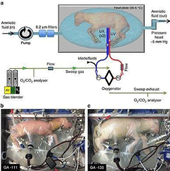
The first patent , for an illustration of an artificial uterus, was issued, in 1955 [20,21], to Greenberg who had started to study and write about its potential use in the future. Greenberg’s design (Figure 3) included a tank to place the fetus filled with amniotic fluid, a machine connecting to the umbilical cord, blood pumps, an artificial kidney, and a water heater. Cooper William, also had his US patent [22], in 1993, for another life support system for a premature baby which remains attached to its placenta through its umbilical cord: the system includes upper and lower chambers separated by a dome-like partition. The lower chamber contains physiological liquid in which the baby is suspended, and the upper chamber contains an oxygen-containing atmosphere and a supply of nutrients for contact with the placenta which rests on the top of the dome-like partition. Already before, in 1987, Kuwabara Y, in Tokyo’s Juntendo University, and his staff , was the first scientist to sustain in an artificial womb for that long; a new extra-uterine incubation system has been developed using 14 goat fetuses. The goat fetus is surrounded by artificial amniotic fluid and is connected to an extracorporeal membrane oxygenator [23,24]. The blood is drained from the umbilical arteries and returned to the umbilical vein. They developed a technique called Extra Uterine Fetal Incubation (EUFI), which successfully supported a 17 weeks goat fetus, for three weeks; EUFI is described as a rectangular transparent plastic box, filled with artificial amniotic fluid at body temperature, connected to devices for vital functions ; the blood was cleaned with a dialysis machine connected to the umbilical cord; next step was suggested for fetal monitoring after extracted from EUFI. In 2017, US scientists from the Center for Fetal Research of Philadelphia [25], developed an artificial device, also called extra-uterine life support system, closely reproduces the environment of the womb; Director Flake, and his group, that kept alive extremely premature lamb fetuses (Figure 4), with biological age equivalent to a human fetus of 24 weeks of gestation, for four weeks, creating a fluid environment from a polyethylene bag, Biobag, which incorporated an oxygen pumpless circuit, in which blood flow is driven exclusively by the fetal heart with a very low resistance, closely mimic the normal fetal-placental circulation, in which lambs maintained a stable circulation of blood and gas. This study was limited for four weeks, aiming at maintaining stable conditions between 23 and 28 weeks of gestation ( barrier period for the premature infants ) and indicated a greater survival of premature lambs, thanks to a bridging system between maternal uterus and extra uterine life. The animals were able to develop brain and lungs for four weeks; during this time they were able to move, open the eyes and swallow. However there were technical difficulties related to the connection via the umbilical cord and the creation of a suitable amniotic fluid. The researchers’ s objective will therefore be to improve the system and to adapt it to a human newborn, even, Director Flake said, “friendly to the parents” [25].
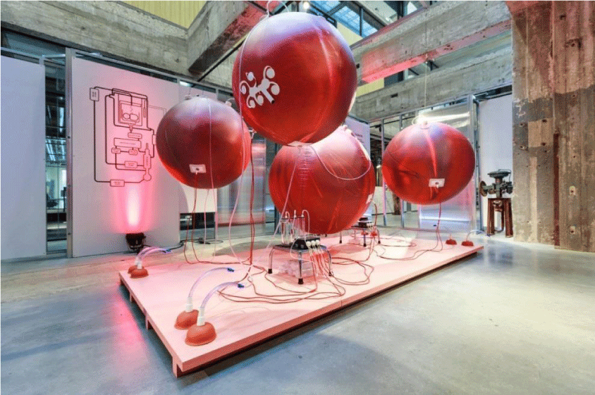
In 2018, Church J. T. , Mychaliska G. B. et al, at University of Michigan, also evaluated whether brain and lungs development continues and injury is prevented during extracorporeal life support, also with an artificial placenta, in preterm lambs , by jugular drainage and umbilical vein reinfusion for seven days [26]. They found that lungs and brains maturation appears to continue normally, and the injuries, observed with mechanical ventilation, were avoided thanks to artificial placenta. At the Dutch Design Week 2018 (Figure 5), was presented a speculative design proposal for an artificial womb , “ a second womb” for premature babies, in close collaboration with researchers of Maxima Medical Centre and Eindhoven University of Technology, who had the assignment, in 2019, to develop an artificial womb within the next decade [6,27]. The device, called Perinatal Life Support System (PLS), for clinical translation, started its planning on October 2019, would provide preterm babies with artificial respiration and a natural environment for the newborn for the transition to the new life, by a similar biological condition of human pregnancy; the babies will receive oxygen through the umbilical cord. The new technology will able to increase the chances of survival , currently very low ( 61% dies with 24 weeks and 43% with 25weeks ), of extremely premature babies in the period of 24-28 weeks of gestation; moreover, the babies that survive often are suffering from brain damage, breathing and retina problems and the risk of blindness. The team, plans to build a prototype, ready for use in clinics within five years and it could be the first in the world; on the opinion of the project’s coordinators, F. van de Vosse and G. Odei ( with other groups ) , it will require the contribution of different technologies. Their goal will be to help extremely premature babies get through the critical period of 24 to 28 weeks.
Conclusions
In the field of human fetal, perinatal and postnatal surveillance, the study of an artificial uterus will improve the outcome and the chances for survival of extremely low birth weight foetuses born before 28 weeks of gestation; if successful, it could a breakthrough. At the moment, for decades, scientists worldwide, have clinical and instrumental control systems, in order to check the normal and risk pregnancy to check the fetal well-being; in particular, fetal and maternal ultrasound studies, helps also in managing the timing of delivery becoming sophisticated and with high diagnostic value and, probably, could be integrated into the monitoring of the preterm infant incubated in a PLS system (probably). That device could save millions of babies who die due to premature births; with PLS solution, the supply of oxygen and nutrients, connected by an artificial placenta, will support fetal cardiorespiratory physiology and will avoid the negative effects of air-based ventilation. Artificial uterus and artificial placenta will be similar to biological conditions.
- World Health Organization and partners (2012) Born too soon: The global action report on preterm birth. Link: https://bit.ly/309B1QK
- Rodriguetz RJ (2003) Management of Respiratory Distress Syndrome: An Update. Respiratory Care 48. Link: https://bit.ly/367ZKIY
- Prospects for neonatal intensive care. Editorial. The Lancet 389: 1582. Link: https://bit.ly/3406y91
- Romanis EC (2018) Artificial womb technology and the frontiers of human reproduction: conceptual differences and potential implications. J Med Ethics 44: 751–755. Link: https://bit.ly/30b4qtJ
- Rosen C (2003) Why not artificial wombs? The New Atlantis 67-76. Link: https://bit.ly/332E0Mz
- The world’s first artificial womb for humans. BBC news 10-15-2019. Link: https://bbc.in/3ic2rvG
- Solerte L. Fetal cardiac (1996) cardiac systolic and diastolic time relationships in normal and growth retarded fetuses. Research Trainig. European Commission.
- Assan WA, Wladimiroff J, Alberry M, Fanelli T, Wladimiroff J, et al. (2013) Cardiac function in early onset small for gestational age and growth restricted fetuses. Eur J Obstet Gynecol Reprod Biol 171: 262-265. Link: https://bit.ly/30byIN8
- Royalcollection.org.uk/exhibitions/leonardo-da-vinci-anatomist/the-drawings
- Anticaginecologia.wordpress.com/2016/05/01/i-disegni-di-anatomia-di-leonardo-da-vinci-il-feto-nelluter
- Hilary G (2008) Leonardo da Vinci’s Embryological Drawings of the Fetus. Embryo Project Encyclopedia. Link: https://bit.ly/2HzNK8X
- Tirard S (2017) JBS Haldane and the origin of life. Journal of genetics 96: 735-739. Link: https://bit.ly/36497JM
- Pinard A (1909) Tarnier 1828 – 1897. Éloge prononcé à l'Académie de Médecine dans sa séance annuelle du 15 Décembre. Annales de gynécologie et d'obstetriques 6: 2-28. Link: https://bit.ly/3i2WRM0
- Fraser MS (1994) Stéphane Tarnier and the origin of incubators for premature babies [Abstract]. Rep Proc Scott Soc Hist Med 1994-1996: 1-2. Link: https://bit.ly/2EDWud8
- Gluck (1960) Conceptualization and initiation of a neonatal intensive care nursery in 1960. Neonatal intensive care: a history of excellence.
- M V Kinney (2012) 15 million preterm births annually: what has changed this year? Reprod Health 9: 28. Link: https://bit.ly/2HztFj6
- Spinillo A, Capuzzo E, Stronati M, Iasci A, Solerte L, et al. (1995) Early neonatal complication after elective preterm delivery in hypertensive pregnancies. J Perinat Med 23: 175-181. Link: https://bit.ly/3mSUjnd
- Solerte L, Ragusa A (2002) Use of Amniotic fluid index and color Doppler in the management of amnioinfusion in preterm premature rupture of membranes. Proceedings of The National Congress SIEOG Gubbio.
- Stoll BJ, Hansen NI, Bell EF, Walsh MC Carlo WA, et al. (2015) Trends in Care Practices, Morbidity, and Mortality of Extremely Preterm Neonates 1993-2012. JAMA 314: 1039-1051. Link: https://bit.ly/2GeoJzf
- Greenberg EM (1955) Artificial Uterus. United States Patent n. 2723660; 11-15-1955
- American Association of Obstetricians and Gynecologists; American Gynecological Society (1920) American Journal of Obstetrics & Gynecology : AJOG. Link: https://bit.ly/3mUbNzK
- Cooper W (1993) Placental chamber - artificial uterus. United States Patent no .5218958;06-15-1993.
- Kuwabara Y, Okai T, Imanishi Y, Muronosono E, Kozuma S, et al. (1987) Development of Extrauterine Fetal Incubation System Using Extracorporeal Membrane Oxygenator". Artificial Organs 11: 224-227. Link: https://bit.ly/30dpcZY
- Kuwabara Y, Okai T, Kozuma S, Unno N, Akiba K, et al. (1989) Artificial placenta: long-term extrauterine incubation of isolated goat fetuses. Artif Organs 13: 527-531. Link: https://bit.ly/2S1szic
- Partridge EA, Davey MG, Hornick MA, McGovern PE, Mejaddam AY, Flake AW, et al. (2017) An extra-uterine system to physiologically support the extreme premature lamb. Nat Commun 8: 15112. Link: https://bit.ly/3j6Lcge
- Mychaliska GB (2018) Effects of artificial placenta on brain development and Injury in premature lambs. J Pediatr Surg 53: 1234-1239. Link: https://bit.ly/3di2SUj
- Van de Vosse F, Guid Oei LF (2019) Multimillion grant brings artificial womb one step closer. Eindhoven University of Technology.
Article Alerts
Subscribe to our articles alerts and stay tuned.
 This work is licensed under a Creative Commons Attribution 4.0 International License.


 Save to Mendeley
