Journal of Gynecological Research and Obstetrics
Are visceral adiposity index and ultrasonographic visceral fat thickness measurements correlated to endometrial thickness in postmenopausal women?
Fatma Eskicioğlu1*, Muhammet Sakıp Eskicioğlu2 and Beyhan Özyurt3
2Çiğli Regional Education Hospital, Department of Radiology, Izmir, Turkey
3Celal Bayar University, School of Medicine, Department of Public Health, Manisa, Turkey
Cite this as
Eskicioğlu F, Eskicioğlu MS, Özyurt B (2019) Are visceral adiposity index and ultrasonographic visceral fat thickness measurements correlated to endometrial thickness in postmenopausal women? J Gynecol Res Obstet 5(1): 009-012. DOI: 10.17352/jgro.000062Objective: During the postmenopausal period, visceral fat tissue is responsible not only for increased cardiometabolic risk, but also for endocrine function leading to increased endometrial thickness (ET). The current study aimed to 1) determine the predictive value of ultrasonographic visceral fat thickness (VFT) and the visceral adiposity index (VAI), which is defined as «an indicator of visceral adipose function,» for the prediction of an ET>5 mm and 2) to search for a correlation between these two measures.
Results: There were no significant differences between the two groups with respect to the VFT and VAI values. Positive correlations were observed between the VFT and VAI, as well as between VFT and ET (r=0.242, p=0.03 and r=0.217, p=0.05).
Conclusion: VFT and VAI values are correlated in postmenopausal women. However, neither VFT nor VAI appears to have a predictive value for an ET>5mm. Additional studies need to be performed on this subject.
Introduction
The risk of cardiovascular diseases (CVD) and metabolic syndrome (MS) increase during the post-menopausal period [1]. Obesity is a component of MS. Increased adipose tissue is not only a storage compartment of unconsumed energy, but also a contributor of several factors involved in endocrine regulation and increased insulin resistance [1,2]. Furthermore, increased adipose tissue is the primary source of circulating estrogen, which is synthesized by its androgen precursors, and is also responsible for increased endometrial thickness (ET) and CVD during the post-menopausal period [3-5].
Body fat distribution plays more of a role in increased ET than tissue fat amount [5]. Visceral fat mass, a component of abdominal fat accumulation that is particularly typical of the postmenopausal period, leads to MS and increased ET [5,6]. Although the measurement of waist circumference (WC) is an easy-to-measure marker of visceral fat tissue, it has a low specificity [7]. WC not only measures visceral fat tissue, but also subcutaneous fat tissue, which is another component of abdominal fat mass [5]. In other words, women with the same WC value may have significantly different visceral fat measurements [7]. Recently, it has been demonstrated that the ultrasonographic measurement of visceral fat thickness (VFT) is an inexpensive, practical, reproducible, easily accessible technique that is correlated with reliable scanning methods such as computerized tomography [8]. In addition, the visceral adiposity index (VAI), a mathematical model that uses body mass index (BMI), WC, triglyceride (TG) and high density lipoprotein (HDL) parameters, is a sensitive marker of visceral obesity that is known to be a valuable indicator of “visceral adipose function” and insulin sensitivity. It is claimed that increased VAI levels are strongly associated with cardiometabolic risk [9].
There exists a positive correlation between ET increase and abnormal endometrial conditions. When an endometrial thickness of 5 mm is accepted as a cutoff value for endometrial pathology in the post-menopausal period, endometrial cancers and atypical endometrial hyperplasias can be detected with a sensitivity of 80.5% and a specificity of 86.2% [10]. Currently, the literature includes certain studies that suggest a correlation between body fat distribution and ET, yet the role of the former in the determination of an ET greater than 5 mm remains unclear [5,7,11].
This study aims to determine whether easily calculable VAI values and VFT measurements, which do not cause as much irritation as transvaginal ultrasonography, possess adequate predictive power in relation to ETs of greater than 5 mm during the postmenopausal period. In addition, this study also seeks to determine if these parameters are interrelated.
Materials and Methods
This prospective study was carried out in Merkezefendi State Hospital with the approval of the Ethical Committee of Celal Bayar University. After obtaining their informed consent, this study was conducted on 79 postmenopausal women admitted in the gynecology outpatient clinic of the hospital for annual gynecological examinations. All participants were at a status of at least one year after natural menopause. Diabetes mellitus, hypertension, uterine bleeding, endometrial polyps, smoking, receiving hormone replacement therapy or any medication known to influence endometrial thickness, including hypolipidemic or hypoglycemic medications, were exclusion criteria. Blood pressures above 140/90mmhg or receiving antihypertensive medication were accepted as hypertension criteria. Receiving insulin, oral antidiabetic medications, or fasting blood glucose (FBG) > 115mg/dL (normal range: 70-115 mg/dL) were accepted as diabetes mellitus. Age, time since menopause (age to age at menopause) were evaluated as demographic data.
Anthropometric and metabolic measurements
The participants’ weights and heights were measured without shoes and with light clothing. BMIs were calculated by the following formula: weight (kg) / height (m)2. WC was detected measuring from the middle point of the border of the iliac crest and the last costa after normal expiration in a straight position. Hip circumference (HC) was measured from where it protruded the most. Blood pressure was measured using an automated sphygmomanometric procedure after resting in a seated position for at least five minutes, with the average of two measurements five minutes apart being considered. FBG, total cholesterol (TC), TG, HDL and low density lipoprotein (LDL) level were determined from pre-prandial blood taken in the morning.
VAI was calculated the following formula: [WC/36.58 + (1.89 × BMI)] × (TG/0.81) × (1.52/HDL). TG and HDL levels were introduced in the formula as mmol/l [9].
Ultrasonographic evaluation
A high resolution ultrasonographic system (Aplio 500; Toshiba, Toshigi, Japan) was used for ultrasonographic measurements. The measurements were conducted using the PLT-704SBT (4.8-11.0 MHz) linear transducer in the abdominal mode of the ultrasonography device for VFT. ET was performed with a PLT-661VT (3.6-8.8 MHz) endovaginal transducer in vaginal mode.
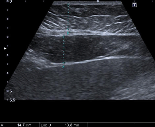
VFT measurement was performed with the patient in a supine position and after respiration to exclude respiration-based abdominal wall tension. The VFT was accepted as the fat thickness between the liver surface and the linea Alba. Maximum preperitoneal VFT were measured from the point at which subcutaneous fat tissue thickness (fat thickness between the skin and linea Alba) is minimal by scanning longitudinally along the linea Alba from the xiphoid to umbilicus using a linear probe [12] (Figure 1).
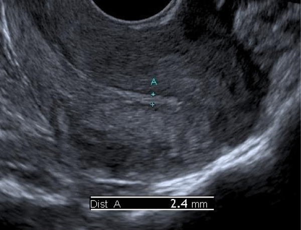
ET measurement was performed with the patient in the dorsal lithotomy position. Double-layer thickness from thickest part of endometrium in the longitudinal plane was accepted as ET (Figure 2).
VFT and SFT measurements were measured by the same radiologist (M.S.E.), and ET was performed by the same gynecologist (F.E.).
Statistical analysis
The statistical package SPSS (Statistical Package for Social Sciences; SPSS Inc., Chicago, IL) for Windows 16.0 was used to analyze the data. A Mann-Whitney U test was used to compare between the EM>5mm and EM≤5mm groups. Mean and standard deviations were used to describe the data. Pearson’s correlation coefficient was used for evaluating the relationships between the metabolic and anthropometric measurements, as well as the ultrasonographic measurements and VAI. A p value of 0.05 or less was considered to be statistically significant.
Results
This study included 79 women in total. Fifteen of the seventy-nine women in the study sample had an ET>5 mm.
The basic characteristics of the ET≤5mm and ET>5mm groups are presented in table 1. The ET>5mm group had higher BMI and HC measurements than ET≤5mm group (p=0.03). The two groups did not differ with respect to VAI and VFT (p=0.95 and p=0.08).
Considering all postmenopausal women, only HC (p=0.02) and VFT (p=0.05) were positively correlated to ET. On the other hand, VAI had a negative correlation with HDL (p<0.001) and a positive correlation with TG (p<0.001) and VFT (p=0.03). VFT showed had a positive correlation with TG (p=0.003), time since menopause (p=0.03), anthropometric measurements BMI (p<0.001), WC (p=0.008) and HC (p=0.01) (Table 2).
Discussion
To our knowledge, we have investigated for the first time in the literature both the feasibility of VAI as a simple mathematical formula and VFT as a measurement of fat tissue thickness, which is thought to be an endocrine function responsible for estrone increase, in the prediction of ET increase, as well as the relationship between these two measurements. As a result, we found that neither of the two measurements had any predictive value for an ET>5mm. As the VAI increased, ultrasonographic VFT measurement similarly increased.
An increase in BMI has been linked to a parallel increase in ET [13]. Abdominal fat accumulation is typical in the postmenopausal period. This type of lipoidosis is thought to be the most effective fat accumulation in increased ET and CVD risk [5,14]. Warming et al. [5], studied the body fat distributions of 531 healthy postmenopausal women using dual energy x-ray absorptiometry and found that visceral fat tissue was the primary factor in the production of estrone via the conversion of androgen precursors, arguing it to be the most effective fat tissue on endometrial thickness. Subsequent studies also support the notion that subcutaneous and visceral fat tissues have separate endocrinological functions [11]. Body fat distribution affects the plasma levels of sex hormones. Compared to other types of lipoidosis, abdominal lipoidosis is characterized by lower levels of sex hormone binding protein (SHBG), which in turn increases free estrogen levels, leading to enhanced ET thickness [15]. Higher insulin levels due to insulin resistance cause reduced SHBG levels [16].
Abdominal lipoidosis, which is typical during menopause, leads to increased CVD and MS risks [14]. The increased concentration of free fatty acids and enhanced adipokine secretion as a result of increased visceral fat content have been shown to lead to insulin resistance [17]. Increased insulin levels not only cause an increase in free estrogen by decreasing SHBG, but also an insulin growth factor (IGF)-1 that activates endometrial cell growth and IGF-binding protein-1 [18]. Hence, fat tissue can alter endometrial thickness not only by enabling the conversion of androgen precursors into estrogen, but also through the insulin resistance it brings about. Especially, visceral fat mass, a component of abdominal fat accumulation that is typical of the postmenopausal period, leads to increased ET [19].
The VAI was identified as an indicator of visceral fat function by the AlkaMeSy Study Group. The group claims that VAI is significantly correlated with all metabolic syndrome factors, such as altered production of adipocytokines, increased lipolysis, and plasma-free fatty acids, which are not signified by BMI, WC, TG, and HDL separately (9). In our study, the VAI was positively correlated with ultrasonographic VFT values. ET was also positively correlated to VFT. However, we failed to detect any correlation between ET and VAI, which has been defined as “a representation marker of adipose tissue dysfunction” (9). This may have stemmed from inability of VAI to specifically measure VFT, which is the actual factor that is responsible for endocrinological functions, using BMI and WC in its formula.
This study is limited by its low number of postmenopausal women with an ET > 5 mm. Considering the positive correlation between VFT and ET, a larger sample size would have provided the statistical significance result for the VFT values between the ET>5 mm and ET≤5mm groups.
In conclusion, an increase in endocrinologically active VFT results in increased ET. The VFT and VAI values that appeared inter-related were not predictive of an ET > 5 mm. Therefore, further studies on this subject with larger sample sizes are needed.
The author(s) received no financial support for the research, authorship, and/or publication of this article.
Conflict of Interest
The authors declare no potential conflicts of interest with respect to the research, authorship, and/or publication of this article.
Author’s contributions
F Eskicioglu: Project development, Data management, Data analysis, Manuscript writing/editing
MS Eskicioğlu: Project development, Data collection, Manuscript writing/editing
B Özyurt: Data analysis, Manuscript editing
- Carr MC (2003) The emergence of the metabolic syndrome with menopause. J Clin Endocrinol Metab 88: 2404-2411. Link: https://goo.gl/nHJpns
- Galic S, Oakhill JS, Steinberg GR (2010) Adipose tissue as an endocrine organ. Mol Cell Endocrinol 316: 129-139. Link: https://goo.gl/C6T8NP
- Carmichael AR (2006) Obesity and prognosis of breast cancer. Obes Rev 7: 333–340. Link: https://goo.gl/DBjDs2
- Gruber CJ, Tschugguel W, Schneeberger C, Huber J (2002) Production and actions of estrogens. NEJM 346: 340–352. Link: https://goo.gl/Sm1GFE
- Warming L, Ravn P, Christiansen C (2013) Visceral fat is more important than peripheral fat for endometrial thickness and bone mass in healthy postmenopausal women. Am J Obstet Gynecol 188: 349-353. Link: https://goo.gl/uHPXTc
- Toth MJ, Tchernof A, Sites CK, Poehlman ET (2000) Effect of menopausal status on body composition and abdominal fat distribution. Int J Obes Relat Metab Disord 24: 226-231. Link: https://goo.gl/4XU336
- Fernández Muñoz MJ, Basurto Acevedo L, Córdova Pérez N, Vázquez Martínez AL, Tepach Gutiérrez N, et al. (2014) Epicardial adipose tissue is associated with visceral fat, metabolic syndrome, and insulin resistance in menopausal women. Rev Esp Cardiol (Engl Ed) 67: 436-441. Link: https://goo.gl/jocZpH
- Ohashi N, Yamamoto H, Horiguchi J, Kitagawa T, Hirai N, et al. (2009) Visceral fat accumulation as a predictor of coronary artery calcium as assessed by multislice computed tomography in Japanese patients. Atherosclerosis 202: 192–199. Link: https://goo.gl/L1XkoY
- Amato MC, Giordano C, Galia M, Criscimanna A, Vitabile S, et al. (2010) Visceral Adiposity Index: a reliable indicator of visceral fat function associated with cardiometabolic risk. Diabetes Care 33: 920-922. Link: https://goo.gl/9KiHMs
- Jacobs I, Gentry-Maharaj A, Burnell M, Manchanda R, Singh N, et al. (2011) Sensitivity of transvaginal ultrasound screening for endometrial cancer in postmenopausal women: a case-control study within the UKCTOCS cohort. Lancet Oncol 12: 38-48. Link: https://goo.gl/9GQzSS
- Oktem O, Kucuk M, Ozer K, Sezen D, Durmusoglu F (2010) Relation of body fat distribution to femoral neck bone density and endometrial thickness in postmenopausal women. Gynecol Endocrinol 26: 440-444. Link: https://goo.gl/4EP6ya
- Hamagawa K, Matsumura Y, Kubo T, Hayato K, Okawa M, et al. (2010) Abdominal visceral fat thickness measured by ultrasonography predicts the presence and severity of coronary artery disease. Ultrasound Med Biol 36: 1769-1775. Link: https://goo.gl/kTvYCm
- Hebbar S, Chaya V, Rai L, Ramachandran A (2014) Factors influencing endometrial thickness in pstmenopausal women. Ann Med Health Sci Res 4: 608-614. Link: https://goo.gl/ELSPkt
- Pi-Sunyer FX (1999) Comorbidities of overweight and obesity: current evidence and research issues. Med Sci Sports Exerc 31: 602-608. Link: https://goo.gl/Qnh4QP
- Heiss CJ, Sanborn C, Nichols DL, Bonnick SL, Alford BB (1995) Association of body fat distribution, circulating sex hormones, and bone density in postmenopausal women. J Clin Endocrinol Metab 80: 1591–1596. Link: https://goo.gl/98yKhT
- Reid IR, Evans MC, Cooper GJ, Ames RW, Stapleton J (1993) Circulating insulin levels are related to bone density in normal postmenopausal women. Am J Physiol 265: 655–659. Link: https://goo.gl/Rd52tY
- Wise BE (2004) The Inflammatory syndrome: the role of adipose tissue cytokines in metabolic disorders linked to obesity. J Am Soc Nephrol 15: 2792–2800. Link: https://goo.gl/u7h59n
- Augustin LS, Dal-Maso L, Franceschi S, Talamini R, Kendall CW, et al. (2004) Association between components of the insulin‑like growth factor system and endometrial cancer risk. Oncology 67: 54‑59. Link: https://goo.gl/zo6Mab
- 19. Eskicioglu F, Eskicioglu MS, Ozdemir AT, Ozyurt B (2016) Carotid intima-media thickness in postmenopausal women is associated with an endometrial thickness greater than 5 mm. Ginekol Pol 87: 116-123. Link: https://goo.gl/aekjTK
Article Alerts
Subscribe to our articles alerts and stay tuned.
 This work is licensed under a Creative Commons Attribution 4.0 International License.


 Save to Mendeley
