Journal of Gynecological Research and Obstetrics
Role of Soluble FMS-Like Tyrosine Kinase (SFLT-1) /Placental Growth Factor (Plgf) Ratio as Prognostic Marker for Cases of Preeclampsia
Gasser M Elbishry, Ihab F Serag Eldin, Ahmed A ElShahawy*, Gihan E Hawwary and Ahmed M Riad
Cite this as
Elbishry GM, Serag Eldin IF, ElShahawy AA, Hawwary GE, Riad AM (2017) Role of Soluble FMS-Like Tyrosine Kinase (SFLT-1) /Placental Growth Factor (Plgf) Ratio as Prognostic Marker for Cases of Preeclampsia. J Gynecol Res Obstet 3(2): 037-045. DOI: 10.17352/jgro.000036Objective: The aim of this study is to identify the role of sFlt-1/PIGF ratio as a prognostic marker for cases of preeclampsia.
Patients and methods: The current study is a case control study that was conducted over 90 cases of primigravida patients, 24-34 weeks of gestation, randomly selected patients from outpatient clinic and ER of Ain Shams Maternity Hospital, they were classified into two groups, first group is preeclampsia group which was 45 preeclamptic pregnancies (preeclampsia patients and cases with severe criteria) and the second group was control group which was 45 normal pregnancies. Each patient was examined by sFlt-1/PlGF ratio immunoassay kits.
Results: In our study we have found that there was statistically significant positive correlation between the sFlt-1/PlGF ratio and blood pressure in 24-34 weeks. The correlations between the sFlt-1/PlGF ratio and other laboratory markers were statistically significant as well. In the 24-34 weeks PE group, AST, ALT were positive meanwhile Platelet count yielded only a highly significant negative correlation to the sFlt-1/PlGF ratio. We analyzed the time to delivery in all 45 patients with PE/HELLP within 2 days (24 patients), 2-7 days (10 patients), and later than 7 days (11 patients). Patients with interval to delivery within 2 days showed a higher sFlt-1/PlGF ratio, the sFlt-1/PlGF ratio in patients delivering within 2 days was (610.85). Patients with interval to delivery within 2-7 days exhibited a sFlt-1/PlGF ratio of (499.7). However, patients delivered later than 7 days had a sFlt-1/PlGF ratio of (230.43). For all PE/HELLP patients group (24-34 weeks), a sFlt-1/PlGF ratio greater than 590.1 (413.7 – 611.1) is associated with an 11.577 folds increased risk for an immediate occurence of delivery. The current study has found that the best cut off point after applying ROC curve between control group and cases group regarding soluble fms like tyrosine kinase/placental growth factor ratio was (> 85) which gave us sensitivity of 100%, specificity of 100% and accuracy of 100%.
Conclusions: We concluded from our study that the important clinical implication for the use of the sFlt-1/PlGF ratio for diagnosis, differential diagnosis, and risk stratification in PE/HELLP patients. Patients with sFlt-1/PlGF ratio above the level of 85 were preeclamptic and should be monitered for upcoming complications, symptoms and signs of severity. It could be used as a prognostic tool regarding maternal and fetal outcomes for patients with Preeclampsia between 24-34 weeks of gestation and patients at risk of having PE.
Introduction
Preeclampsia refers to the new occurrence of elevated blood pressure and either proteinuria or end-organ damage or both after 20 weeks of gestation in a previously normotensive primigravida. Severe hypertension and clinical picture of end-organ dysfunction are considered the severe spectrum of the syndrome. In 2013, the American College of Obstetricians and Gynecologists excluded proteinuria as an essential criterion for diagnosis of severe preeclampsia as massive proteinuria has a poor correlation with the outcome. They also excluded fetal growth restriction as possible feature of severe cases because fetal growth restriction is managed similarly whether or not preeclampsia is diagnosed. Oliguria was also excluded as a characteristic of severe disease [1].
The antiangiogenic and angiogenic factors as soluble fms- like tyrosine kinase (sFlt-1) as well as placental growth factor (PIGF) have been involved in the pathogenesis of preeclampsia [2]. sFlt-1 are naturally occurring circulating antagonists to vascular endothelial growth factor (VEGF) and antagonize the pro-angiogenic activity of circulating VEGF and PlGF by binding to them and hindering their interaction with their endogenous receptors. Enhanced placental expression and secretion of sFlt1 appear to play a major role in the development of preeclampsia [3]. SFlt-1 administered to pregnant rats can cause albuminuria, hypertension, and the pathognomonic renal pathologic changes of glomerular endotheliosis. In vitro, removal of sFlt-1from supernatants of preeclamptic tissue culture regains endothelial function and angiogenesis to normal values. Conversely, exogenous intake of VEGF and PlGF reverses the antiangiogenic state produced by excess sFlt-1 [4].
A nested case control study utilizing banked sera to estimate serum sFlt-1, as well as PlGF and VEGF, across gestation found that alterations in sFlt-1 were predictive of the subsequent pathogenesis of preeclampsia. SFlt-1 values enhanced during pregnancy in all women But, compared to normotensive controls, women who developed preeclampsia began this elevation earlier in gestation (at 21- 24 weeks versus 33 -36 weeks) and reached higher values. A considerable difference in the serum sFlt-1 concentration between the two groups was clear five weeks before the clinical onset of the disease. PlGF and VEGF levels fell simultaneously with the increase in sFlt-1 which may had been related, in part, to binding by sFlt1 [1].
The sFlt-1/PIGF ratio might be of value in the prediction of PE as well as in the differential diagnosis of women with atypical presentations of preeclampsia, and chronic hypertension prior to development of superimposed preeclampsia [5,6].
Studies had demonstrated that sFlt-1, soluble Endoglin (sEng) and PIGF might be suitable biomarkers for preeclampsia before the onset of the disease. Second trimester sFlt-1/PIGF ratio was proved to be helpful as an aid in the prediction and diagnosis of pregnancy induced hypertension. The addition of sFlt-1/PIGF to Doppler ultrasound was proved to increase the sensitivity and specificity of Doppler alone [7].
Recent studies on the utilization of angiogenic markers in the prediction of preeclampsia have demonstrated that: the sFlt-1/PlGF ratio is a more helpful predictor than either of these parameters alone. Sequential alterations in the level of sFlt-1, PlGF and sEng might be more informative than single measurements, the addition of angiogenic marker estimates can increase the predictive value of Doppler ultrasound alone. There is a more pronounced change of angiogenic factors in early-onset compared with late-onset pregnancy induced hypertension. In the second trimester the sFlt-1/PlGF ratio might be an ideal parameter to diagnose early-onset preeclampsia (< 34 weeks) or preterm preeclampsia (< 37 weeks) [8]. The aim of the current work is to identify the role of sFlt-1/PIGF ratio as a prognostic marker for cases of Preeclampsia.
Materials and Methods
Study design
Case Control Study.
Study setting
This study was conducted at aim shams university maternity hospital in the period from February 2016 till January 2017.
Population of the study
Pregnant women were recruited from those attending outpatient clinic and Emergency Room, A total of 90 pregnant females were enrolled in the study: 45 singleton normal pregnancies (Control Group), 45 singleton pregnancies with Preeclampsia (Mild cases and Severe Cases).
Inclusion criteria:
1) Primigravida (singleton).
2) Diagnosed as preeclampsia by one of the following criteria:
• Hypertension: diastolic blood pressure > 90 mmHg, systolic blood pressure >140 mmHg based on at least 2 measurements taken at least 4 hours apart and Proteinuria.
• Severe hypertension (systolic blood pressure ≥160 mm Hg or diastolic ≥110 mm Hg on two occasions at least four hours apart or only once if treated) with Proteinuria.
• In absence of proteinuria abnormal laboratory tests besides any of the following signs:
• Persistent and/ or severe headache
• Visual abnormalities (scotomata, photophobia, blurred vision, or temporary blindness [rare])
• Upper abdominal or epigastric pain
• Nausea, vomiting
• Dyspnea, retrosternal chest pain
• Altered mental status
3) Gestational age 24- 34 weeks.
The following patients were excluded from the study:
1) Pre-mature rupture of membranes (PROM).
2) Chorioamnionitis.
3) Multiple gestation.
4) Rh isoimmunization.
5) Fetal anomalies.
6) Intra uterine fetal death (IUFD).
7) Maternal auto immune diseases.
8) Chronic inflammatory diseases.
9) History of D.M.
Ethical and legal issues
Informative consent: Permission was obtained from patients to perform a specific test and that the study involves research and explanation of the purposes of the research was done.
All participants were counseled and give a written informed consent. All patients had the right to withdraw from the research anytime they desire.
The study protocol and patients informed consent were reviewed and approved by the ethics committee of obstetrics and gynecology department Ain Shams University.
Group 1 (Preeclampsia group): Singleton pregnancies, 45 primigravida patients were examined and tested. All patients diagnosed with preeclampsia according to American College of Obstetrics and Gynecology (HTN+Proteinuria) (ACOG, 2013).
Group 2 (Control group): Singleton pregnancies, 45 healthy primigravida patients.
All patients were subjected to the following:
1) Detailed history taking:
• Detailed history of present pregnancy e.g persistent headache, visual disturbances or epigastric pain.
• Past history of chronic hypertension, diabetes mellitus or autoimmune diseases.
• Family history of similar conditions.
2) Physical examination:
• General: Blood pressure: it was measured to obtain hypertension as it was defined as the repeated measurement of systolic blood pressure of 140mmHg or greater (Korotkoff I) and diastolic blood pressure of 90mmHg or greater (Korotkoff V). It was measured by mercury sphygmometer with suitable cuff size by residents, house officers and training doctors only in semi sitting position. It was repeated 4 hours apart to confirm the condition.
• Examination for pallor, jaundice, chest and heart auscultation and lower limb oedema.
• Abdominal examination:
∗ Fundal level ± gestational age.
∗ F. H.S by U/S.
∗ Lie and presentation of fetus.
3)Investigation:
• Lab investigations:
∗ C.B.C.
∗ Blood grouping and Rhesus factor.
∗ Urine analysis.
∗ ALT, AST (affected in preeclampsia) & Uric acid.
∗ S.Albumin, S.Creatinine.
∗ Blood glucose test.
∗ Co-agulation profile: PT, PTT & INR.
Samples and immunoassays for measurement of sFlt/PlGF ratio:
• Serum samples were collected by Nurses, junior doctors or residents by venipuncture in tubes without anticoagulant.
• After clotting, the samples were centrifuged with 2000g and serum was pipetted and stored at -8C until testing at Ain Shams Maternity Hospital Laboratory.
• Samples were tested each for sFIt-1/PIGF ratio by the specialists using sFlt-1/PlGF ratio kits.
• The kits for sFIt-1/PIGF ratio were imported from United States of America as Elecsys® Preeclampsia (sFlt-1 & PlGF) immunoassay kits.
Primary Outcome:
Time to delivery in correlation with level of sFlt-1/PIGF ratio.
- Delivery within 48 hours.
- Delivery within 2-7 days.
- Delivery later than 7 days.
- Delivery at term.
Secondary Outcome:
1. Fetal complications: e.g IUGR, pathological cardiotemography (CTG) and Neonatal death. Nonreassuring fetal testing (nonreassuring nonstress test or biophysical profile score, estimated fetal weight less than the fifth percentile for gestational age, oligohydramnios with amniotic fluid index <5.0 cm or maximal vertical pocket <2.0 cm, and/or persistent absent or reversed diastolic flow on umbilical artery.
2. Maternal complications:
• HELLP syndrome (Hemolysis, Elevated Liver enzymes, Low Platelet count) probably represents a severe form of preeclampsia.
• Eclampsia refers to the development of grand mal seizures in a woman with preeclampsia, in the absence of other neurologic conditions that could account for the seizure.
• Multi organ failure: Women with preeclampsia are at an increased risk for life threatening events, including placental abruption, acute kidney injury, cerebral hemorrhage, hepatic failure or rupture, pulmonary edema, disseminated intravascular coagulation.
Sample Size: It was calculated using PASS 11.0 program using findings from previous study carried out by Verlohren et al. (2011). The total sample of 6 subjects achieves 85% power to detect differences among the means versus the alternative of equal means using an F test with a 0.05000 significance level. The size of the variation in the means is represented by their standard deviation which is 53.79. The common standard deviation within a group is assumed to be 21.89.
Statistical analysis
Data were collected, revised, coded and entered to the Statistical Package for Social Science (IBM SPSS) version 20. Qualitative data were presented as number and percentages while quantitative data were presented as mean, standard deviations and ranges when parametric and median with interquartile range (IQR) when non parametric. The comparison between two groups regarding quantitative data with parametric distribution was done by using Independent t-test wile with non-parametric distribution was done by using Mann-Whitney test. The comparison between more than two independent groups regarding quantitative data with parametric distribution was done by using One Way Analysis of Variance (ANOVA). Spearman correlation coefficients were used to assess the correlation between two quantitative parameters in the same group. Receiver operating characteristic curve (ROC) was used to assess the best cut off point for sFlt-1/PlGF ratio in differentiation between cases group and control group with its sensitivity, specificity, positive predictive value (PPV), negative predictive value (NPV) and area under curve (AUC). The confidence interval was set to 95% and the margin of error accepted was set to 5%. So, the p-value was considered significant as the following:
P > 0.05: Non significant
P < 0.05: Significant
P < 0.01: Highly significant.
Results
This is a case control study that was conducted at Ain Shams University in the time between February 2016 till February 2017. This study included 45 patients (PET, case group) and 45 pregnant women with normal pregnancy (Table 1).
The table showed there was no statistical significance between preeclampsia group and control group regarding any of the demographic data (Table 2).
The previous table showed that there was statistical significance difference between control group and preeclampsia group regarding ALT, AST, systolic and diastolic blood pressure, platelets, proteinuria, fetal outcomes and delivery (Table 3).
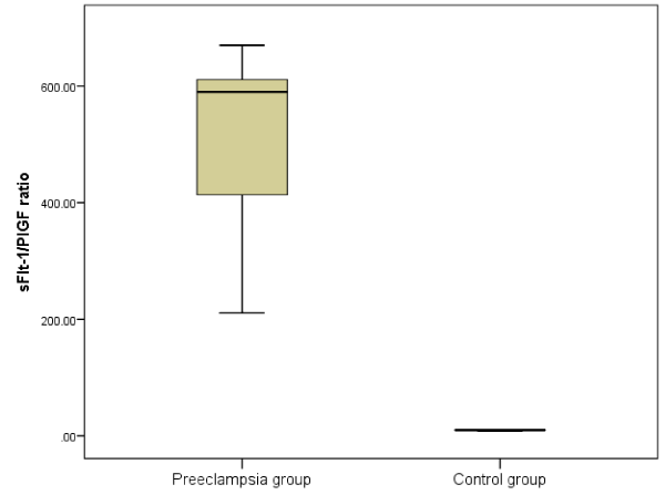
The previous table shows that there was highly statistically significant increase in preeclampsia group than control group regarding sFlt-1/PIGF ratio (Figure 1), (Table 4).
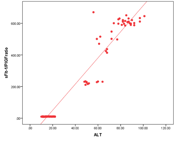
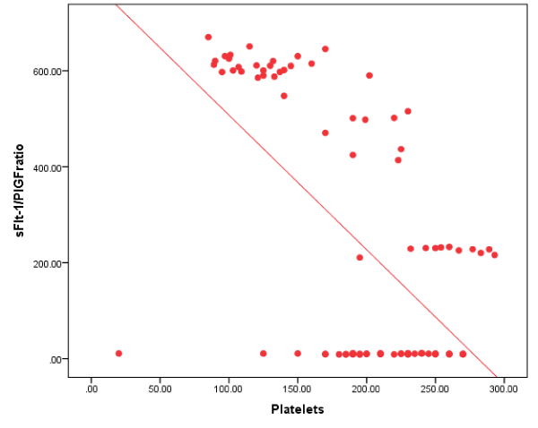
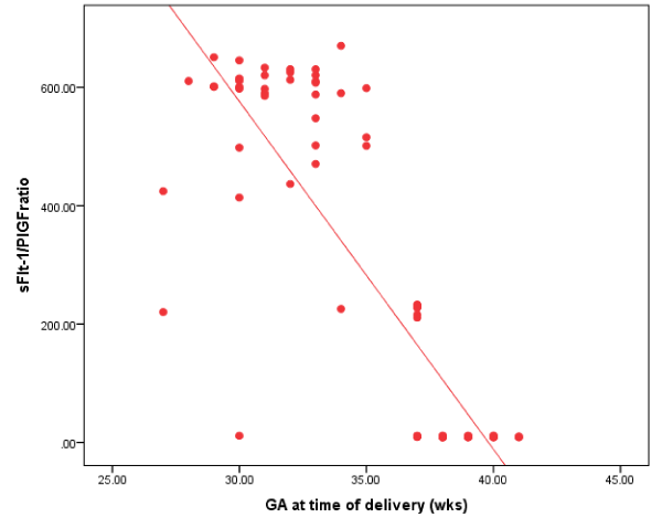
The previous table showed that there was statistical significance positive correlation found between sFlt-1/PlGF ratio and blood pressure, ALT and AST. There was negative correlation between sFlt-1/PlGF ratio and platelets and gestational age at time of delivery among the whole population of study group. (Figures 2-7), (Table 5).
The previous table showed that there was statistically significant relation found between interval to delivery with ALT, AST, systolic blood pressure, platelet level with time to delivery while no statistically significant relation found with Diastolic blood pressure, proteinuria (Figure 8), (Table 6).
The previous table showed that there was statistically significant negative correlation found between time to delivery and sFlt-1/PlGF ratio (Figure 9,10).
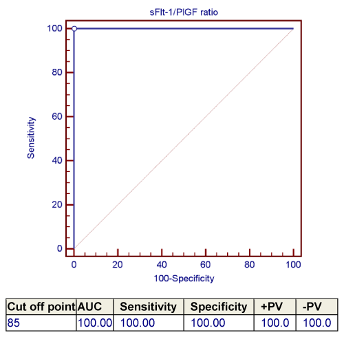
The previous table showed that the best cut off point between preeclampsia group and control group regarding sFlt-1/ PIGF ratio was found > 85 which gave us sensitivity of 100%, specificity of 100% and accuracy of 100%.
Discussion
Preeclampsia (PE) is a multisystem disease in pregnancy it is still the second most common cause of maternal and fetal morbidity and mortality. The disease incidence is 2-8% worldwide, and it represents one of the main contributors of preterm birth, accounting for 15% of all preterm births [9]. The reliable diagnosis of high-risk PE women is crucial because close monitoring and referral to specialized perinatal care centers substantially decline maternal and fetal morbidity [10].
Elevated serum levels of sFlt-1 and dropped levels of PlGF, thereby leading to an elevated sFlt-1/PlGF ratio, could be detected in the second half of pregnancy in patients diagnosed to have not only PE but also fetal jeopardy, i.e. placenta-related diseases. These changes are clearer in early-onset rather than late-onset disorders and are linked to severity of the clinical disease. Moreover, the disturbances in angiogenic substances are demonstrated to be detectable before the onset of clinical disease, so allowing discrimination of patients with normal pregnancies from those at high risk for developing PE [11].
Plasma levels of angiogenic/antiangiogenic substances are of prognostic value in obstetric triage: similar to a progressively worsening clinical course detected in women with early-onset PE, alterations in the angiogenic profile linked to a more antiangiogenic state could be found [12].
We examined the hypothesis that the level of the sFlt-1/PlGF ratio is predictive for a risk for imminent delivery and has thereby putative eligibility as a prognostic marker. Furthermore, we performed correlation analysis between the sFlt-1/PlGF ratio and clinical and laboratory markers of PE/HELLP (hemolysis, elevated liver enzymes, and low platelet count) syndrome. We aimed at this study to identify the role of sFlt-1/PIGF ratio as a prognostic marker for cases of Preeclampsia. Our study is a case control study that was conducted over 90 cases of primigravida patients 24-34 weeks of gestation patients were enrolled from outpatient clinic and ER of Ain Shams Maternity Hospital, they were classified into two groups, first group is preeclampsia group which was 45 preeclamptic pregnancies and the second group is control group which was 45 singelton pregnancies. Serum samples were collected from the patients alongside other laboratory tests within the period 24-34 weeks of gestation.Maternal and fetal outcomes were denoted to observe the statistical relationship between laboratory tests and sFlt-1/PlGF ratio. The PE/HELLP group consisted of 45 patients, 19 with PE, 26 with severe criteria of PE, 9 with HELLP.There was no statistical significance between age, gestational age at time of sampling and BMI (demographic data) and sFlt-1/PlGF ratio. We have analyzed sFlt-1/PlGF ratio in patients with PE and healthy controls at 24-34weeks. Patients with PE/HELLP had a significantly increased sFlt-1/PlGF ratio as compared with controls 34 weeks (590.1 vs 9.9 pairwise comparison, P .001).
In our study we had found that there was statistically significant positive correlation between the sFlt-1/PlGF ratio and blood pressure in 24-34 weeks. The correlations between the sFlt-1/PlGF ratio and other laboratory markers were statistically significant as well. In the 24-34 weeks PE group, AST, ALT were positive meanwhile Platelet count yielded only a highly significant negative correlation to the sFlt-1/PlGF ratio.
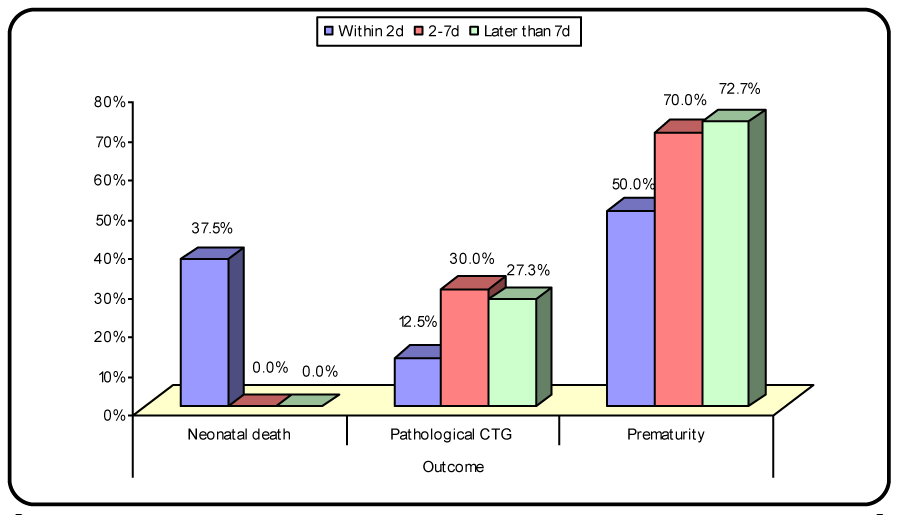
We analyzed the time to delivery in all 45 patients with PE/HELLP within 2 days (24 patients), 2-7 days (10 patients), and later than 7 days (11 patients). Patients with interval to delivery within 2 days showed a higher sFlt-1/PlGF ratio, the sFlt-1/PlGF ratio in patients delivering within 2 days was (610.85). Patients with interval to delivery within 2-7 days exhibited a sFlt-1/PlGF ratio of (499.7). However, patients delivered later than 7 days had a sFlt-1/PlGF ratio of (230.43).
For all PE/HELLP patients group (24-34 weeks), a sFlt-1/PlGF ratio greater than 590.1 (413.7 – 611.1) is associated with an 11.577 folds increased risk for an immediate occurence of delivery.
The current study had reported that the best cut off point after applying ROC curve between control and cases groups regarding soluble fms like tyrosine kinase/placental growth factor ratio was (> 85) which gave us sensitivity of 100%, specificity of 100% and accuracy of 100%.
In a previous study, that was done to determine whether the levels of soluble fms-like tyrosine kinase-1 (sFlt-1) and placenta growth factor (PlGF) ratio are changed during the second trimester in the plasma of women who subsequently develop PE. They had done a case-control study to compare the levels of sFlt-1 and PlGF in the preeclamptic (n=46) and normal pregnant women (n=100). The plasma levels of sFlt-1 and PlGF were measured by ELIZA. sFlt-1 levels were significantly elevated in the preeclamptic women than in normal controls (p<0.001), while the PlGF values were significantly lower (p<0.001).b
This was in agreement with our study in denoting that the significance of the soluble fms like tyrosine kinase/placental growth factor ratio increases with cases of preeclampsia and declines in normal pregnancies, but that was against our study as they conducted their study with mean maternal age was 34, 35 years for participants while our study the maternal age was 23 years. They have found the maternal plasma log [sFlt-1/PlGF] ratio with the cut-off value of 1.4 gave the best combination with 80.4% sensitivity and 78% specificity [13].
In another study that was designed to illustrate the value of utilizing the sFlt-1:PlGF ratio for the prediction of preeclampsia. In consistency with our results, they conducted their study with time of sampling (24-34 weeks) and it was useful to utilize the soluble fms like tyrosine kinase/Placental growth factor ratio as a prognostic factor for detecting preeclampsia. In contrast with our results, they had made their study on 1000 women, 500 cases and 500 controls [14].
Alpoim et al. 2013 had conducted their study according to uterine artery Doppler and sFlt-1/PlGF ratio whether they were helpful for the diagnosis of preeclampsia or not. In early onset PE, mPI-UtA at diagnosis was abnormal in 100% of cases with fetal jeopardy and in 91% without fetal affection, while sFlt-1/PlGF was abnormal in 100% and 96%, respectively. In late onset PE, mPI-UtA was abnormal in 50% and 37% of cases with and without fetal distress while the sFlt-1/PlGF ratio was abnormal in 50% and 26%, respectively. In agreement with our study, the sFlt-1/PlGF ratio was 100% specific [15].
Lamminpää et al. 2012, they aimed at their study to identify critical evaluation of the clinical validity of the prenatal diagnosis of sFlt-1/PlGF for preeclampsia (PE), pregnancy-induced hypertension, and proteinuria. Their analysis was based on a specificity of 95 % and a sensitivity of 82 % for the prediction of preeclampsia, as described by Elecsys (Roche). They had found although the specificity of the sFlt-1/PlGF ratio was high for PE, the sensitivity was low (only 59.4 %), thus producing unsatisfying results for PE. The sensitivity only elevated to 62.5 % for the early-onset PE group. Interestingly, a high ratio was detected for the combination of IUGR and PE in the early-onset PE group (8 cases). In the control group, 4 cases exceeded the cut-off level of 85 but showed no clinical picture of PE and the birth was unremarkable [16].
Discussion
We had found that soluble fms like tyrosine kinase/placental growth factor ratio was increased in cases of PE. The cut off level of 85 gave us high sensitivity 100% and specificity 100%. The sfms like tyrosine kinase/placental growth factor ratio was increased in cases of severe criteria of preeclampsia with poor obstetric outcome and might be used as a valuable prognostic marker for women with preeclampsia.
Disclosure
All authors report no conflict of interest
- American College of Obstetricians and Gynecologists (2013) Hypertension in Pregnancy-Induced Practice Guideline WQ 244. Link: https://goo.gl/M6ANOu
- Verlohren S, Galindo A, Schlembach D, Zeisler H, Herraiz I, et al. (2010) An automated method for the determination of the sFlt-1/PGF ratio in the assessment of preeclampsia. Am J Obstet Gynecol. Link: https://goo.gl/MPFEfg
- Staff AC, Johnsen GM, Dechend R, Redman CW (2013) Preeclampsia and uteroplacental acute atherosis: immune and inflammatory factors. J Reprod Immunol; 101-102: 120-126. Link: https://goo.gl/EXoW7M
- Steegers EA, Von Dadelszen P, Duvekot JJ, Pijnenborg R (2010) Preeclampsia. Lancet 376: 631-644. Link: https://goo.gl/pWzREa
- Roberts JM, Bodnar LM, Patrick TE (2011) The role of obesity in preeclampsia. Pregnancy Hypertens 1: 6–16. Link: https://goo.gl/hPIQjD
- Perni U, Sison C, Sharma V, Helseth G, Hawfield A, et al. (2012) Angiogenic factors in superimposed preeclampsia: a longitudinal study of women with chronic hypertension during pregnancy. Hypertension 59: 740–746. Link: https://goo.gl/qi9xcf
- Kuc S, Wortelboer EJ, van Rijn BB, Franx A, Visser GH, et al. (2011) Evaluation of 7 serum biomarkers and uterine artery Doppler ultrasound for first-trimester prediction of preeclampsia: a systematic review. Obstet Gynecol Surv 66: 225-239. Link: https://goo.gl/42K7Ro
- Illanes S, Parra M, Serra R, Pino K, Figueroa-Diesel H, et al. (2009) Increased free fetal DNA levels in early pregnancy plasma of women who subsequently develop preeclampsia and IUGR. Prenat Diagn 29: 1118-22. Link: https://goo.gl/mgxaH4
- Huppertz B (2008) Placental origins of preeclampsia: challenging the current hypothesis. Hypertension 51: 970. Link: https://goo.gl/eq5VWU
- Yildiz A, Yetimalar H, Kasap B, Aydin C, Tatar S, et al. (2012) Preoperative serum CA 125 level in the prediction of the stage of disease in endometrial carcinoma. Eur J Obstet Gynecol Reprod Biol 164: 191-195. Link: https://goo.gl/BZcUqJ
- Yin BW, Lloyd KO (2001) Molecular cloning of the CA-125 ovarian cancer antigen: Identification as a new mucin, MUC16. J. Biol. Chem 276: 27371–27375. Link: https://goo.gl/vwer1c
- Yagmur E, Bastu E, Karamustafaoglu-Balci B, Akhan SE, Buyru F (2013) Non-invasive diagnosis of endometriosis based on a combined analysis of four plasma biomarkers. Centr Eur J Immunol 38: 154-158. Link: https://goo.gl/47FWZr
- Kim JH, Lim SY, Park SY (2007) Effective prediction of preeclampsia by a combined ratio of angiogenesis-related factors. Obstet Gynecol; 111: 1403. Link: https://goo.gl/40KdDe
- Wathen KA, Tuutti E, Stenman UH, Alfthan H, Halmesmäki E, et al. (2006) Maternal serum-soluble vascular endothelial growth factor receptor-1 in early pregnancy ending in preeclampsia or intrauterine growth retardation. J Clin Endocrinol Metab 91: 180-184. Link: https://goo.gl/Ft9L3E
- Alpoim PN, de Barros Pinheiro M, Junqueira DR, Freitas LG, das Graças Carvalho M, et al. (2013) Preeclampsia and ABO blood groups: a systematic review and meta-analysis. Mol Biol Rep 40: 2253-2261. Link: https://goo.gl/bZYg5v
- Lamminpää R, Vehviläinen-Julkunen K, Gissler M, Heinonen S (2012) Preeclampsia complicated by advanced maternal age: a registry-based study on primiparous women in Finland 1997-2008. BMC Pregnancy Childbirth 11: 12:47. Link: https://goo.gl/Z8UzCK
Article Alerts
Subscribe to our articles alerts and stay tuned.
 This work is licensed under a Creative Commons Attribution 4.0 International License.





 Save to Mendeley
