Journal of Dental Problems and Solutions
Chemical surface modification of lithium disilicate needles of a silica-based ceramic after HF-etching and ultrasonic bath cleaning: impact on the chemical bonding with silane
A Poulon-Quintin1*, F Rouzé l’Alzit1, E Ogden1, A Large1,2, C Bertrand1,2 and M Bartala2
2University of Bordeaux, UFR d’odontologie, 146 rue Léo Sagnat, F-33076, Bordeaux, France
Cite this as
Poulon-Quintin A, l’Alzit FR, Ogden E, Large A, Bertrand C, et al. (2022) Chemical surface modification of lithium disilicate needles of a silica-based ceramic after HF-etching and ultrasonic bath cleaning: impact on the chemical bonding with silane. J Dent Probl Solut 9(2): 035-041. DOI: 10.17352/2394-8418.000115Copyright License
© 2022 Poulon-Quintin A, et al. This is an open-access article distributed under the terms of the Creative Commons Attribution License, which permits unrestricted use, distribution, and reproduction in any medium, provided the original author and source are credited.Recommendations to obtain the best bonding to silica-based ceramics are to prepare its surface by hydrofluoric-acid HF etching and regular application of a silane. This study investigated how the HF-etching followed by ultrasonic water bath cleaning or by an additional phosphoric acid treatment impacts the adhesion properties of a resin (G-CEM LinkForce®) with a lithium disilicate glass-ceramic (IPS Emax Press, Ivoclar Vivadent). Comparison is based on results obtained with HF etching and direct silane application. After HF-etching, a water ultrasonic bath (4 minutes), and a final air drying, the scratch test critical load increases (+ 46%) thanks to chemical bonding. Additional tests are presented including heat treatments (at 85 °C before and after silanization). If HF-etching is followed by phosphoric acid treatment and drying of silane at 85 °C, scratch test critical load increases (+ 42%) due to mechanical bonding. Similar adhesion properties are obtained with two opposite protocols.
Introduction
The clinical success of restoration does not only depend on the intrinsic properties of the ceramic used. It is strongly associated with the quality and the durability of the ceramic-cement resin interface [1], even if each restoration has two interfaces: tooth/resin and resin/ceramic. Preclinical testing of dental materials is crucial to validate their mechanical capability and compatibility to service in the oral cavity. Such studies are numerous and many challenging are existing due to the oral environment conditions (humidity, pH, strength, and cyclic loading) [1,2]. Laboratory tests show that a lack of testing standardization still exists leading to impractical direct comparisons between the studies for similar types of restorations.
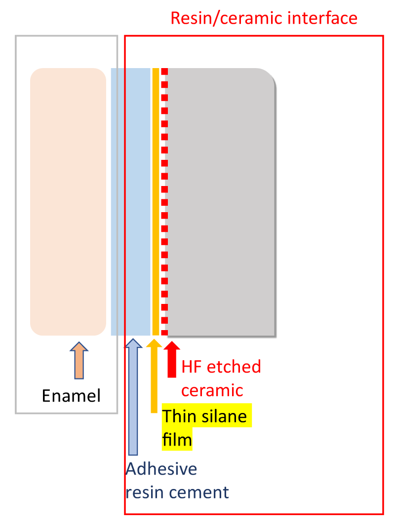
This study is focused on a lithium disilicate glass-ceramic (Li2Si2O5 crystals and glassy matrix; IPS Emax Press, Ivoclar Vivadent) bonded with an adhesive resin cement thanks to the MPS silane. The most used in dentistry is the γ-methacryloxypropyl trimethoxysilane (C10H20O5)Si. The lithium disilicate glass ceramic presents good translucency and is recommended for inlays, veneers, and anterior/posterior crowns supported by implants or teeth [3]. Figure 1 localizes the resin-ceramic interfaces of interest for a schematic bonding system in dental restoration when the ceramic surface is prepared by hydrofluoric acid (HF-etching) followed by a silane treatment and finished with the adhesive resin cement layer. In the literature, it is well known that ceramic surface pre-treatment helps to improve the bonding properties at the resin-ceramic interface [1-11]. Silanes are hybrid functional monomers able to form chemical bonds with organic and inorganic surfaces. They are used as surface primer agents for adhesion promotion increasing the critical surface energy of a substrate because they are chemically bifunctional (composed of non-hydrolyzable groups such as methacrylate, and hydrolyzable groups such as ethoxy). They bond dissimilar materials together by forming a branched 3D siloxane (-Si-O-Si-) film (i.e. chemical bonding). The stability and effectiveness of this adhesion are also supposed to be improved by heat treatment [11-17]. Bruzi, et al. [16] based on the evaluation of the shear bond strength of composite resin to CAD/CAM lithium disilicate ceramics recommends a regular application of silane. They advise applying silane for 20 s, air dry for 20 s, and hot drying at 60 °C for 20 s. Others authors advocate drying only with hot air [18-20] or washing with hot water [21,22]. Yavuz [20] focuses its attention on the necessity of post-silanization heat treatment to an HF etching first step, to improve the shear bond strength (SBS). It concludes that the heat treatment is not sufficient to achieve high SBS values compared with HF-etching.
HF-etching is known to provide the dissolution of the glassy and/or crystalline phase [23]. However, some residual salts of silicofluoride negatively influence the resin bond strength [24,25]. Several cleaning methods are suggested in the literature after HF-etching. The most supported post-etching protocol is the use of an ultrasonic distilled water bath for 2 min [25-28]. Poulton-Quintin et al. have proved the formation of LiSi2F6 nano-precipitates on the Li2Si2O5 needles even with this post-etching protocol [29]. Others proposed cleaning techniques are including the use of an ultrasonic bath [30], a 37% phosphoric acid smear [31] rinsing under running water, or a combination of a 37% phosphoric acid smear and an ultrasonic bath [27].
Surface treatments in dentistry aim to increase roughness and therefore promote the phenomena of mechanical bonding adhesion. They also create an increase in the available surface area and contact with the crystalline grains for the infiltration of the adhesive bonding modes. If a chemical bonding adhesion is present, this should normally increase its effectiveness. It is also seeking to increase the wettability of the products used for bonding. Common surface treatment option according to their purely mechanical or chemical action or their combined mechanochemical action are grinding, air-particle abrasion or sandblasting acid etching, and combinations of any of these methods [1,4].
In this article, three different types of surface preparation for the lithium disilicate glass-ceramic are compared. To assess the efficiency of the adhesion at the resin/ceramic interface, scratch tests are selected as a quantitative method.
Materials and methods
The lithium disilicate glass-ceramic (IPS Emax Press, Ivoclar Vivadent) samples were pellets (diameter: 1.9 cm; thickness: 4 mm) with parallel faces and a global composition (in wt%): SiO2 57-80, LiO2 11-19, K2O 0-13, P2O5 0-11, H3PO40-8, ZnO 0-8 and other oxides as Al2O3 0-10 (supplier data).
A mirror-polished face of each tested sample was first HF-etched applying 9% hydrofluoric acid (Porcelain Etch®, Ultradent) for 20 s (recommended action time by Ivoclar-Vivadent), then rinsed 1 min with water (no distilled water) and then air-dry as dentists done. To improve the reproducibility of our results, the quantities of HF acid deposited were weighed (100 mg). The acid application is done by a professional dentist with the same gesture as in its dental office for the restauration surfaces preparation. The second batch of samples was prepared as previously but after being water-rinsed (1 min), they were immersed in a water ultrasonic bath (WUS bath) for 4 minutes, water-rinsed (1 min) and dry again. The last batch of the sample was prepared as the first batch and then for 30 seconds etched with 80 mg of H3PO4 phosphoric acid (37% phosphoric acid etching gel, GC etchant). Also known as orthophosphoric acid, it is regularly used in dental practices usually in a concentration of 30% to 40% and water-rinsed again for 1 minute. WUS or H3PO4 post-treatments were supposed to eliminate the fluoride salts formed during HF-etching. However, no study has investigated the quality of resin bonding depending on the initial surface treatment.
Because the application of silane coupling agents is known to be very efficient in promoting adhesion for silica-based materials, for all the studied samples, the liquid silane was applied over the etched ceramic surfaces. Silane 3-methacryloxypropyltrimethoxysilane (MPS) in a pre-hydrolyzed state is deposited. For this step, as for the previous step, the quantities of silane applied were systematically weighed (2 mg) and applied by a professional dentist.
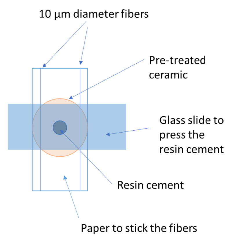
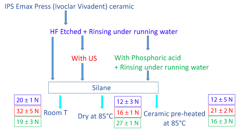
The final step for all the samples was the application of adhesive resin cement. It was a universal dual composite glue (chemo and photopolymerizable), the G-CEM LinkForce® (Paste A: bis-GMA, urethane dimethacrylate, dimethacrylate, barium glass…; Paste B: bis-MEPP, urethane dimethacrylate, barium glass…). In a real protocol in dentistry, this step is done directly on the patient teeth. Resin is applied on the pre-treated ceramics surface and pressed onto the tooth to restore. In this study, a glass slide is pressed against the resin cement (G-CEM LinkForce®) to obtain a thin layer of resin (homogeneous in thickness). This was a necessary condition for the scratch testing. A weighted quantity of resin (5 mg) was applied on the pre-treated ceramic using the mixing tip, pressed with a glass slide on which two fibers of 10 µm diameters were glued. The resin was cured using a blue phase-style lamp from Ivoclar-Vivadent delivering an intensity of 1100 mW/cm2. The glass slide was removed after photopolymerization. The final thickness of the polymerized resin is always close to 10 µm ± 1 µm. A schematic representation of this protocol just before applying a load to press the resin cement is presented in Figure 2. A synoptic representation of all the protocols used is done in Figure 2.
The critical load Lc measured when the coating-substrate interface decohesion occurred was interesting because adhesion failure was often a failure mechanism in dentistry restoration caused by the masticatory forces for example and limiting its performance and lifetime. Indeed, when performing a progressively increasing load stripe, it was customary to observe a succession of damages, the ultimate damage being generally the perforation and the complete delamination of the coating over the entire width of the striation groove. The Lc value, therefore, depended on the adhesion of the coating to the substrate. During the scratch test, mechanical solicitation is less complex (shear friction) but still makes it possible to test the bonding efficiency. For each sample, three scratch tests were performed using a Tribotechnic Millenium 100 scratch-tester under Standard ISO/EN 1071-3. A Rockwell C diamond tip indenter with a 200 µm radius of curvature was drawn during the scratch test over the coated surface for 5 mm length with an applied normal load increasing continuously (100 N/min) up to 50 N. The tip wear was controlled between each measurement according to the supplier's procedure. SEM images were performed with a conventional JSM6360A JEOL, 10kV. Samples beforehand metalized with a thin gold layer were used to follow the evolution of the morphology of the surface, to study the scratch track with the localization of the cracks or the spalling beginning (EDS-SEM analyses performed), and to make a precise evaluation of the critical load Lc.
Results
Evolution of the surface morphology after etching
Figure 3 shows the surface morphology evolution of the IPS Emax Press lithium disilicate glass-ceramic pellet depending on the three different surface treatments:
1) Aqueous HF-etching (20 s) followed by 1 min water rinsing,
2) Aqueous HF-etching (20 s) followed by 1 min water rinsing followed by an additional cleaning step using WUS for 4 min,
3) Aqueous HF-etching (20 s) followed by 1 min water rinsing and then 30 s etched with phosphoric acid and rinsed again with water for 1 min.
The appearance of the surface porosity is at two different scales (Figure 3). Pores are observable even when the surface is mirror-polished (Figure 3a). For low magnification levels (× 250), the presence of large pores containing particles (identified as ZrO2 or Al2O3 particles by EDS) for water treatment or phosphoric acid is noticeable (resp. (Figure 3b and d) whereas WUS seems removed them (Figure 3c).
When the magnification is increased (× 10000), noticeable differences appear between the three types of surface treatment. In the matrix containing the Li2Si2O5 needles, the glassy part is still present in various quantities (Figure 4) unless when WUS protocol is used (Figure 4b). Existing porosity is always present at this magnification but finer than the previous one observed in Figure 3.
Modification of the surface morphology after silanization
After applying the adhesive liquid (G-Premio BOND) containing MDP molecule and natural drying, the sample surfaces were observed by SEM (Figure 5). No more fine porosity is noticed (Figure 5, magnification × 2500). Small pores are full of MDP. The previous large porosities existing are still observable even if MDP covers them and the surface of the entire sample (Figure 5, magnification × 500).
Scratch test results
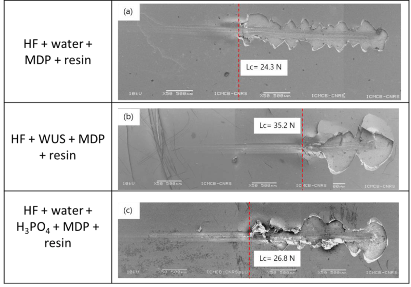
The surface roughness of the resin after photopolymerization is similar for all samples because correlated with the surface roughness of the glass side used to guarantee flatness and is the thickness of the resin layer. Figure 6 presents three examples of the scratch track depending on the initial ceramic surface treatment. An evaluation of the corresponding critical load Lc is noticed on the images. For all samples, spallation and chipping are easily observable (Figure 6).
EDS-SEM point analyses at the first spallation on the three scratches of the different types of samples reveal the presence of the barium element only when WUS is used. This means that resin is still present at the surface whereas when potassium is found, a crack occurs at the interface with the ceramic. These two elements are used as witnesses because are respectively only present in the resin or the ceramic.
Table 1 presents the average values of the critical loads when MDP is used or not as a function of the process. The use of WUS without MDP treatment shows no significant interest. Whereas when WUS is combined with the use of MDP, it helps to increase the critical load from 30% to 60%.
Discussion
Comparison of the critical load values Lc obtained for HF-etched and HF-etched with WUS treatment samples are in agreement with results from literature based on shear bond strength SBS tests. First, HF-Etching combined with WUS improves the shear bond strength SBS (up to time 13 for 60 s HF-etching time with 9.5% solution [28]). The HF concentration as the etching time influences the results (reduced to time 10 for 20 s HF-etching time with 5% solution [30]). Second, the use of MDP helps to increase the adhesion properties independently of the ceramic surface treatment before silanization. The Lc values increase by around 20% for HF-etched samples and 46% for HF+WUS treated samples. As previously published [29], the improvement of the bonding for WUS could be explained by the creation of additional nano-roughness (protrusion or excrescence) on the Li2Si2O5 needles at the extreme free surface. The direct access to the clean surface of interlocked needles is responsible also for micro-roughness and densification of the 3D siloxane network (i.e. chemical bonding). The absence of large particles (identified as ZrO2 or Al2O3 particles) surrounded by pores for WUS treatment is also favorable to adhesion. When particles are removed, MDP is in direct contact with the ceramic surface. WUS treatment also helps to eliminate all the material on the surface not well bonded, leading to its morphology change with flat crater formation (observable Figure 6b; × 250). Authors characterizing adhesion thanks to SBS testing find the same tendency. Without HF-etching pre-treatment, SBS results illustrate that silanization improves bonding properties (up to time 7 [30]). When combine with HF-etching the increase in bonding properties is even more significant [26,30,32-33].
As mentioned, WUS or H3PO4 post-treatments are supposed to eliminate the fluoride salts formed during HF-etching. The Lc values estimated when a combination of HF-etched surface treatment and a 37% phosphoric acid smear, are similar and comparable to the HF-etched value of 21 N. Compared to the WUS benefic effect on bonding, a 37% phosphoric acid smear effect is negligible.
Heat treatment is known to improve the silane performance during the bonding process by eliminating by-products and water from the surface to treat [34-36], improving the wettability of the etched surfaces, and providing a covalent bond with the methacrylate groups in the resin and the ceramic surface [35,36]. However, some authors [37,38] have also concluded that additional silane heat treatments (post-treated from 60 to 100 °C) are not as efficient as HF acid treatment. Or as for Ergun-Kunt, et al. [39], authors concluded that the silane heat treatment was similar to HF etching and silane treatment. When the resin–ceramic bond strength was significantly improved after silane heat treatment, authors mentioned that heat treatment can increase the strength of the bond between feldspathic porcelain and composite resin [18,38-40]. However, no use of US bath was mentioned in these studies nor details explanations of the involved mechanisms. Using different test protocols, heat treatments demonstrate a significant resin–ceramic bond strength increase in certain studies [26,38-40], whereas they did not in others [18,34,37,41-44].
Additional samples were prepared to quantify the effect on the Lc values when the ceramic is pre-heated at 85 °C for 10 min in an oven before silane application (i.e. application on a “hot” sample surface) or when silane is dry at 85 °C in an oven after its surface application. The resin layer is added following the protocol previously described. Results are presented in Figure 7. For HF-etched and the HF etched + WUS samples, the heat treatments affect are damageable for the bonding properties: - 40% compared to results at room temperature (in blue color in Figure 7) and – 50% when dry at 85 °C or – 35% when ceramic is pre-heated, respectively. However, for samples HF-etched and treated with phosphoric acid, if silane is dry at 85 °C, an increase of + 42% is observed on the critical load value when a reduction of – 15% occurs with a pre-heating of the ceramic.
Change of viscosity is detrimental when HF and HF+WUS are used. It is probably due to the impact of the temperature on the densification of the 3D siloxane network which reduces the chemical bonding responsible for the good final properties. Heating after silane deposit and double acid etching (HF and then H3PO4) leads to an improvement of the properties thanks to a better repartition of the silane on the roughened surface and mechanical bonding increase (better interlocking of adhesive resin cement between ceramic grains due to roughness).
Conclusion
In the literature, authors agree to the silane benefic effect for the resin–ceramic bond strength. About the benefic effect of heat treatment on feldspathic ceramics and composite resin bonding, answers vary. In this study, using a reproductive protocol of sample preparation and scratch-testing method to quantify the adhesion properties depending on the surface samples preparations, 3 conclusions can be drawn:
- After the first HF-etched and drying step, the use of an additional water ultrasonic bath (WUS) step (for 4 minutes) before water-rinsed (1 min) and drying again samples, helps to increase significantly the critical load value found (+ 46%). Chemical bonding is mostly responsible for the improvement of adhesion properties.
- Ceramic pre-heating before silane application is damageable for adhesion properties whatever the surface sample preparation.
- Adhesion properties are also improved without the use of a WUS bath if HF-etching is followed by phosphoric acid treatment and silane drying at 85 °C (critical load increase: + 42%). Mechanical bonding is mostly responsible for the improvement of adhesion properties.
Two opposite protocols are shown to allow similar adhesion properties with different mechanisms responsible for bonding.
The authors want to thanks Ivoclar Vivadent for the supply of the lithium disilicate glass-ceramic IPS Emax Press pellets.
- Blatz MB, Sadan A, Kern M. Resin-ceramic bonding: a review of the literature. J Prosthet Dent. 2003 Mar;89(3):268-74. doi: 10.1067/mpr.2003.50. PMID: 12644802.
- Nawafleh N, Hatamleh M, Elshiyab S, Mack F. Lithium Disilicate Restorations Fatigue Testing Parameters: A Systematic Review. J Prosthodont. 2016 Feb;25(2):116-26. doi: 10.1111/jopr.12376. Epub 2015 Oct 27. PMID: 26505638.
- Li RW, Chow TW, Matinlinna JP. Ceramic dental biomaterials and CAD/CAM technology: state of the art. J Prosthodont Res. 2014 Oct;58(4):208-16. doi: 10.1016/j.jpor.2014.07.003. Epub 2014 Sep 22. PMID: 25172234.
- Gonzalez ACC, Mejia ED. Alternatives of surface treatments for adhesion of lithium disilicate ceramics. Rev. Cubana Estomatol. 2018; 55(1): 59-72.
- Peumans M, Hikita K, De Munck J, Van Landuyt K, Poitevin A, Lambrechts P, Van Meerbeek B. Effects of ceramic surface treatments on the bond strength of an adhesive luting agent to CAD-CAM ceramic. J Dent. 2007 Apr;35(4):282-8. doi: 10.1016/j.jdent.2006.09.006. Epub 2006 Nov 7. PMID: 17092625.
- Monteiro JB, Riquieri H, Prochnow C, Guilardi LF, Pereira GKR, Borges ALS, de Melo RM, Valandro LF. Fatigue failure load of two resin-bonded zirconia-reinforced lithium silicate glass-ceramics: Effect of ceramic thickness. Dent Mater. 2018 Jun;34(6):891-900. doi: 10.1016/j.dental.2018.03.004. Epub 2018 Mar 24. PMID: 29588077.
- Moro AFV, Ramos AB, Rocha GM, Perez CDR. Effect of prior silane application on the bond strength of a universal adhesive to a lithium disilicate ceramic. J Prosthet Dent. 2017 Nov;118(5):666-671. doi: 10.1016/j.prosdent.2016.12.021. Epub 2017 Apr 3. PMID: 28385437.
- Monteiro JB, Oliani MG, Guilardi LF, Prochnow C, Rocha Pereira GK, Bottino MA, de Melo RM, Valandro LF. Fatigue failure load of zirconia-reinforced lithium silicate glass ceramic cemented to a dentin analogue: Effect of etching time and hydrofluoric acid concentration. J Mech Behav Biomed Mater. 2018 Jan;77:375-382. doi: 10.1016/j.jmbbm.2017.09.028. Epub 2017 Sep 23. PMID: 28988143.
- Matinlinna JP, Lung CYK, Tsoi JKH. Silane adhesion mechanism in dental applications and surface treatments: A review. Dent Mater. 2018 Jan;34(1):13-28. doi: 10.1016/j.dental.2017.09.002. Epub 2017 Sep 29. PMID: 28969848.
- Lung CY, Matinlinna JP. Aspects of silane coupling agents and surface conditioning in dentistry: an overview. Dent Mater. 2012 May;28(5):467-77. doi: 10.1016/j.dental.2012.02.009. Epub 2012 Mar 15. PMID: 22425571.
- Almiro M, Marinho B, Delgado AHS, Rua J, Monteiro P, Santos IC, Proença L, Mendes JJ, Gresnigt MMM. Increasing Acid Concentration, Time and Using a Two-Part Silane Potentiates Bond Strength of Lithium Disilicate-Reinforced Glass Ceramic to Resin Composite: An Exploratory Laboratory Study. Materials (Basel). 2022 Mar 10;15(6):2045. doi: 10.3390/ma15062045. PMID: 35329495; PMCID: PMC8950098.
- Roulet JF, Söderholm KJ, Longmate J. Effects of treatment and storage conditions on ceramic/composite bond strength. J Dent Res. 1995 Jan;74(1):381-7. doi: 10.1177/00220345950740011501. PMID: 7876433.
- Monticelli F, Toledano M, Osorio R, Ferrari M. Effect of temperature on the silane coupling agents when bonding core resin to quartz fiber posts. Dent Mater. 2006 Nov;22(11):1024-8. doi: 10.1016/j.dental.2005.11.024. Epub 2005 Dec 20. PMID: 16364422.
- Shen C, Oh WS, Williams JR. Effect of post-silanization drying on the bond strength of composite to ceramic. J Prosthet Dent. 2004 May;91(5):453-8. doi: 10.1016/S0022391304001301. PMID: 15153853.
- Fabianelli A, Pollington S, Papacchini F, Goracci C, Cantoro A, Ferrari M, van Noort R. The effect of different surface treatments on bond strength between leucite reinforced feldspathic ceramic and composite resin. J Dent. 2010 Jan;38(1):39-43. doi: 10.1016/j.jdent.2009.08.010. PMID: 19744537.
- Filho AM, Vieira LC, Araújo E, Monteiro Júnior S. Effect of different ceramic surface treatments on resin microtensile bond strength. J Prosthodont. 2004 Mar;13(1):28-35. doi: 10.1111/j.1532-849X.2004.04007.x. PMID: 15032893.
- Sakrana AA, Al-Zordk W, El-Sebaey H, Elsherbini A, Özcan M. Does Preheating Resin Cements Affect Fracture Resistance of Lithium Disilicate and Zirconia Restorations? Materials (Basel). 2021 Sep 27;14(19):5603. doi: 10.3390/ma14195603. PMID: 34640000; PMCID: PMC8509625.
- Colares RC, Neri JR, Souza AM, Pontes KM, Mendonça JS, Santiago SL. Effect of surface pretreatments on the microtensile bond strength of lithium-disilicate ceramic repaired with composite resin. Braz Dent J. 2013;24(4):349-52. doi: 10.1590/0103-6440201301960. PMID: 24173254.
- Abduljabbar T, AlQahtani MA, Jeaidi ZA, Vohra F. Influence of silane and heated silane on the bond strength of lithium disilicate ceramics - An in vitro study. Pak J Med Sci. 2016 May-Jun;32(3):550-4. doi: 10.12669/pjms.323.9851. PMID: 27375687; PMCID: PMC4928396.
- Yavuz T, Eraslan O. The effect of silane applied to glass ceramics on surface structure and bonding strength at different temperatures. J Adv Prosthodont. 2016 Apr;8(2):75-84. doi: 10.4047/jap.2016.8.2.75. Epub 2016 Apr 21. PMID: 27141250; PMCID: PMC4852270.
- Tian T, Tsoi JKH, Matinlinna JP, Burrow MF. Evaluation of microtensile bond strength on ceramic-resin adhesion using two specimen testing substrates. Int J Adhes. 2014; 54:165-171. doi: 10.1016 /j.ijadhadh.2014.06.003
- Baratto SS, Spina DR, Gonzaga CC, Cunha LF, Furuse AY, Baratto Filho F, Correr GM. Silanated Surface Treatment: Effects on the Bond Strength to Lithium Disilicate Glass-Ceramic. Braz Dent J. 2015 Oct;26(5):474-7. doi: 10.1590/0103-6440201300354. PMID: 26647931.
- Della Bona A, van Noort R. Ceramic surface preparations for resin bonding. Am J Dent. 1998 Dec;11(6):276-80. PMID: 10477978.
- Belli R, Guimarães JC, Filho AM, Vieira LC. Post-etching cleaning and resin/ceramic bonding: microtensile bond strength and EDX analysis. J Adhes Dent. 2010 Aug;12(4):295-303. doi: 10.3290/j.jad.a17709. PMID: 20157658.
- Peumans M, Van Meerbeek B, Yoshida Y, Lambrechts P, Vanherle G. Porcelain veneers bonded to tooth structure: an ultra-morphological FE-SEM examination of the adhesive interface. Dent Mater. 1999 Mar;15(2):105-19. doi: 10.1016/s0109-5641(99)00020-2. PMID: 10551102.
- Bruzi G, Carvalho AO, Giannini M, Maia H P, Magne P. Post‑etching cleaning influences the resin shear bond strength to CAD/CAM lithium‑disilicate ceramics. Appl Adhes Sci. 2017; 5:17. doi: 10.1186/s40563-017-0096-6.
- Magne P, Cascione D. Influence of post-etching cleaning and connecting porcelain on the microtensile bond strength of composite resin to feldspathic porcelain. J Prosthet Dent. 2006 Nov;96(5):354-61. doi: 10.1016/j.prosdent.2006.09.007. PMID: 17098499.
- Jones GE, Boksman L, McConnell RJ. Effect of etching technique on the clinical performance of porcelain veneers. Quintessence Dent Technol. 1986 Nov-Dec;10(10):635-7. PMID: 3538215.
- Poulon-Quintin A, Ogden E, Large A, Vaudescal M, Labrugère C, Bartala M, Bertrand C. Chemical surface modification of lithium disilicate needles of a silica-based ceramic after HF-etching and ultrasonic bath cleaning: Impact on the chemical bonding with silane. Dent Mater. 2021 May;37(5):832-839. doi: 10.1016/j.dental.2021.02.006. Epub 2021 Feb 25. PMID: 33640173.
- Leite FP, Özcan M, Valandro LF, Moreira CHC, Amaral R, Bottino MA. Effect of the Etching Duration and Ultrasonic Cleaning on Microtensile Bond Strength Between Feldspathic Ceramic and Resin Cement. J Adhes. 2013; 89:159-173. doi:10.1080/00218464.2013.739024.
- Quaas AC, Yang B, Kern M. Panavia F 2.0 bonding to contaminated zirconia ceramic after different cleaning procedures. Dent Mater. 2007 Apr;23(4):506-12. doi: 10.1016/j.dental.2006.03.008. Epub 2006 Aug 7. PMID: 16893563.
- Yoshihara K, Nagaoka N, Sonoda A, Maruo Y, Makita Y, Okihara T, Irie M, Yoshida Y, Van Meerbeek B. Effectiveness and stability of silane coupling agent incorporated in 'universal' adhesives. Dent Mater. 2016 Oct;32(10):1218-1225. doi: 10.1016/j.dental.2016.07.002. Epub 2016 Jul 25. PMID: 27461880.
- El-Damanhoury HM, Gaintantzopoulou MD. Self-etching ceramic primer versus hydrofluoric acid etching: Etching efficacy and bonding performance. J Prosthodont Res. 2018 Jan;62(1):75-83. doi: 10.1016/j.jpor.2017.06.002. Epub 2017 Jun 23. PMID: 28651905.
- de Carvalho RF, Martins ME, de Queiroz JR, Leite FP, Ozcan M. Influence of silane heat treatment on bond strength of resin cement to a feldspathic ceramic. Dent Mater J. 2011;30(3):392-7. Epub 2011 May 20. PMID: 21597220.
- Hooshmand T, van Noort R, Keshvad A. Bond durability of the resin-bonded and silane treated ceramic surface. Dent Mater. 2002 Mar;18(2):179-88. doi: 10.1016/s0109-5641(01)00047-1. PMID: 11755598.
- Filho AM, Vieira LC, Araújo E, Monteiro Júnior S. Effect of different ceramic surface treatments on resin microtensile bond strength. J Prosthodont. 2004 Mar;13(1):28-35. doi: 10.1111/j.1532-849X.2004.04007.x. PMID: 15032893.
- Yavuz T, Eraslan O. The effect of silane applied to glass ceramics on surface structure and bonding strength at different temperatures. J Adv Prosthodont. 2016 Apr;8(2):75-84. doi: 10.4047/jap.2016.8.2.75. Epub 2016 Apr 21. PMID: 27141250; PMCID: PMC4852270.
- Aguiar TR, de Sá Barbosa WF, Francescantonio MD, Giannini M. Effects of ceramic primers and post‑silanization heat treatment on bond strength of resin cement to lithium disilicate‑based ceramic. Appl. Adhes. Sci. 2016; 4:20. doi 10.1186/s40563-016-0078-0
- Ergun-Kunt G, Sasany R, Koca MF, Özcan M. Comparison of Silane Heat Treatment by Laser and Various Surface Treatments on Microtensile Bond Strength of Composite Resin/Lithium Disilicate. Materials (Basel). 2021 Dec 16;14(24):7808. doi: 10.3390/ma14247808. PMID: 34947402; PMCID: PMC8706105.
- Tetè G, Sacchi L, Camerano C, Nagni M, Capelli O, Giuntoli Vercellin S, La Rocca G, Polizzi E. Management of the delicate phase of the temporary crown: an in vitro study. J Biol Regul Homeost Agents. 2020 Nov-Dec;34(6 Suppl. 3):69-80. PMID: 33412782.
- de Figueiredo, VMG, Corazza PH, Lepesqueur LSS, Miranda GM, Pagani C, de Melo RM, Valandro LF. Heat treatment of silanized feldspathic ceramics: Effect on the bond strength to resin after thermocycling. Int. J. Adhes. Adhes. 2015; 63:96-101.
- Roulet JF, Söderholm KJ, Longmate J. Effects of treatment and storage conditions on ceramic/composite bond strength. J Dent Res. 1995 Jan;74(1):381-7. doi: 10.1177/00220345950740011501. PMID: 7876433.
- Jardel V, Degrange M, Picard B, Derrien G. Surface energy of etched ceramic. Int J Prosthodont. 1999 Sep-Oct;12(5):415-8. PMID: 10709522.
- Corazza PH, Cavalcanti SC, Queiroz JR, Bottino MA, Valandro LF. Effect of post-silanization heat treatments of silanized feldspathic ceramic on adhesion to resin cement. J Adhes Dent. 2013 Oct;15(5):473-9. doi: 10.3290/j.jad.a29592. PMID: 23593645.
Article Alerts
Subscribe to our articles alerts and stay tuned.
 This work is licensed under a Creative Commons Attribution 4.0 International License.




 Save to Mendeley
