Journal of Dental Problems and Solutions
Anatomic Endodontic Technology: Listen to the needs of the tooth
E Soujanya*, Chandrakanth Majeti
Cite this as
Soujanya E, Majeti C (2020) Anatomic Endodontic Technology: Listen to the needs of the tooth. J Dent Probl Solut 7(1): 056-058. DOI: 10.17352/2394-8418.000085Anatomic Endodontic Technology (AET) is a minimally invasive endodontic preparation system. The evolution of reciprocating AET has advanced the efficiency and predictability of the enlargement of root canal system. This new advancement utilizes stainless steel shaping files to anatomically instrument the coronal and middle third of the tooth, followed by nickel titanium files to instrument the critical zone towards the apex. It eliminates or minimizes the degree and types of procedural errors that occur during shaping.
Introduction
The main objective of root canal therapy is thorough cleaning and shaping of all pulp spaces and its complete obturation with an inert filling material. Although successful endodontic therapy depends on many factors, one of the most important steps in any root canal treatment is canal preparation. Root preparation includes mechanical debridement, total elimination of remaining pulp tissue, microorganisms from the root canal system, creation of space for delivery of medicaments, and the creation of optimal canal geometries for adequate obturation [1]. The aim of root canal preparation is to achieve a progressive and uniform conical shape within the canal. However, this may not always be possible in canals that do not have a circular morphology.
Latrou (1980) classified root canals based on their cross-sectional shape as: laminar or tubular. Laminar canals can be further divided into semi lunar, figure of 8 and straight, while tubular canals may be circular, triangular or oval. Variation in canal cross sectional shape & the presence of anatomic irregularities together with canal curvature lead to procedural errors such as ledge, zip, elbow formation, canal transportation and perforation. [2-6].
This anatomical reality demonstrates the need for a reciprocating system that address each canal according to its unique anatomy. Recently, an innovative concept of mechanical root canal preparation, AET, Anatomic Endodontic Technology has been introduced.
Basic concepts of the aet technique
The AET instruments, consistent with the crown-down technique permits a perimetric or circumferential preparation of the coronal and middle canal thirds. The dentin is selectively removed and weakening of the walls of the canal or perforation in those areas where they are thinner is avoided [7-9].
Description of system
The AET technique is composed of shaping files, apical files and Endo eze handpiece.
Shaping files
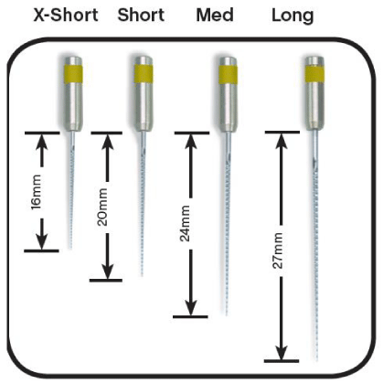
Shaping files consists of four stainless steel instruments with thin, flexible, non-cutting tips. The standard series comprises of three instruments & an additional 4th file (auxiliary) designed to instrument calcified and/or curved canals. (Table 1) (Figure 1).
The blades of the instrument extend throughout its length from the tip almost up to the handle so it can cover the coronal portion of the operating canal. The flexible tip of the instrument is rounded. Shaping files are available in lengths of X-short - 16 mm, Short -20 mm, Medium - 24 mm and Long -27 mm.
Apical files
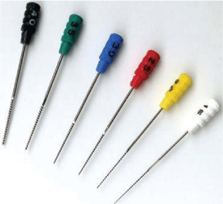
Apical files are available in lengths of 19, 23, 27 and 30 mm; they have an active part 12mm in length. Apical files have tip diameters of 0.15-0.30mm and tapers 2.0% (sizes 15,20 and 25) and 2.5% (size 30), respectively (Figure 2).
Endo-Eze AET shaping files are used in a special reciprocating/ oscillating handpiece for instrumentation of the coronal part of the root canal about 3 mm short of the apex. The apical files are hand files with shortened cutting flutes to cut only in the apical region of the canal and are used in a clockwise turn and pull motion.
Endo-Eze hand piece
The Endo-Eze handpiece is a dedicated handpiece for the Shaping files with a 30 º right 30º left reciprocating action. It is possible to vary the insertion depth of the instrument within the head of the handpiece, via a push button collett. Minimal (only 30º) reciprocating motion facilitates rapid, complete, uniform instrumentation of all the walls in an irregularly shaped canal. It has “milling” action rather than the “drilling” action of traditional rotary systems [10] (Figure 3).
Operative procedure
Coronal Phase: In this phase, access opening is done and canal orifices are identified. Round and tapered diamond are used to prepare the access cavity; Non end-cutting bur is used to remove the roof of the chamber without damaging the pulpal floor and Safe-point diamond bur is used to refine the walls without dentine interferences.
Coronal-middle Phase: In this phase, middle third of canal is prepared along with refinement of coronal third of root canal. For this , 4 Shaping files S1,Sc,S2,S3 and Endo Eze hand piece is used.
Methodology: Tooth length is determined on diagnostic radiograph; 3mm is subtracted from the obtained measurement. Ex: tooth length in the x-ray = 21 mm - 3 mm = 18 mm.
Shaping 1 (yellow) oscillating instrument is used to shape the canal to previously determined length, and then Shaping 2 (blue), Shaping 3 (green) is used. The Shaping 1 (yellow) oscillating instrument is inserted manually into the root canal to determine the working length (WL) then it is attached to the handpiece for the shaping of the canal till working length.
Apical phase
Safe NiTi hand instruments are used to achieve the goal of preserving apical curvature and respecting anatomy. Apical Working Length (AWL) is determined by subtracting 0.5 mm from the electronically determined canal length. In most canals, having completed preparation with Shaping files, the size 25 hand apical NiTi used in manual rotary motion will easily reach the AWL. If this is not possible, use stainless steel apical files with diameters ranging from size 08 up to size 20 and with ¼ turn and withdrawal movements until reaching the AWL with the size 25 Apical NiTi. Continue with the Apical NiTi (size 30, 35, 40, etc.) until reaching the final Diameter of Apical Preparation (DAP); Ethylenediamine tetracetic acid (EDTA) and sodium hypochlorite (NaOCl) should be used as irrigants after each instrumentation.
Advantages
• Reciprocating contra-angle greatly reduces the risk of file separation
• Relies on a minimally invasive approach
• Addresses each canal according to its unique anatomy
• Follow the natural canal shape
• Prepares irregular shaped canals less aggressively in comparison with nickel-titanium rotary motion systems.
• Minimize apical transportation and ledging
• Results in a simplified procedure
• More affordable than other systems
• Can be hybridized with current rotary techniques and systems [11].
Conclusion
Anatomic Endodontic Technology is an innovative concept of mechanical root canal preparation. It is an oscillating (reciprocating) instrumentation system, which uses flexible stainless steel instruments driven by the 4:1 reduction Endo-Eze AET contra-angle handpiece. It offers simplicity, reliability and profitability and promotes better results with fewer risks regardless of anatomical variations of tooth system.
- O Zmener (2005) Effectiveness in cleaning oval-shaped root canals using Anatomic Endodontic Technology, ProFile and manual instrumentation: a scanning electron microscopic study. Int Endod J 38: 356-363. Link: https://bit.ly/2XVDcW2
- Latrou A (1980) Abrege d’Anatomie Dentaire. Paris, France: Masson. Link: https://bit.ly/2XR6tkH
- Kerekes K, Tronstad L (1977a) Morphometric observations on root canals of human premolars. J Endod 3: 24–29. Link: https://bit.ly/3gLUQUQ
- Kerekes K, Tronstad L (1977b) Morphometric observations on root canals of human molars. J Endod 3: 74–79. Link: https://bit.ly/3gHtYoZ
- Kerekes K, Tronstad L (1977c) Morphometric observations on root canals of human anterior teeth. J Endod 3: 114–118. Link: https://bit.ly/2ZWAQsM
- Wu M, R’oris A, Barkis D, Wesselink PR (2000) Prevalence and extent of long oval canals in the apical third. Oral Surg Oral Med Oral Pathol Oral Radiol 89: 739-743. Link: https://bit.ly/2XoKliT
- Wu MK, van der Sluis LW, Wesselink PR (2003) The capability of two hand instrumentation techniques to remove the inner layer of dentine in oval canals. Int Endod J 36: 218-224. Link: https://bit.ly/3dnKwR2
- Barbizam JVB, Fariniuk LF, Marchesan MA, Pecora JD, Sousa-Neto MD (2002) Effectiveness of manual and rotary instrumentation techniques for cleaning flattened root canals. J Endod 28: 365-366. Link: https://bit.ly/3eJasqe
- Abou-Rass M, Frank AL, Glick DH (1980) The anticurvature filing method to prepare the curved root canal. J Am Dent Assoc 101: 792-794. Link: https://bit.ly/2U0NOC9
- Fischer D (2005) Root canal preparation with Endo-Eze AET: changes in root canal shape assessed by micro-computed tomography. Int Endod J 38: 456-464. Link: https://bit.ly/3ck4hHK
- Riitano F (2005) Anatomic Endodontic Technology (AET)-A crown-down root canal preparation technique: basic concepts, operative procedure and instruments. Int Endod J 38: 575-587. Link: https://bit.ly/3eHb3ZH
Article Alerts
Subscribe to our articles alerts and stay tuned.
 This work is licensed under a Creative Commons Attribution 4.0 International License.


 Save to Mendeley
