Journal of Dental Problems and Solutions
Temporomandibular Joint Movements during Rigid Bronchoscopy and Laryngoscopy under General Anesthesia and Pre-Post Intervention Comparisons
Heinrich D Becker1*, Karl-Ludwig Sattler1,2 and Rudolf Slavicek2*
2Center for Interdisciplinary Dentistry, Danube University, Krems, Austria
Cite this as
Becker HD, Sattler KL, Slavicek R (2020) Temporomandibular Joint Movements during Rigid Bronchoscopy and Laryngoscopy under General Anesthesia and Pre-Post Intervention Comparisons. J Dent Probl Solut 7(1): 024-029. DOI: 10.17352/2394-8418.000081Recently rigid bronchoscopy with metal tubes has gained increasing importance, namely for interventional procedures. The objective was to examine condyle movement during rigid bronchoscopy under general anesthesia. A method to record the tracings was developed using the CADIAX III system (GAMMA AG, Klosterneuburg, Austria). The purpose of our efforts was to find out whether rigid bronchoscopy harms the temporomandibular joint. To this end, we recorded mandible movements before, during and after operation. We also conducted a brief comparison between bronchoscopy and laryngoscopy. The majority of Heidelberg Thoraxklinik patients have a past or present history of lung cancer and as such were under considerable mental stress; this extraordinary mental situation was a factor. Extreme movements were recorded, with motion probably limited by the ligaments. We found no evidence of harm to the temporomandibular joint system.
Introduction
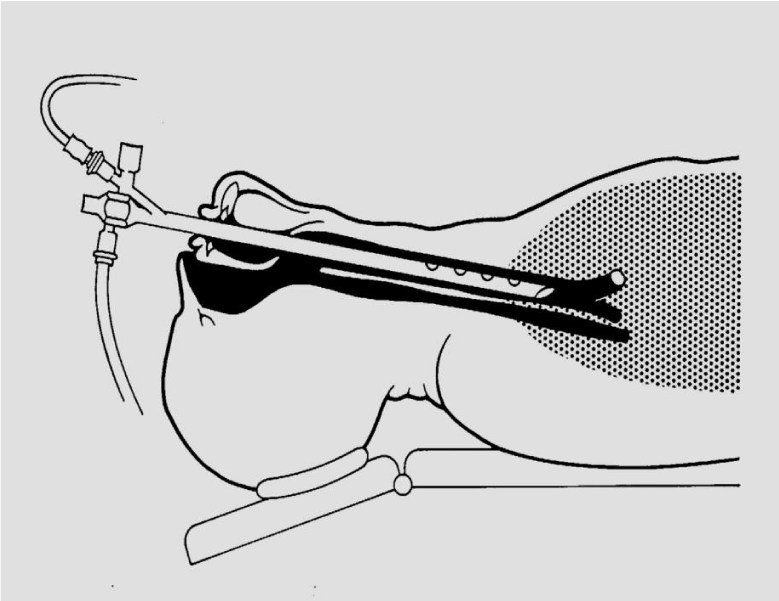
Rigid bronchoscopy, introduced by G. Killian in 1898, prevailed until it became largely replaced by the flexible fiber bronchoscope introduced by S. Ikeda in 1966. In recent decades it experienced a renaissance as new interventional techniques require general anesthesia with safe ventilation and sufficient access for instrumentation. In contrast to intubation via laryngoscopy where the floor of the mouth is lifted by inserting the scope above the epiglottis, the rigid bronchoscope is introduced by lifting the base of the tongue and the epiglottis with the scope and entering the larynx (Figure 1). In both procedures the temporomandibular joint undergoes a considerable passive rotational and translational forward movement.
The temporomandibular joint (TMJ) is unique in the human musculoskeletal system and different in its phylogenetic origin. Quadratum and mandibulare (Meckel´s cartilage) derive from the first visceral arch. Between them the primary TMJ is positioned, which is converted into the hammer-anvil-joint. The bigger, frontal part of Meckel´s cartilage delivers the mandible. Between mandible and skull a new joint is formed. This secondary joint is to be found in mammals only.
Anatomy and physiology of the temporomandibular joint
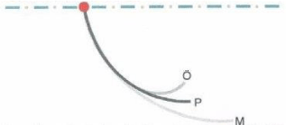
The short and pertinent definition of the temporomandibular joint presented by the Viennese anatomist Harry Sicher still applies: The temporomandibular joint is a synovial gliding joint with a flexible socket [1]. The bony parts are the fossa and eminentia articulare on the temporal bone and the caput mandibulae (Figure 2). These bony parts are completed by the articular disc located on top of the caput mandibulae and the extensive articular capsule surrounding the system. The articular disk separates an upper from a lower compartment. Clearly distinguishable ligamentary structures are embedded in the capsule which form rational boundaries of terminal positions [2]. The activity of the temporomandibular muscles is required for guiding and positioning the joint. This function was demonstrated inter alia in 1973 by the "group of four" led by R. Slavicek. Infusion of curare to conscious subjects caused a slackening and downward motion of the mandible [3].
Temporomandibular joint and anesthesia
A free range of movement of the temporomandibular joint is a basic requirement for intubation by laryngoscope and rigid bronchoscope tube. At the same time, the intubation process or insertion of the bronchoscope may harbor a hidden risk. Wide opening of the mouth starts with rotation, followed by protrusion (Figure 2). It is hard for the operator to identify when the patient's joint has reached a rotational or translational borderline position and when a risk of imposing excessive strain on soft tissue structures begins. According to Piehslinger, et al. the angle of maximum rotation is 7.15° in the reference position and 35° with the patient's mouth wide open (highest reading) [4]. From a dental surgery point of view, it is essential that the anesthetist should establish the range of mandibular movement when examining a patient by assessing the visibility of the palate and uvula (Malampati score). This at least gives him or her a rough idea whether any restrictions are present [5,6]. There are very many possible reasons for an inability to open the mouth to its fullest extent, but a description would be beyond the scope of this study.
Method for Measuring the Movements of the Temporomandibular Joint
Mechanisms to record lower jaw movement meanwhile are common in dentistry. Before the introduction of electronic registration systems, mechanical devices were long in use. However, as the motion oft he condyles is hardly more than 10mm, for interpretation one needs an 8x magnifying glass. With the Helkimo-Index the maximum opening can be approximately assessed by the distance of the cuting edges of the teeth. However measuring of lateral movements and protrusion can not be precisely measured, bacause this and other methods ar static. Electronic movement registration is user independent and creates an objective storable image.
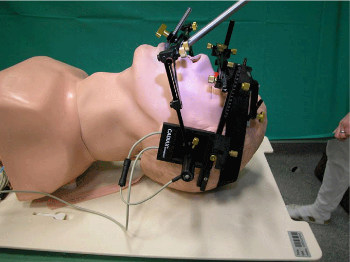
The CADIAX III system (GAMMA AG, Klosterneuburg, Austria) allows the user to present translation and rotation separately and in temporal progression. To establish the basic feasability of the project, a preliminary test was performed in a dummy (Figure 3,4).
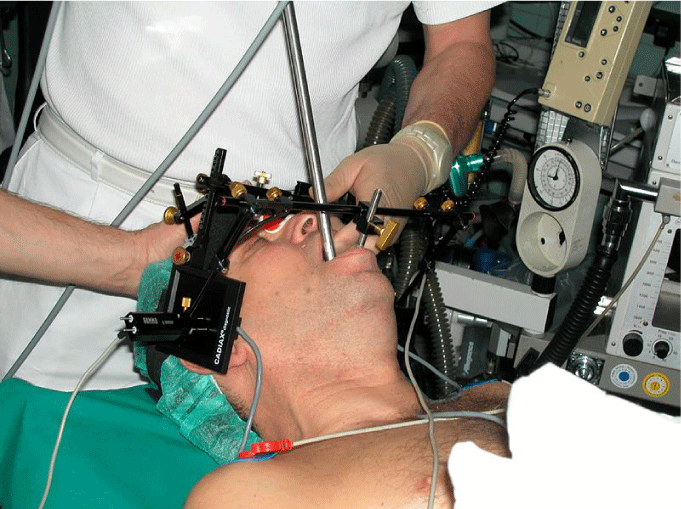
1. Upper arch, fixed with ear olives, nose support and elastic band around head, with electronic pads, on which the movements of the mandible are recorded
2. A mandible clutch, fixed on the lower teeth
3. Lower arch, parallel to the upper arch and fixed to the clutch
4. The styli fixed by arms to the lower arch. They record the movement of the mandible on the electronic pads in translational and transversal direction (insert)
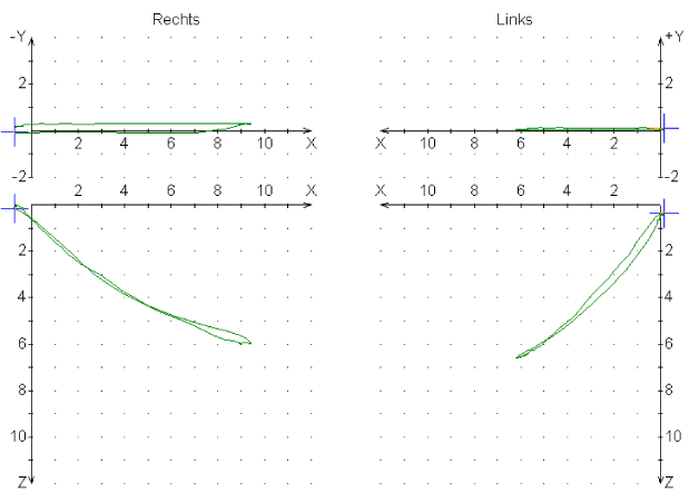
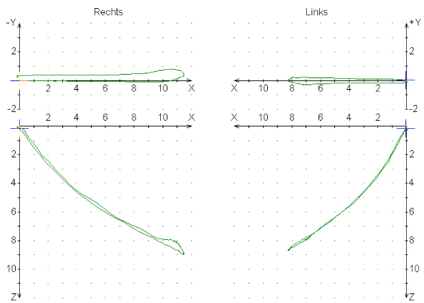
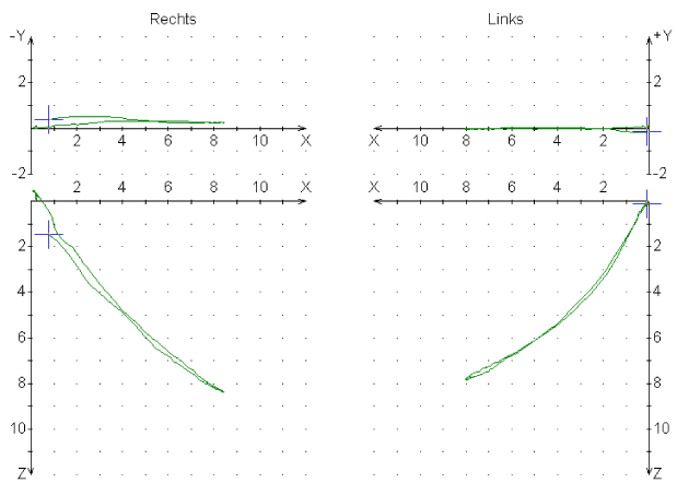
An uncompromised function curve of the temporomandibular joint shows concavity, is long enough, excursion and incursion are identical, both sides are equal and there is no side shift.
In an anesthesized and relaxated patient it is possible that by laryngoscopic intubation or insertion of a rigid bronchoscope passive stretching of the capsule and ligaments may cause serious damage to the joint. After stretching of the ligaments, in extreme movements even luxation may occur. The aim of the study was to investigate, whether rigid bronchoscopy and laryngoscopy are causing extreme excursions of the joint and may lead to persisting trauma Table 1.
Answers were sought to the following questions:
- What movements take place in the temporomandibular system during rigid bronchoscopy?
- Are these movements within physiologically normal limits?
- Is there a correlation between the anesthetic process (anesthesia record, degree of relaxation) and the curves traced?
- What is the ratio of translational to rotational components?
- Does a before and after comparison suggest that irritation or trauma has occurred?
Patients and Procedures
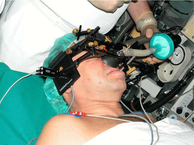
Fifteen patients took part in the study. We selected patients over 18y from the Thoraxklinik who gave their informed consent. Patients were eligible who understood the procedure and had sufficient dentition in their lower jaw to allow a paraocclusal clutch to be put in place. A paraocclusal clutch is an adjustable metal tray with a synthetic matrix that is affixed laterally to the teeth of the lower jaw such that the upper and lower teeth almost touch, whilst at the same time allowing lower jaw movement to be plotted on a recording system. No other inclusion or exclusion criteria were implemented. After clinical examination, especially with regard to the denture, a first recording was performed on the awake patient in the preparation room. After transfer to the bronchoscopy suite and induction of general anesthesia, preoxygenation was performed via nasal mask with the lower arch elevated (Figure 5). After sufficient ventilation the lower arch was attached to the dental clutch and the bronchoscope was introduced (Figure 6). This was not always easy via the midline due to the volume of the CADIAX III system or due to limitations of the mouth opening. Then, the bronchoscope was introduced via the side of the mouth and sometimes it needed several attempts to lift the epiglottis and enter the larynx. After conclusion of the bronchoscopy the patient was transferred to the recovery room and after fully awakening a repeat recording was performed. Pre-, intra- and post-procedural recordings were analyzed and compared.
Results
Comparison of condylography tracings before and after bronchoscopy was possible in 12 cases. The condyle trajectories during the bronchoscopy we observed were extremely wide in some instances. However, it is important to note that there were no signs of dislocation or overextension at follow-up in any patient. Review of the data showed that four of the bronchoscopy tracings describe a curve that approximates condylar movement along the eminence. The rotation component exceeds 50% in all these cases. Almost all the curves traced during bronchoscopy are within ranges that might be seen in association with continued conscious action. In contrast, the two laryngoscopy cases produced extreme trajectories with a significantly greater translation component. It is possible that the width of the joint capsule imposes limits on condylar excursion. Nine of 12 comparable cases show post-intervention curves which are definitely more physiological than the pre-intervention tracings. The frequency of the phenomena is striking but any interpretation could only be speculative. For statistical analysis the number is too low. Neither in bronchoscopy nor in Laryngoscopy we detected resultinge damages of the joint. The smoothening of the condylography curves may be due to anesthesia, which temporarily eradicates trained avoiding effects.
Pardigmatic recording samples
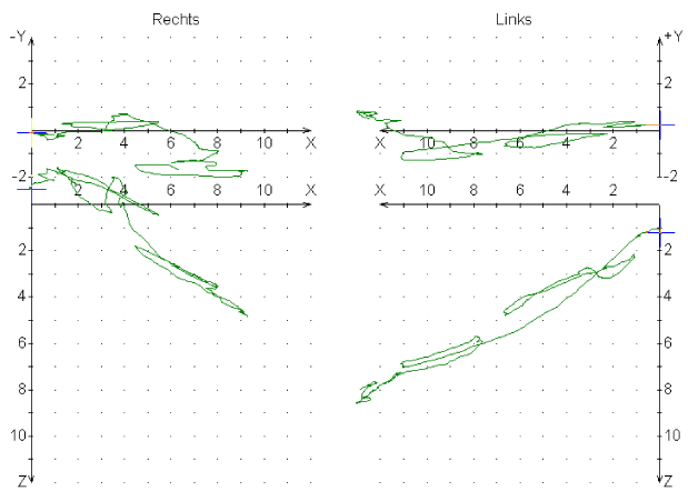
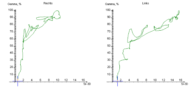
Case 3: R. K.-J. (*March 3, 1948)
D: Male patient looking older than stated age, ill and poorly nourished appearance, diabetes, obstructive pulmonary disease
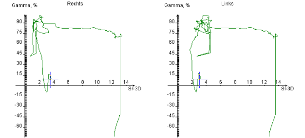
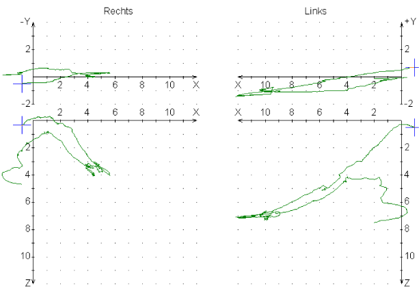
prior cases. The anesthesia record gives no indication of unusual events in the waking phase Graphs 1-3.
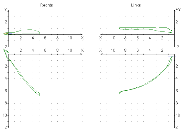
Case 5: N.D. (*October 12, 1949)
D: Unspecified neoplasm in the right main bronchus. Normal overall appearance, slightly reduced nutritional condition Graphs 4-7.
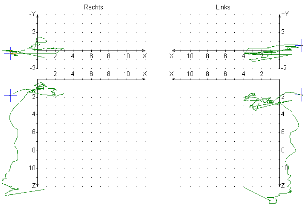
In the next case the curves were traced during laryngoscopy. The purpose of this experiment was to identify any differences between bronchoscopy and laryngoscopy. Therefore, no before-after comparison was attempted. For laryngoscopy, the anesthetist advances the instrument to the base of the tongue and raises the floor of the mouth to allow inspection of the larynx. Accordingly, the condylar trajectories produced are likely to be different from those seen in association with bronchoscopy. The condylography equipment was set up in the following cases in a manner identical to that already described.
Case 15: K.F. (*March 19, 1934)
D: Metastatic carcinoma of the thymus with multiple obstruction of the bronchial tree. Acute dyspnea Graphs 8-10.
Results
Statistical analysis needs to produce results that can be generalized. To this end levels of statistical significance need to be defined prior to initiating a study. That was not the intention of the study described in this paper, and in any case would not have been possible in such a small patient population. It made sense, nevertheless, to conduct a case comparison, as the identification of a trend may provide a basis for future statistical analysis in a larger study. The variation of tracings that we found in our patients is very wide compared to normal mandibular movements. The movements traced during rigid bronchoscopy and laryngoscopy are passive as they are induced by the instrumentation under relaxation. In bronchoscopy only the base of the tounge is lifted, whereas in laryngoscopy the whole floor of the mouth is elevated. Thus, the capsule of the joint is not controlled by any muscular activity. Rigid bronchoscopy is associated with a definite potential risk of injury to the temporomandibular joint, but in our study, we found no persistant effect. In contrast the majority of patients show post-intervention curves which are definitely more physiological than the pre-intervention tracings. A lasting relaxation effect of the drugs used could be excluded based on pharmacokinetics and half-lives. The frequency of the phenomenon is striking. As mentioned above, most patients were under psychologial tension before bronchoscopy, either because of the intervention itself or of fearing the results. It is well known that such stresss can have significant effects on the mastication system (“clenching one’s teeth“) Probably the relief of anxiety afterwards and some lingering anxiolytic action had an effect. In order to minimize the risks for the temporomandibular joint in rigid bronchoscopy, it is absolutely crucial to ensure that the patient has achieved a deep enough degree of anesthesia. In addition, the bronchoscopist must have sufficient skills and acquired enough technical expertise in performing the procedure. If these criteria are met, iatrogenic damage of the temporomandibular joint in association with bronchoscopy is highly unlikely.
- Sicher H (1929) On the mechanics of the temporomandibular joint. Stomatol 27: 27-33.
- Bauer A, Gutowski A (1984) Gnathologie.
- Slavicek R (2002) Oral communication.
- Piehslinger E, Celar RM, Horejs T, Slavicek R (1993) Orthopedic jaw movement observations. Part II: The rotational capacity of the mandible. Cranio 11: 206-210. Link: https://bit.ly/35ckGw9
- Aiello G, Metcalf I (1992) Anesthetic implications of temporomandibular joint disease. Can J Anaesth 39: 610-616. Link: https://bit.ly/2yEXJ8B
- Bellmann MH, Babu KVR (1978) Jaw dislocation during anesthesia. Anesthesia 33: 844.
- Kowalski K (1960) Endotracheal anesthesia and trauma incident to endotracheal intubation. Oral Surg 13: 543-551. Link: https://bit.ly/2VzvQYE
- Rosenberg H (1987) Trismus is not trivial. Anesthesiology 64: 453-455. Link: https://bit.ly/2S4m66f
- Schwartz L, Rockoff MA, Koka BV (1984) Masseter spasm with anesthesia. Incidence and implications. Anesthesiology 61: 772-775. Link : https://bit.ly/3bC17zx
- Liu YH, Wang JJ, Chang CF, Jin CH (2000) Difficult tracheal intubation as a result of unsuspected abnormality of the temporomandibular joint. Anesth Analg 92: 783-784. Link: https://bit.ly/2VzJryM
- Becker HD (1990) Atlas of bronchoscopy. Stuttgart
- Gysi A (1916) The new simple Simplex articulator Schw. F. f. ZHK H3.
- Slavicek R, Mack H (1979) Functional analysis measures in the current. System Dentist practice.
- Klett R (1982) Electronic registration procedure for TMJ diagnostics German Dentist Z 37: 991.
- Kett R (1989) Computer-aided registration diagnostics with the String-Condylocomp LR2, ZWR 98: 976.
- Meyer G (1986) Development and application of an electronic procedure for the three-dimensional hinge axis-related registration of lower jaw movements for the functional diagnosis of the stomatognathic system. Med Habil Göttingen.
- Slavicek R (1988) Clinical and instrumental functional analysis for diagnosis and treatment planning. Part 7. Computer- aided axiography. J Clin Orthod 22: 776-787. Link: https://bit.ly/2XXV44p
- Lipp MDW (1993) Studies on the motility of the temporomandibular joints in humans during oral and nasal laryngoscopic intubation and their importance for post-operative disorders of joint function. Med Habil Mainz.
- Dick W (1992) Emergency and Intensive Care Medicine de Gruyter Verlag Berlin-New York.
- Grange Cs (1993) Intubation with propofol Europ. J Anesthesiol 10: 9.
- Larsen R (1987) Anesthesia Urban and Schwarzenberg Verlag Munich.
- Lipp M, von Domarus H, Daubländer M, Leyser KH, Dick W (1987) Effects of intubation anesthesia on the temporomandibular joint. Anaesthesist 36: 442-445. Link: https://bit.ly/2VvOXCC
- Lipp M1, Daubländer M, Ellmauer ST, von Domarus H, Stauber A, et al. (1988) Changes in temporomandibular joint functions in various general anesthesia procedures. Anaesthesist 37: 366-373. Link: https://bit.ly/2Vx1BkQ
- Donlon JV, Newfield P, Sreter F, Ryan JF (1978) Implications of masseter spasm after succinylcholine. Anesthesiology 49: 298-301. Link: https://bit.ly/2x1moUa
Article Alerts
Subscribe to our articles alerts and stay tuned.
 This work is licensed under a Creative Commons Attribution 4.0 International License.



 Save to Mendeley
