Journal of Dental Problems and Solutions
Influence of three different dentin bonding agents on the adhesion of composite resin to dentine–An in vitro study
Christina Hoferichter, Stefan Rüttermann, and Susanne Gerhardt-Szep*
Cite this as
Hoferichter C, Rüttermann S, Szep SG (2019) Influence of three different dentin bonding agents on the adhesion of composite resin to dentine–An in vitro study. J Dent Probl Solut 6(2): 056-060. DOI: 10.17352/2394-8418.000074The aim of this randomized in vitro study is to clarify if there are differences between the dentin adhesives as well as their adhesion and fracture behaviour. The study design was based on the ISO standard TS 11405 (2003). For this purpose, ninety extracted, caries-free human molars were embedded in a plastic block, and the dentinal surfaces were dissected free. Teeth were divided into three groups (n = 30) and each treated with a different adhesive system (SYC: Syntac Classic; CSE: Clearfil SE, CS3: Clearfil S3). All trial teeth were coated with a cylindrical standardized test specimen (diameter: 3.0 mm, height: 3.0 mm) on buccal surfaces with an ISOconforming application aid. The aging of the samples was achieved by thermocycling (500 cycles). Thereafter, the adhesive forces (MPa) of the various dentin adhesive systems were determined by means of shear bond testing with a universal testing machine from Zwick. The samples were examined under a scanning electron microscope at 20x and 2000x magnification for their fracture modes (adhesive, cohesive, mixed). Furthermore, parametric Weibull regression models were applied to evaluate whether there was a significant association between shear bond strength and the used adhesive system. Weibull analysis was performed using R software (version 2.11.1, R Foundation for Statistical Computing, Vienna, Austria). Differences between groups were tested using Wald tests, and p-values below 0.05 were considered significant.
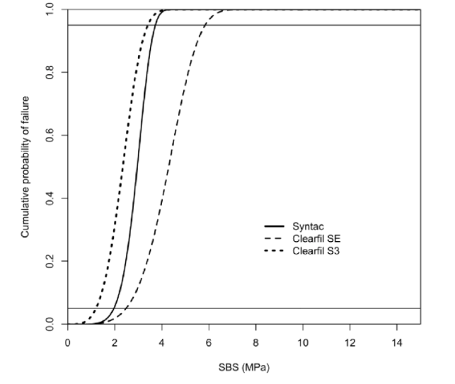
CSE (3.97 MPa), followed by SYC (3.02 MPa), and CS3 (2.16 MPa) had the highest bond strength averages. The result of the statistical evaluation shows statistical differences between the different groups. Mostly mixed fractures were observed. SYC (m = 6.35) was most reliably followed by CSE (m = 4.75). The lowest Weibull module scored CS3 (m = 3.91).
Under the limitations of this study, it can be stated that the differences between the groups were significant and the One-Step Self-Etch-system (CS3) showed the lowest values. Additionally one major problem was the acrylic application aid (split mould according to ISO/TS 11405), which may have transferred adverse forces to the specimen and which cannot be recommended for further studies.
Introduction
The demand for aesthetic, durable and quality restorations has increased significantly. Teeth should be unobtrusive and ideally minimally invasive reconstructed. It is a problem to get a permanent bond between the hydrophilic dentin and the hydrophobic composite [1,2]. Dentin adhesives play a key role in this [3-6]. In order to have feedback on the bond between composite materials and hard tooth substance, in vitro scientific studies are carried out. The experimental setups differ in many parameters in the studies and are therefore often not comparable. This variance variety influences the results [5]. Standardization of the parameters as in the ISO paper [4], is necessary to obtain comparable results. To our knowledge, no previous study has investigated the shear bond strength of the adhesive interface created by conventional Four-Step Etch-and Rinse and Self-etching Adhesives under the ISO/TS 11405 protocol.
The aim of this study was to compare three different dentin adhesives (control group: Four-Step Etch-and-Rinse versus experimental groups: Two-Step Self-etching and One-Step Self-etching) conforming to ISO (ISO TS11405: 2003) in terms of adhesive strength and fracture behaviour [5,6].
The following research questions interested us:
1. Is there a difference between the adhesion values obtained by the control group (Four-Step Etch-and-Rinse) on the one hand and the experimental groups on the other?
2. Are there differences between the fractures of the control group (Four-Step Etch-and-Rinse) and those of the experimental groups?
Materials and Methods
Specimen preparation based on the ISO specification
One hundred and twenty extracted human molars were completely embedded in a cold polymer (Technovit 4004, Kulzer, Hanau, Germany). All teeth were extracted for not longer than six months, were caries-free, unrestored and not root filled. After embedding, the numbering of the teeth was done to allow an exact assignment. Subsequently, the teeth were freed on the buccal surface except for the dentin. For this they were hand-worked on a polishing unit (Jean Wirz, Düsseldorf, Germany) with grinding wheels of different grain sizes (320-600 grit) at 6000 rpm with constant water cooling against overheating. The ISO standard TS11405 (2003) [4], requires a dentinal surface to be finished with a 600 grit abrasive paper. The teeth were rotated during the entire grinding process to achieve a uniformly ground surface. The plane parallelism was checked with a spirit level (Mercateo, Köthen, Germany). The samples were stored in distilled water at room temperature all the time until the experiment, which was changed every three days. The freedom from melting of the test surfaces was checked under a microscope (Wild Heerbrugg, Heerbrugg, Germany). It was also checked that there was no opening of the pulp and caries freedom. Teeth with these criteria were discarded. The number of teeth was reduced to ninety. These molars were randomized to three groups.
Restoration materials and application aid
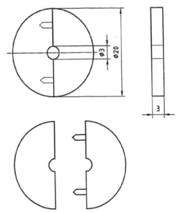
The materials used are listed in Table 1. The different groups were treated with the appropriate adhesive. The manufacturer's recommendations for the individual adhesives were strictly followed. The application of the individual components per adhesive was done with a disposable brush, which was discarded after each application. The required exposure times were monitored by means of a stopwatch. The Syntac Classic group (SYC), which includes three components, initially applied Syntac Prime for 15 seconds. Surpluses were then blown and Syntac Adhesive was applied for 10 seconds. Surpluses were again blown and the order was made by Heliobond. Thereafter, polymerization was carried out with a polymerization lamp (Elipar Trilight, 3M ESPE, Seefeld, Germany) for 10 seconds. The Clearfil SE (CSE) group, a two-component adhesive, initially applied Cleafil Primer was allowed to act for 20 seconds and then excesses were fused. Clearfil Bond was afterwards polymerized for 10 seconds. The Clearfil S3 (CS3) group applied the bonding for 20 seconds. Thereafter, the excesses were removed with oil-free air for at least 5 seconds. The mixture was then polymerized for 10 seconds. The test specimens were transferred from a light-curing composite to the test area using an ISO-compliant application aid (Figure 1) [4]. This consisted of a transparent plastic and was constructed by the Technical University (TU) in Darmstadt (Germany). The design allowed an exact size of the adhesive surface (7,069mm2). The application aid consisted of two parts which could be opened via a screw thread by means of a hexagon. The application aid was isolated against the composite probe for five seconds with a release wax (Glorex, Füllingsdorf, Germany). The composite probe was filled in 2mm layers and cured for 20 seconds. After each operation, the samples were stored in distilled water. Subsequently, all samples were subjected to a thermal fatigue process by cyclic thermal cycling. This process took place over 500 cycles (Haake Technik GmbH, Wehrheim, Germany) in 5 and 55 °C water baths. The residence time per water bath was 30 seconds, the transfer time between the baths was 10 seconds.Methodology of shear bond strength and fracture mode
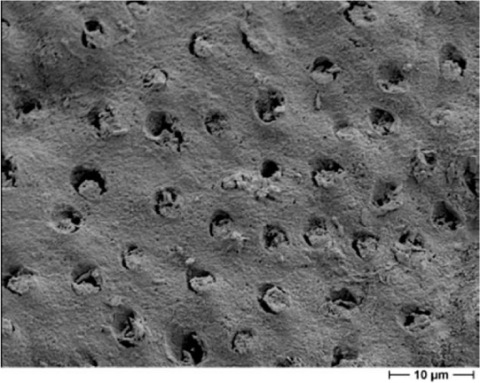
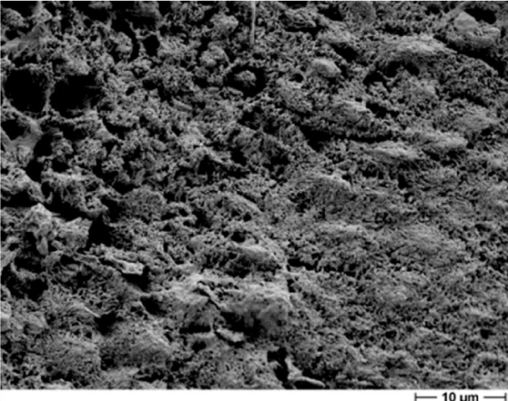
The ISO standard TS11405 (2003) [4], allows different test methods for measurement of bond strength. In this study, we chose shear bond testing. One sample each was clamped vertically into the universal testing machine (Zwick, Ulm, Germany). A shear blade moved at a constant speed (0.5 mm/min) toward the composite cylinder until the composite cylinder broke off from the dentin. Force transmitted to the load cell during the test was recorded by a computer and converted to voltage by the testXpert software (ZwickRoell, Ulm, Germany). A voltage drop of more than 30% was rated as a fraction of the sample and the maximum voltage measured so far registered as shear bond strength. The shear force was determined in Newton, which was converted to MPa for standardization. After the adhesion force measurement, the samples for the determination of the fracture type were evaluated by scanning electron microscopy (Hitachi, Tokyo) at 20-fold and 2000-fold magnification. The fracture types were differentiated into cohesive, adhesive and mixed fractures with cohesive fraction above or below 10%. Samples with a cohesive or mixed fraction exceeding 10% cohesive were not included in the statistical evaluation. Since only the other forms of fracture provide information about the bond [5].
Statistics
For the statistical analysis of the results, the Kruskal-Wallis Multiple-Comparison Z-Value Test with a significance level of p ≤ 0.05 was applied. The test was performed using the statistical analysis package NCSS (Version 6.0.2.1. Kaysville, Utah). The strength values of the samples were evaluated by a Weibull distribution.
Weibull analysis was performed using R software (version 2.11.1, R Foundation for Statistical Computing, Vienna, Austria). Differences between groups were tested using Wald tests, and p-values below 0.05 were considered significant.
Results
The individual results are shown in Tables 2-4. Most fractions are mixed forms with up to 80%. With the exception of CS3, there were otherwise more adhesive fractures (Table 2, Figures 2-4). CSE (3.97 MPa), followed by SYC (3.02 MPa), and CS3 (2.16 MPa) had the highest bond strength averages (Table 3). Significant differences were evaluated between SYC and CSE and between CSE and CS3 (Table 3). SYC (6.35) was most reliable followed by CSE (4.75). The lowest Weibull module scored ClS3 (3.91), (Table 4, Figure 5).
Discussion
To date, no publications based on ISO TS 11405 2003 [4], have been published according to the investigated adhesives in our study. However, there exist many studies on the adhesive strength in the literature, and the dispersion of the results between different dentine adhesives is very large (Table 5). There are some studies that have achieved higher adhesive forces than our study [7-15], others reported from comparable [16-18], some also showed lower values (Table 5) [19-21]. These differences are mostly related to different study designs.
Since 2015 there exists a new revised specification of the ISO [22], which is unchanged in most points compared with the 2003 version. However, it seems be clear that it is not the absolute value of bond strengths that are decisive, but the relation between the investigated groups. As stated, it may be more appropriate to compare the ranking of materials [22]. It is also known, that while bond strength cannot predict exact clinical behaviour, they may be useful for batch quality control [4], or for comparing adhesive materials [22].
In some circumstances, bond strength tests are only useful for screening. They may allow only rough guidance with respect to the clinical performance of an adhesive system. Low values are more likely correlated with poor clinical performance namely retention in adhesive cavities. However, bond strength values above a certain threshold value might not indicate better clinical performance. This value is unfortunately not further clarified in the actual ISO version [22].
Many studies have found similar relations between the different types of adhesives as in the present study [8,10,13,15]. Cause of the differences may be the origin of teeth [23,24] bovine [8,13] or human even in human selection of areas [11,14,15]. It may cause differences in the preparation of the samples [11,14], whole teeth are embedded, the pulp chamber is blocked out [11-13,15], only toothed discs are used. An important point is also the influence of the application aids [10,12,16,17] and the choice of the aging process [14,18], for example only storage in water for days or months as well as thermocycling [8,10,15,17]. The methods of determining the adhesion [5,15,25,26] may differ even all in choice of the debonding speed [18,19,25]. In the review by Scherrer [5] 147 studies with six different adhesives were summarized. Depending on the test method used, the results of the 147 studies showed a very broad variation of the bond strength values, with the shear strength of the micro tensile strength test producing the higher adhesion values [5,15]. Many studies do not take into account the fracture modes [5,15-17], which are critical in determining shear bond strength and the inclusion of cohesive fractures may also influence the results. In the study by Makowski, a correlation was found between the occurrence of low adhesion values and adhesive fractures as well as high adhesion values and mixed fractures [11]. In that study, however, not the mixed fractions, as required by Scherrer, were subdivided again [5].
Most experimental values are not clinically relevant because they do not allow any information to the material reliability. Therefore, a Weibull analysis is strictly recommended [5,22], which was also calculated in our study. A major problem however was probably the recommended ISO application aid, which may have transferred adverse forces on the specimen (shear, bend or rotation). This split mould can be formed of polytetrafluoroethylene (PTFE) or other suitable materials, as stated in the ISO [5]. Our experiences with an acrylic mould material however cannot be recommended for further studies. Interestingly this split mould is no longer described in the actual ISO-version [22].
Conclusion
The differences between the groups were partly significant and the One-Step Self-Etch dentin adhesive system showed the lowest values. These were however not significantly different to the values of the Four-Step Etchand-Rinse adhesive. The Two-Step Self-Etching system has achieved the best results in both adhesion and reliability. Furthermore, the acrylic split mould according to ISO/TS 11405 cannot be recommended for further studies.
- Bounocore MA (1955) Simply method of increasing the adhesion of acrylic filling materials to enamel surfaces. J Dent Res 34. 849-853. Link: https://tinyurl.com/yxp4jwh4
- Schmalz G (2002) Materials science: Biological aspects. J Dent Res 81: 660-663. Link: https://tinyurl.com/yxnw4o8a
- Lutz F, Krejci I, Schüpbach P (1993) Adhesive systems for tooth-colored restorations. A review. Schweiz Monatsschr Zahnmed 5: 537-549. Link: https://tinyurl.com/y43ar7a8
- Technical Specification ISO/TS 11405: 2003 (E), Dental materials – Testing of adhesion to tooth structure. Geneva, Switzerland. Link: https://tinyurl.com/y4tr9eb7
- Scherrer S, Cesar PF, Swain MV (2010) Direct comparison of the bond strength results of the different test methods: A critical literature review. Dental Mater 26: e78-e93. Link: https://tinyurl.com/y4f6mkzg
- Olio G (1993) Bond strength testing - what does ist mean? Int Dent J 43: 492-498. Link: https://tinyurl.com/yybnm3mj
- Ansar AA (2004) An evaluation of strength of composite resin restoration using different bonding agents- A In-vitro study. J Indian Soc Pedod Prev Dent 4: 162-167. Link: https://tinyurl.com/y45kje25
- Ishii T, Ohara N, Oshima A, Koizumi H, Nakazawa M, et al. (2008) Bond Strength to bovine dentin of a composite core build-up material combined with four different bonding agents. J Oral Sci 50: 329-333. Link: https://tinyurl.com/yya4v67a
- Khursan T (2011) Basic physical data of a newly developed composite material. Erlangen Nürnberg: University.
- Latta MA (2007). Shear Bond Strength and Physiochemical Interactions of XP Bond. J Adhes Dent 9: 245-248 Link: https://tinyurl.com/y37opsug
- Makowski M (2011) Comparison of the micro-adhesion of five dentin adhesion promoters in combination with flowable composites - an in vitro study. Halle-Wittenberg Martin Luther University.
- Sadr A, Ghasemi A, Shimada Y, Tagami J (2007) Effects of storage time and temperature on the properties two self-etching systems. J Dent 35: 218-225. Link: https://tinyurl.com/y3vptnxm
- Tamura Y, Tsubota K, Otsuka E, Endo H, Tabubo C, et al. (2011) Dentin bonding: influence of bonded surface area and crosshead speed on bond strength. Dent Mater J 30: 206-211. Link: https://tinyurl.com/y3ub52hy
- Ursu S (2010) Fatigue of the dentin-compsite complex in the tensile test. Erlangen-Nürnberg, Friedrich Alexander University.
- Wimmer S (2011) To compare different methods of adhesive force measurement of dentin adhesives after artificial aging. Erlangen-Nürnberg, Friedrich Alexander University. Link: https://tinyurl.com/y4apxmtb
- Gernhardt CR, Bekes K, Hahn P, Schaller HG (2008) Influence of Pressure Application Before LightCuring on the Bond Strength od Adhesive Systems to Dentin. Braz Dent J 19: 62-67. Link: https://tinyurl.com/y4lvk5kx s
- Szober D (2013) Influence of Dentin Adhesives G-Bond, One-UP-Bond and Clearfil SE on the Adhesion of Composite Samples to Extracted Teeth - An ISO-Compliant In Vitro Study. Frankfurt am Main, Johann Wolfgang Goethe University. Link: https://tinyurl.com/y3am8v5m
- Zorzin JI (2011) On the influence of chlorhexidine on dentin bonding with Syntac in a micro tensile test. Erlangen-Nürnberg, Friedrich Alexander University. Link: https://tinyurl.com/yxpw2awx
- Borger GA, Spohr AM, de Oliveira WJ, Correr-Sobrino L, Correr AB, et al. (2006) Effect of refrigeration on bond strength of self-etching adhesive systems. Braz Dent J 17: 186-119. Link: https://tinyurl.com/y2u7dgzf
- Cehreli ZC (2005) Effect of self-etching primer and adhesive formulations on the shear bond strength of orthodontic breackets. AM J Orthod Dentofacial 127: 573-579. Link: https://tinyurl.com/y2u7dgzf
- Szep S, Kessler B, May N, Langner N, Gerhardt T, et al. (2001) Adhesion and marginal closure behavior of modern dentin adhesion mediating systems with simulated CSF pressure In vitro. Dtsch Zahnärztl. Z 56: 532-538.
- Technical Specification ISO/TS 11405 (2015) Dentistry - Testing of adhesion to tooth structure. Geneva, Switzerland. ISO. Link: https://tinyurl.com/yyb7nwp2
- Pashley DH, Pashley EI (1991). Dentin permeability and restorative dentistry: A status report for the American Journal of Dentistry. Am J Dent 4: 5-9. Link: https://tinyurl.com/y43574yd .
- De Munck J, Van Landuyt K, Peumans M, Poitevin A, Lambrechts P, et al. (2ßß5). Critical review of the durability of adhesion to tooth tissue: Methods and Results. J Dent Res 84: 118-132. Link: https://tinyurl.com/y2pqjrmc .
- Courson F, Bouter D, Ruse ND, Dagrande M (2005) Bond strengths of nine current dentine adhesive systems to primary and permanent teeth. J Oral Rehabil 2: 296-303. Link: https://tinyurl.com/y2xe93jm
- Cengiz T, Ünal M (2019) Comparison of microtensile bond strength and resin-dentin interfaces of two self-adhesive flowable composite resins by using different universal adhesives: Scanning electron microscope study. Microsc Res Tech 82: 1032-1040. Link: https://tinyurl.com/yxhtq4oy
Article Alerts
Subscribe to our articles alerts and stay tuned.
 This work is licensed under a Creative Commons Attribution 4.0 International License.

 Save to Mendeley
