Journal of Dental Problems and Solutions
Case report on management of oral mucocele in paediatric patients using cryosurgery and surgical excision
Nandini R Katta1*, Shruthi Arekal2, Sudheesh Kakkunath Mani3 and Jeevan Mathad Basavarajaiah4
2Department of Paediatric Dentist, CODS, Davangere, Karnataka, India
3Department of Oral Surgeon, CODS, Davangere, Karnataka, India
4Department of Oral Pathologist, Aimst University, Malaysia
Cite this as
Katta NR, Arekal S, Mani SK, Basavarajaiah JM (2018) Case report on management of oral mucocele in paediatric patients using cryosurgery and surgical excision. J Dent Probl Solut 5(1): 016-019. DOI: 10.17352/2394-8418.000057Mucoceles are considered to be the most common oral lesion, with an approximate prevalence of around 2.4 cases per 1,000 people. The exact prevalence in children is not reported, but they are thought to occur more frequently in younger individuals when compared to adults. The age old treatment for mucocele involves surgical excision, which is associated with mental trauma and discomfort to the patient. Other treatment options that can be considered include cryosurgery, CO2 laser ablation, micro marsupialization, intralesional corticosteroid injection, marsupialization and electrocautery. Cryosurgery is the procedure where there is deliberate destruction of tissue by freezing, using liquid nitrogen. This treatment is well received by patients, as minimal or no local anesthesia is used, relative lack of discomfort, no bleeding and minimal to no scarring after healing. This paper discuses two cases of oral mucocele treated via surgical excision and cryosurgery.
Introduction
Mucocele is a mucus filled cyst that occurs in the oral cavity, paranasal sinuses, lacrimal sac, appendix or gall bladder [1]. Oral mucoceles represent an estimated 2% to 8% of all mucoceles [2]. Based on the histologic features of the cyst wall, there are two types of mucous cysts. The more common is a mucous extravasation cyst which is formed by a mucous pool surrounded by granulation tissue, and accounts for 92% of these lesions. The other is a retention cyst, which has an epithelial lining, and accounts for 8% of these lesions [2,3]. Salivary mucoceles are more commonly seen on the lower lip, but can also occur on the cheek, retromolar fossa, floor of the mouth, palate, and the dorsal surface of the tongue [3]. Mucoceles on the anterior lingual salivary glands, and the glands of Blandin and Nuhn are relatively uncommon [4]. The exact prevalence of mucoceles in children is unknown, but they are thought to occur more frequently in younger individuals than in adults. Trauma to the oral cavity or lip biting habits are considered to be the most common cause for mucoceles in the lower lip [2]. Obstruction to the ducts of minor salivary glands due to calculus or any injury may also be a cause of the mucous cyst [5]. The clinical appearance of a mucocele is a distinct, fluctuant, painless swelling of the mucosa. Most of these lesion are smaller than 1 cm in diameter; However, the size can vary from a few millimeters to several centimeters. In most cases clinical examination is enough however, histopathological reports are required for final diagnosis. The appearance of mucoceles is pathognomonic [2,5], with the following outstanding features; bluish colour and soft consistency, lesion location, rapid appearance, variations in size, and history of trauma [6,7]. Surgical excision is the most frequently recommended treatment for these lesions, but inadequate excision can lead to recurrence [8,9].
In addition to surgical intervention other treatment options also exist including them are; cryosurgery, CO2 laser ablation, micro marsupialization, intralesional corticosteroid injection, marsupialization and electrocautery [6]. Cryosurgery is the procedure where there is deliberate destruction of tissue by freezing using liquid nitrogen. Currently most popular treatment strategy is liquid nitrogen. When sufficient amount of liquid nitrogen is applied via spray or probe temperatures ranging from -25°C to -50°C (-13°F to -58°F) can be easily achieved within 30secs. The resulting inflammation in 24h after treatment, further contributes to destruction of the lesion, through immunologically mediated mechanisms [8,9]. This treatment is well received by patients, as minimal or no local anesthesia is used, relative lack of discomfort, no bleeding and minimal to no scarring after healing. It has wide applications in all the branches of dentistry, and is extremely helpful in patients for whom surgery is contra-indicated either cause of age, behavioral problems or medical history [8,9]. This paper describes the surgical technique and cryosurgery in management of oral mucocele. Based on the size of the oral mucocele, patient age and cooperation levels, the clinician can decide the better treatment options such as cryosurgery or surgical excision.
Case Report
Case 1
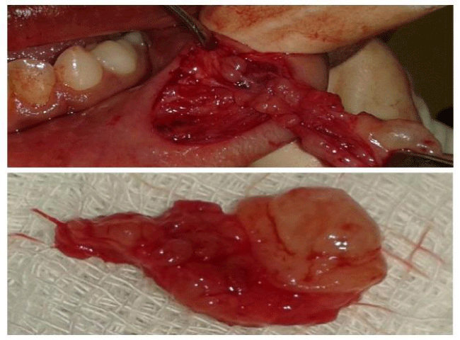
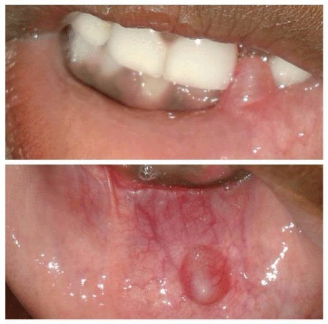
A 9 year old male patient reported to the department of pedodontics and preventive dentistry with a chief complaint of painless swelling on the left side of lower lip (Figure 1A). Patient noticed the swelling two months back which increased in size with passage of time, to the present size (2cm x 2cm). The patient had a lip biting habit and his medical, dental and family history was not contributory. There was history of bursting of the swelling which recurred after few days. On examination, a well circumscribed, transparent, slight bluish coloured, swelling was seen. The swelling was flaccid and painless, with a smooth surface. A provisional diagnosis of mucocele of lower lip was made and surgical excision of the mucocele was planned (Figure 1B). Blood investigations of the patient were within normal limits and past medical history was noncontributory. Surgical excision of the mucocele was done under local anesthesia after obtaining informed and written consent from the patient’s parents. The specimen was sent for histopathological examination, which confirmed it as mucocele. Sutures were removed after 7 days with uneventful healing and follow up was done for 6 months (Figure 1C). At 6 months follow up the healing was satisfactory and no signs of recurrence were present.
Case 2
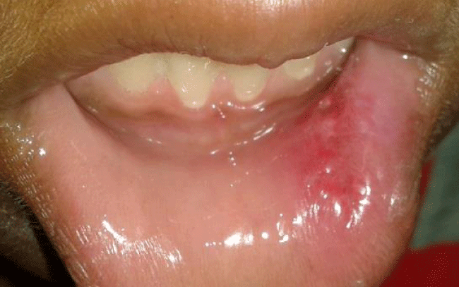
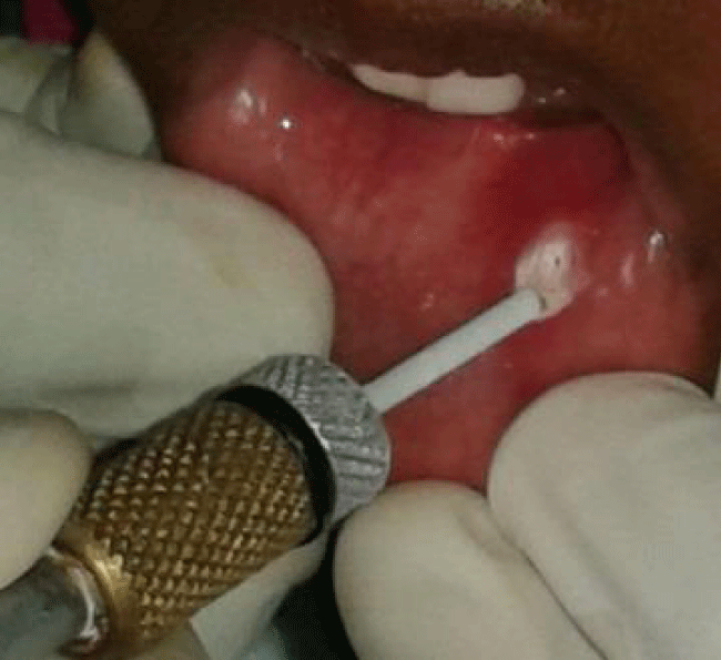
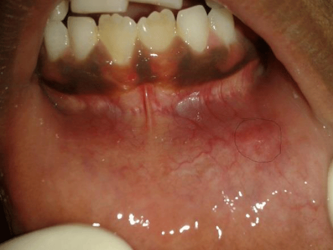
An 8yr old girl reported to the department of pedodontics and preventive dentistry for regular dental checkup. On examination, a localized intra oral swelling was noticed on the left side of lower lip, measuring around 0.5cm x 0.5cm (Figure 2A). Patient had a lip sucking habit (Figure 2A). Based on the history and clinical features, the lesion was provisionally diagnosed as mucocele. Cryosurgery was considered for treatment based on patient’s unwillingness to undergo surgery and the size of the lesion as well. Signed informed consent was obtained from the parents. Treatment consisted of direct application of liquid nitrogen with a nozzle approximate to the size of the lesion. After application of topical anesthetic, the lesion was exposed directly to five to six consecutive freeze-thaw cycles, with each cycle lasting for about 5 to 10 sec, beginning from the center of the lesion, and progressing towards the borders, until the lesion showed white frozen appearance (Figure 2B). During the first 10 mins after the freezing cycles, mild erythema and swelling was noted (Figure 2B). Patient was recalled every week for subsequent sessions of the freeze thaw cycles, which lasted for three consecutive weeks. During the fourth week the lesion disappeared completely without any scar, bleeding, or infection. At the 6-month follow-up, no recurrence was noted (Figure 2C).
Discussion
Mucoceles are common oral pathological conditions. Though not associated with severe complications, they can cause discomfort, especially in the children. The most common treatment modality is excision of the lesion along with the offending minor salivary glands [10]. In our first clinical case, surgical excision was considered, as the lesion was recurrent in nature. There was no recurrence 6 months after the excision. The recurrence rate according to the studies is reported around 14% [10]. Excising the mucocele along with surrounding glandular acini and removing the lesion down to the muscle layer, avoiding the adjacent gland and duct damage while placing the suture will reduces the chances of recurrence [1]. With the advances in paediatric dentistry, it is utmost important to wisely plan the treatment of mucocele. Surgical excision of the mucocele, is associated with postoperative discomfort, which is unavoidable. Especially, when treating young children, it’s necessary to come out with a treatment plan with minimum discomfort. Surgical excision does not require extensive equipment, has negligible cost, and can be performed by most trained dentists. The procedure demands great precision and detailed knowledge of the mucocele and the surrounding anatomy as well. Administration of local anesthesia is required, and this can be more challenging in children, particularly those with behavior management issues. The postoperative bleeding is also greater when compared to other treatment modalities such as the cryosurgery and possibly a longer healing period too [11,12].
Non invasive approaches for treatment of mucocele include laser ablation (CO2, Er,Cr:YSGG), cryosurgery, electrosurgery, micromarsupialization, medication (gammalinolenic acid [GLA]), and “watchful waiting” if the lesion is superficial and not problematic for the patient. In our second case cryosurgery was preferred over surgery. In cryosurgery rapid freezing is used for destruction of lesion. The frozen lesion results in a necrotic tissue which is allowed to slough spontaneously. The two possible ways of performing this procedure involve open and closed cryosurgical systems. In open systems, there is direct placement of a freezing agent (ie, liquid nitrogen) onto the lesion via a cotton swab [12]. In the closed cryosurgical system, sophisticated equipment is required. It can be tedious to obtain/store liquid nitrogen and/or closed system equipment if one is not in a hospital environment [12]. In our second case clinical case open cryosurgery system was used. Advantages of cryosurgery include no intraoperative or postoperative bleeding, minimal surgical defects, no need for suture placement and minimal scarring. Therefore this procedure is considered in areas of aesthetic concern, such as the vermillion border. In addition, no local anesthesia is required in most cases, so has an additional advantage in the pediatric population [12].
In the present case series the cryosurgery resection displayed less postoperative discomfort and more favorable clinical healing compared to surgical excision. The acceptance rate of cryosurgery in children is high based on its advantages but surgical excision has its own merits. Hence, treatment of mucocele in children should be carefully planned based on the age of the patient, level of cooperation and size of the lesion with knowledge of recurrence rates with each treatment option.
Conclusion
Mucocele is the most common oral lesion. Most mucoceles are diagnosed clinically however histopathological reports are mandatory to rule out any other pathology. Mucoceles can be treated with invasive or non invasive methods.
- Baurmash HD (2003) Mucoceles and ranulas. J Oral Maxillofac Surg 61: 369-378. Link: https://goo.gl/EU5bWv
- Bodner L, Tal H (1991) Salivary gland cysts of the oral cavity: Clinical observation and surgical management.Compendium 12: 150–156. Link: https://goo.gl/jqr6fq
- Yamasoba T, Tayama N, Syoji M, Fukuta M (1990) Clinicostatistical study of lower lip mucoceles. Head Neck 12: 316-320. Link: https://goo.gl/1b7v6s
- Sugerman PB, Savage NW, Young WG (2000) Mucocele of the anterior lingual salivary glands (Glands of Blandin and Nuhn): report of 5 cases. Oral Surg Oral Med Oral Pathol Oral Radiol Endod 90: 478-482. Link: https://goo.gl/7HLaCs
- Chaudhry AP, Reynolds DH, Lachapelle CF, Vickers RA (1960) A clinical and experimental study of mucocele (retention cyst) J Dent Res 39: 1253–1262. Link: https://goo.gl/UdwgKL
- Tran TA, Parlette HL III (1990) Surgical pearl: removal of a large labial mucocele. J Am Acad Dermatol 40: 760-762. Link: https://goo.gl/QzVJsx
- Crysdale WS, Mendelsohn JD, Conley S (1988) Ranulas – mucoceles of the oral cavity: experience in 26 children. Laryngoscope 98: 296-298. Link: https://goo.gl/hPAhHh
- Bodner L, Tal H (1991) Salivary gland cyst of the oral cavity: clinical observation and surgical management. Compendium 12:150, 152, 154-156. Link: https://goo.gl/jj89v4
- Poswillo DE (1986) Cryosurgery of benign and orofacial lesions. In: Cryosurgery of the Maxillofacial Region. Bradley PF, ed. Boca Raton, FL: CRC Press 1: 153-175.
- López-Jornet P (2006) Labial mucocele: a study of eighteen cases. The Internet Journal of Dental Science 3. Link: https://goo.gl/eJfasP
- Harris DM, Gregg RH, McCarthy DK (2004) Laser-assisted new attachment procedure in private practice. Gen Dent 52: 396-403. Link: https://goo.gl/HsQkY5
- Cobb CM (2006) Lasers in periodontics: A review of the literature. J Periodontol 77: 545–564. Link: https://goo.gl/afmxTp
Article Alerts
Subscribe to our articles alerts and stay tuned.
 This work is licensed under a Creative Commons Attribution 4.0 International License.


 Save to Mendeley
