Journal of Dental Problems and Solutions
Solitary Median Maxillary Central Incisor Syndrome: An esthetic solution in a child
Rafael de Lima Pedro1*, Erika Calvano Kchler2, Laura Guimarães Primo3 and Marcelo de Castro Costa3
2PhD in Sciences of Health of Federal Fluminense University, Brazil
3Adjunt Professor of Pediatric Dentistry and Orthodontic Departament of Federal University of Rio de Janeiro, Brazil
Cite this as
de Lima Pedro R, Kchler EC, Primo LG, de Castro Costa M (2017) Solitary Median Maxillary Central Incisor Syndrome: An esthetic solution in a child. J Dent Probl Solut 4(4): 072-075. DOI: 10.17352/2394-8418.000053Solitary median maxillary central incisor syndrome (SMMCI) is a complex disorder consisting of multiple, mainly midline, defects of development resulting from unknown factors and an uncertain etiology. SMMCI tooth itself is chiefly an esthetic problem. This case report is an esthetic resolution for a 9- year-old girl with SMMCI as suggested and diagnosed for the first time by a team of dentists. The patient presented the philtrum of her upper lip indistinct, the maxillary labial frenum absent and a single median maxillary central incisor, with symmetrical crown form. Thus, the dentist group contacted her pediatrician reporting these findings and suggesting the diagnosis of SMMCI. The proposed dental treatment was the movement of the central incisor and the right lateral incisor for a better alignment by using some mobile orthodontic appliances and recontouring this lateral tooth with composite. The esthetical finish result pleased the patient as well as her parents.
Introduction
The “Solitary Median Maxillary Central Incisor” Syndrome or SMMCI syndrome was initially described by Hall et al. in 1997 [1], with an incidence estimated in 1:50,000 live births [1].
Its etiology is still unclear, however, it is known that SMMCI is a genetically heterogeneous condition with complex variable expressivity [2]. In some instances, the SMMCI is considered as part of the spectrum, as one of the most minimal expressions, of Holoprosencephaly (HPE), which the brain does not separate into distinct hemispheres [3,4].
SMMCI is characterized by an error of midline facial development, including the cranial bones, the maxilla, the teeth (specifically the central incisor tooth germs) and the nasal airways [1,5]. The most remarkable feature of this syndrome is the presence of a single maxillary incisor tooth situated in the midline region with a unique symmetrical form [5,6], and despite the several findings of this syndrome, it may possibly occur as isolated trait [7].
Identification of SMMCI is difficult due to the extreme variability in the phenotype and its component features [5]. Early diagnosis of this is important because it may be a sign of more severe congenital malformations [8]. Usually, the management needs to be interdisciplinary where dentist plays an important role in orthodontic and oral aesthetic treatment.
Therefore, the aim of this case was to report an aesthetic solution for a child with SMMIC who had her first suspicion of an unusual syndrome suspect by dentists.
Case Report
A Child with 9-year-old female sought to a Pediatric Dentistry Clinic of a Public University with chief complains of the absence of an anterior tooth, leading to problems of low self-esteem and socialization.
In medical history, her mother had reported a serious cardiac problem detected at birth, requiring heart surgery and implantation of a permanent pacemaker. Her mother denied any dental trauma and reported that in primary dentition she also presented only one maxillary central incisor. Her younger sibling, as other relatives were healthy with unremarkable medical histories. Currently, she is still accompanied by a pediatrician and cardiologist, without any other medical complication.
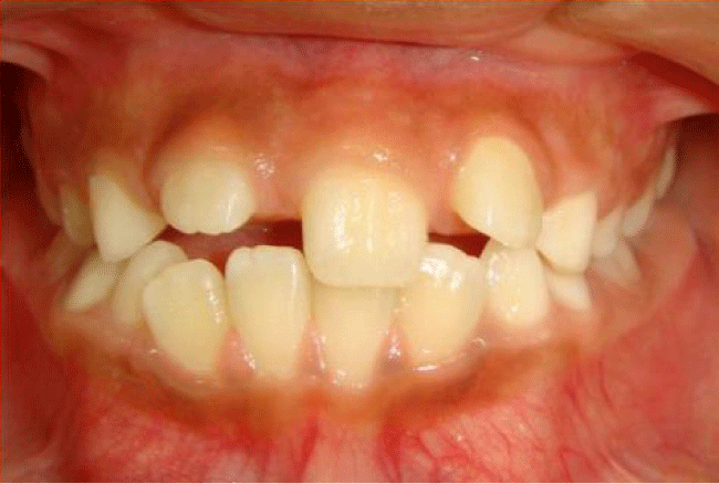
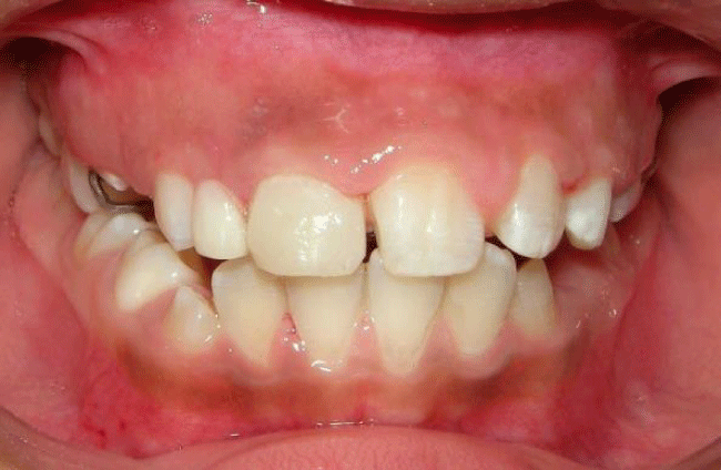
On oral examination, the patient presented a Class I of Angle and there was no sign of nose block or mouth-breath. In the upper labial segment, there was a single median maxillary central incisor, with apparent symmetrical crown form (Figure 1).
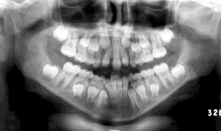
The philtrum of her upper lip was indistinct; the maxillary labial frenum was absent. This factor is pathognomonic for the diagnosis of SMMCI. The orthopantomogram confirmed a solitary maxillary permanent central incisor and all other permanent teeth were developing normally, except the third molars (Figure 2).
In the presence of these oral findings, the dentist team contacted her pediatrician reporting the findings and suggesting the diagnosis of SMMCI. This diagnosis was confirmed and her parents were referred to counselling geneticist service.
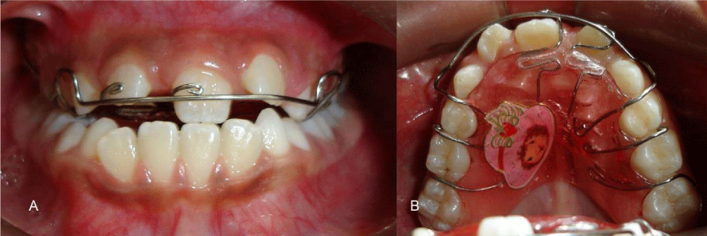
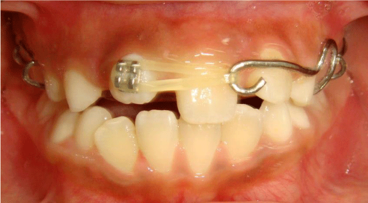
After evaluation by pediatric dentists and orthodontists, the proposed treatment consisted in movement the maxillary central incisor for left side for better dental alignment using a removable orthodontic appliance with a spring (Figure 3). After that, a bracket was bonding in the permanent right lateral incisor and this was movement mesially with a use of elastic and a modified Hawley retainer (Figure 4).
When these teeth were in the desired position, the right lateral incisor was recontouring with composite using a matrix as reference guide as described by dos Santos & Maia [9], and finishing with an appearance looks like central incisor (Figure 5). To finish this part of the treatment a removable space maintainer with a stock tooth in right lateral incisor region (Figure 6) was chosen.
Both the patient and her family were extremely pleased with the outcome of treatment and also reported an improvement in her self-esteem. This patient was followed-up regularly until her permanent dentition erupted completely and the definite treatment as use of the implants or fixed dental prostheses could be done.
Discussion
SMMCI presence may be a clinical indicator of potential HPE in the subsequent generations [10]. The HPE represents one of the most common causes of embryonic lethality, it has found in 1:250 spontaneously aborted fetuses. Its incidence occurs 1:16,000 live births [11,12], with severe sequelae for the patients [10,11].
Thus the presence of unexplained SMMCI is therefore considered a risk factor even in the absence of any other clinical signs [3,4,10]. As SMMCI may present with a variable phenotypic, it may go unnoticed during the life of the individuals. Early diagnosis of SMMCI is important because it may be a sign of more severe congenital malformations [2,8,10].
Some conditions tended to confuse the correct diagnoses of SMMCI where only one central incisor tooth is present as a traumatic loss, presence of mesiodens, fusion of a central incisor with a supernumerary tooth or rarely agenesis of maxillary central incisor [5,13,14]. Nevertheless, an accurate clinical and radiographic examination is indispensable for a correct and reliable diagnosis.
In our case, despite the patient receiving a medical care since birth, the diagnosis of SMMCI was never done appropriately before. Only with the investigation of oral manifestations by a dentist group, the hypothesis of SMCCI syndrome was suggested, despite the presence of a single central maxillary incisor and absence of upper lip frenum of the upper lip, indistinct philtrum and absent incisive papilla [8,15] are pathognomonic for this syndrome, as found in our patient.
Over 70 systemic anomalies have been reported in patients with SMMCI without a recognized syndrome [16]. Among these, short stature, pituitary dysfunction, microcephaly, hypotelorism and congenital nasal obstruction were more commonly reported [1, 5,16-18]. Nevertheless, our patient did not show any of these anomalies, which might have been difficult to diagnose earlier. Although, in literature it is not uncommon that the individuals with SMMCI do not have any systemic disorder [7,8].
Only a congenital cardiac abnormality was found in our patient since birth. This condition may affect up to 25% of SMMCI individuals [5], Early diagnosis of this syndrome is important because it may be a sign of more severe congenital malformations [2,8,10].
Thus, in cases where SMMCI cannot be explained on the basis of the clinical history, it is suggested that the subject should be referred for further genetic analysis. In our case, the patient presented a less severe phenotypic of SMMCI, only presented oral findings and previous history of cardiac abnormally, therefore the performance of the dentist group was decisive for correct diagnosis of the syndrome and was referred to medical staff for evaluation of other possible alterations that may have gone unnoticed.
Usually, no treatment is carried out in the primary dentition [5] and the patients were followed until their permanent dentition completely erupted, then orthodontic and prosthetic treatment was performed for correction of malocclusion, for which tooth implant or prosthesis could be used [5,8]. However, no treatment was related to mixed dentition in the literature yet. In this case, we were performed sought to resolve the esthetical complaint of the patient with a combined orthodontic and operative dentistry interventions. This approach was chosen because it was relatively simpler with quick resolution and it was extremely important for the self-esteem of patients and her socialization issue.
Despite the fact that a definite treatment can only be done when all permanent teeth present a stable occlusion, this initial management provided essentials for improvement of the quality of life of the patient her family.
Conclusion
Thus, the dentist should be aware of oral manifestations of SMMCI syndrome, since these findings can be crucial for the correct diagnosis of this syndrome that may go undetected during the individual’s life. Furthermore, it is possible to carry out the necessary treatment to seek a better aesthetics and satisfaction of the patient.
- Hall RK, Bankier A, Aldred MJ, Kan K, Lucas JO, et al. (1997) Solitary median maxillary central incisor, short stature, choanal atresia/midnasal stenosis (SMMCI) syndrome. Oral Surg Oral Med Oral Pathol Oral Radiol Endod 84: 651-662. Link: https://goo.gl/1PP6zD
- Lazaro L, Dubourg C, Pasquier L, Le Duff F, Blayau M, et al. (2004) Phenotypic and molecular variability of the holoprosencephalic spectrum. Am J Med Genet A 129A: 21-24. Link: https://goo.gl/qqgSXC
- Cohen MM Jr (2001) Problems in the definition of holoprosencephaly. American journal of medical genetics 103: 183-187. Link: https://goo.gl/DEXata
- Cohen MM Jr (1989) Perspectives on holoprosencephaly: Part III. Spectra, distinctions, continuities, and discontinuities. Am J Med Genet 34: 271-288. Link: https://goo.gl/KMpBcv
- Hall RK (2006) Solitary median maxillary central incisor (SMMCI) syndrome. Orphanet journal of rare diseases 1:12. Link: https://goo.gl/5TgQfC
- DiBiase AT, Elcock C, Smith RN, Brook AH (2006) A new technique for symmetry determination in tooth morphology using image analysis: application in the diagnosis of solitary maxillary median central incisor. Archives of oral biology. 51: 870-875. Link: https://goo.gl/Rc692a
- Youko K, Satoshi F, Kubota K, Goto G (2002) Clinical evaluation of a patient with single maxillary central incisor. The Journal of clinical pediatric dentistry 26: 181-186. Link: https://goo.gl/kWjEUZ
- Cho SY, Drummond BK (2006) Solitary median maxillary central incisor and normal stature: a report of three cases. International journal of paediatric dentistry / the British Paedodontic Society [and] the International Association of Dentistry for Children 16: 128-134. Link: https://goo.gl/GhAozm
- Alves dos Santos MP, Maia LC (2005) The reference guide: a step-by-step technique for restoration of fractured anterior permanent teeth. Journal (Canadian Dental Association) 71: 643-646. Link: https://goo.gl/XNfiDL
- Johnson VP (1989) Holoprosencephaly: a developmental field defect. American journal of medical genetics 34: 258-264. Link: https://goo.gl/CciyNq
- Cohen MM Jr, Jirasek JE, Guzman RT, Gorlin RJ, Peterson MQ (1971) Holoprosencephaly and facial dysmorphia: nosology, etiology and pathogenesis. Birth defects original article series 7: 125-135. Link: https://goo.gl/KDt2cJ
- Matsunaga E, Shiota K (1977) Holoprosencephaly in human embryos: epidemiologic studies of 150 cases. Teratology 16: 261-272. Link: https://goo.gl/fCiSe4
- DiBiase AT, Cobourne MT (2008) Beware the solitary maxillary median central incisor. Journal of orthodontics 35: 16-19. Link: https://goo.gl/GMnUx7
- Graber LW (1978) Congenital absence of teeth: a review with emphasis on inheritance patterns. Journal of the American Dental Association (1939) 96: 266-275. Link: https://goo.gl/TeQu8R
- El-Jaick KB, Fonseca RF, Moreira MA, Ribeiro MG, Bolognese AM, et al. (2007) Single median maxillary central incisor: new data and mutation review. Birth defects research 79: 573-580. Link: https://goo.gl/nEFL2d
- Nanni L, Ming JE, Du Y, Hall RK, Aldred M, et al. (2001) SHH mutation is associated with solitary median maxillary central incisor: a study of 13 patients and review of the literature. American journal of medical genetics 102: 1-10. Link: https://goo.gl/jbGhVy
- Rappaport EB, Ulstrom RA, Gorlin RJ, Lucky AW, Colle E, et al. (1977) Solitary maxillary central incisor and short stature. The Journal of pediatrics. 91: 924-928. Link: https://goo.gl/y1qw9k
- Wesley RK, Hoffman WH, Perrin J, Delaney JR (1978) Solitary maxillary central incisor and normal stature. Oral surgery, oral medicine, and oral pathology 46: 837-842. Link: https://goo.gl/aJy71g
Article Alerts
Subscribe to our articles alerts and stay tuned.
 This work is licensed under a Creative Commons Attribution 4.0 International License.


 Save to Mendeley
