Journal of Dental Problems and Solutions
Oral Rehabilitation of patient with Cleft Lip and Palate- A Case Report
Vesna Ambarkova1*, Biljana Djipunova2, Manu Batra3 and Slagana Trajkova4
2Associate Professor, PhD, MSc, DDS, University St. Cyril and Methodius, Faculty of Dental Medicine, Department of orthodontics, Skopje, Republic of Macedonia
3Senior Lecturer,MDS,BDS, Department of Public Health Dentistry, Surendera Dental College & Research Institute, Sri Ganganagar, Rajasthan, India
4Logoped, Institute for Rehabilitation of Hearing, Speech and Voice, Skopje, Republic of Macedonia
Cite this as
Ambarkova V, Djipunova B, Batra M, Trajkova S (2017) Oral Rehabilitation of patient with Cleft Lip and Palate- A case report. J Dent Probl Solut 4(4): 061-065. DOI: 10.17352/2394-8418.000051Cleft lip and palate (CLP) is the most common congenital facial anomaly. About 70% of all the CLP cases and 50% of cleft palate only fall within non-syndromic pathologies. The purpose of this report was to show the clinical management of initial obturator therapy from birth to 3 months. The obturator was fabricated from the conventional orthodontic acrylic materials with cold polymerization (OrtoPoli, Polident, Slovenia). For successful treatment of cleft lip and palate patients, during the planning of prosthetic therapy one should take into consideration the deformation of maxillary segments\, as well as the disproportion between the upper and lower jaw alveolar ridge. Well planned prosthetic therapy will result in satisfactory function and aesthetics of a cleft palate patient.
Introduction
Cleft lip and palate (CLP) is the most common congenital facial anomaly. Its incidence varies according to epidemiologic studies but is usually between 1 and 1.82 for each 1000 births [1]. CLP is a congenital deformity that is associated with maxillary sagital and transverse discrepancies [2,3]. In addition to skeletal discrepancies; this deformity is often accompanied by dental abnormalities, such as hypodontia, hyperdontia, and transpositions [4,5].
About 70% of all the CL/P cases and 50% of cleft palate only fall within non-syndromic pathologies. All the other cases are linked to cardiac, limb, ophthalmological syndromes and other. The manifestation of the disease (syndromic and non-syndromic) have been linked to some defects of grow factors and their receptors [6], such as FGF8 and FGFR1 genes. TGFβ is another family gene involved in the formation of the oral cleft, in particular: TGFβ3, with the inactivation of its receptor TGF3βR2 [7], and the inactivation of BMP7 [8,9].
CLP patients might suffer from unfavourable smile aesthetics and low self-esteem, leading mainly to difficulties in social interactions. The treatment for patients with CLP is challenging because of the difficulties inherent in the deformity, the necessity of interdisciplinary involvement, and the need for good patient cooperation. The results might still be limited even if all of these challenges can be overcome [10].
The purpose of this report was to show the clinical management of initial obturator therapy from birth to 3 months.
Case Report
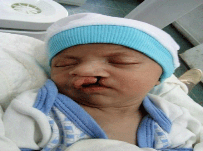
Baby with female gender and diagnosis Cheilognathopalatoschisis was one and half month when her mother and her grandparents come to the Department for preventive and pediatric dentistry and Department for orthodontics, University Dental Clinic Centar- Skopje (Figure 1). They were sent to our department from the University Clinic for facial, jaws and neck surgery. Girl was the firstborn of related, healthy young parents with an unmarkable family history. Baby was born after a normal pregnancy by caesarean section at the 37th gestational week and delivery with birth weight 2060 gr and body length 45 cm. Apgar score was 8/9 at 5/10 minutes, respectively. During the visit of department, the baby was accompanied by the mother and grandparents. The parents were young and of Gipsy origin and without proper medical documentation. The mother is younger than 18 years old. Her pregnancy was ended by caesarean section at the 37th gestational week. The mother was born in Podujevo, Republic of Kosovo and along with her juvenile daughter have been granted legal asylum for humanitarian or subsidiary protection.
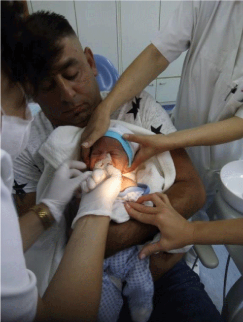
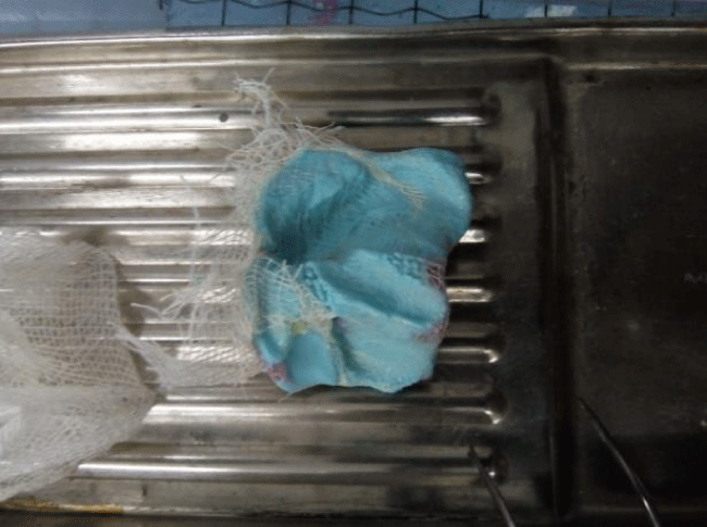
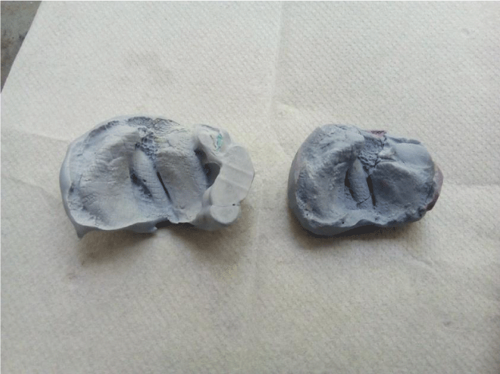
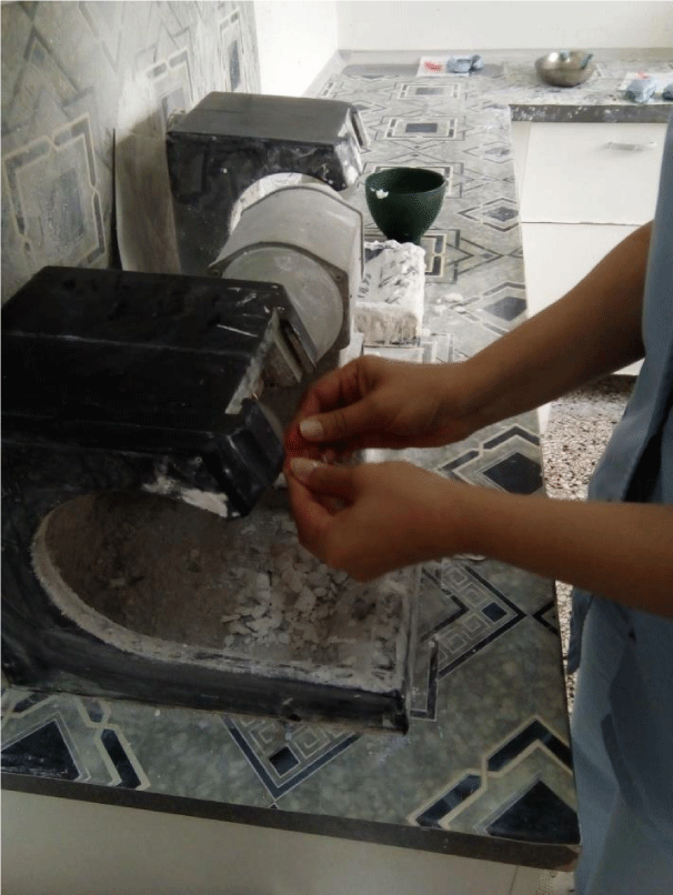
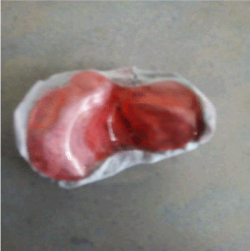
Individual tray was made by wax plate (Dentaurum, Germany). Impression was made with appropriate tray and alginate irreversible hydrocolloid material (Hydrogum soft, Zhermack, Lot. 246967, Italy) (Figures 2,3). The infant was held upright during the impression process to prevent aspiration of excess material. A stone model is then produced (Figure 4).Casts were prepared using hard dental stone. Тhe technician who makes orthodontic appliances, including the obturators must block out excessive undercuts with modeling dough or wax. Modeling dough is preferred because it is easy to remove from the finished prosthesis. The obturator was fabricated in the conventional orthodontic acrylic materials with cold polymerization (OrtoPoli, Polident, Slovenia). Тhe technician pour a mixture of soft, autopolymerizing acrylic resin into the cleft to level of the palate. This will provide retention for the prothesis by gently contouring into the available undercuts. Model has to be place in a warm, moist environment to cure for 20 minutes. Clinical doctor have to add autopolymerizing acrilic resin to the palate using a “salt and pepper” method, making sure the acrylic resin extends well into the mucobuccal fold area. In the final stage the appliance was trimmed and polished (Figure 5). The mother and grandparents were told to wait several hours, and the obturator was given and placed on the baby’s mouth the same day (Figure 6).
Disscusion
Today it is very important to understand that that cleft of lip and palate always results in a deficiency of tissue and not in a mere displacement of normal tissue and is followed by an important anatomical disruption of the lip muscles, especially orbicularis muscle. Nowadays despite significant advances in cleft lip and palate treatment, anatomical controversies still remain. Some authors proposed that the width of the cleft is due to alveolar segmental displacement, while other suggest that the width is due to a palate-alveolar hypoplasia [11,12].
In unilateral cleft lip and palate, the orbicularis muscle is interrupted. Its fibers are atrophic and run upwards and parallel to the edge of the cleft, and are inserted at the base of the columella medially and at the nasal wing laterally (Figure 1). During oral function feeding, crying, smiling etc., the columella on the non-affected side and the nasal wing on the cleft side are stretched in opposite directions, increasing so the width of the cleft. The columella is displaced to the normal side and the nasal wing on the cleft side is displaced laterally, posteriorly, and inferiorly, thus increasing nasal deformity.
The aim of the plate is to stabilize this ideal relationship to prevent segments displacement. Left untreated, the asymmetrical action of the muscles on the two segments ends in the “collapse” of the lateral alveolar segment. During the time the interposition of the tongue in the cleft worsens their reciprocal displacement and have a severe impact on arch form and facial growth.
In principle, the lip and palate clefts occur more frequently on the left side of the face. In our case, baby was with lip and palate cleft of the left side. Cleft lip and palate together account for approximately 50% of all cases, whereas isolated cleft lip and isolated cleft palate each occur in about 25% of cases. An alginate impression of the infant’s maxillary arch is made with a modified stock tray, ideally, this is accomplished as soon after birth as possible. Appropriate emergency equipment, including forced oxygen, suction, and standard airway management equipment, should be available during the impression process. The impression should exhibit good anatomic detail with coverage of the entire maxillary arch.
Pediatric problems of cleft lip and palate
Based on previous research it has been established that children with clefts of lip and palate are more susceptible to some diseases than children without them.
Nutrition
The first problem occurs as soon as the baby is born when it needs to be fed. These babies cannot suck, because they are not able to vacuum during a sucking act. Mothers are advised that if it is possible to keep their milk as long as possible and feed the baby with their milk through a spoon or cucumber. Then the baby should be kept as upright as possible and kept slower, otherwise if the child lays more air, the food returns through the nose, more often vomits, and progresses poorly.
Also, children with lip and palate cleft are with increased risks to have more infections of the upper and lower respiratory tract due to inhalation of unheated and unfiltered air.
Diseases of the middle ear are more common among the children with lip and palate cleft. Infections are transmitted from the throat.
Therapy of cleft of lip and palate
Multidisciplinary approach or team work is essential for success in the treatment of those patients. Who enters the team?
The therapy of cleft lip and palate is very serious, complex, long-lasting. It starts immediately after birth within the largest number of children, and ends mostly with complete cessation of growth and development, after all therapeutic and corrective interventions. The implementation of therapy must be multidisciplinary, which means that a specialized consultation is needed for the treatment of inborn and acquired deformities, which guarantees adequate and timely observation and treatment of such patients. There should be a whole team of experts of various specialties in which the dentist is also involved: phoniatrist , audiologist, speech therapist, psychologist, social worker.
Dentists of various specialties
• Radiologist
• Pediatric dentist
• Endodontist
• Periodontologist
• Orthodontics
• Oral surgeon
• Maxillofacial surgeon
• Plastic surgeon
• A prostodontist
For the parents consciousness that they received a child with a lip and palate cleft is very shocking and pose a serious psychological blow to parents. This can negatively affect the relationship of the parent to the newborn and, consequently, the outcome of the treatment. The duty of the surgeon, as the head of the whole team, is to discern parents in relation to their sense of guilt over the existing anomaly, to explain to the parents the goal and the way of treatment, in order to establish adequate cooperation with parents, which ultimately improves the outcome of the treatment.
Orthodontic therapy starts in the first 48 hours after the birth of the child with making an acrylic orthodontic device. This device has a multiple role:
- facilitates the feeding of a child by making a defect of the defect and separating the nose from the mouth of the cavity, and thus allows sucking;
- brings the language into a normal position, allowing normal development of speech at a later age;
- Helps in the orientation of the alveolar segments of the upper jaw to the optimum position before surgery,
- Contributes to achieving a beneficial psychological effect in parents, positive communication between the child and the parent.
In our case report, parents relatively late brought their baby to make an acrylic orthodontic device, mostly and probably because they did not have proper medical documents.
As far as pre-surgical orthopedic therapy is concerned, there are contradictory opinions:Тhе opponents believe that primary surgery should give an anatomical basis for orthodontic therapy, not vice versa. However, often the scar tissue that remains after surgery precisely prevents normal maxilla growth.
The supporters highlight the following benefits:
• bringing maxillary segments into a better position for each other
• narrowing the split and reducing tissue deficiency helps feed, and therefore the progression of the baby
• Proper voice formation
Conclusion
The effect of infant orthopaedics on maxillary arch has been a subject of debate for many years, but controversy about justification of the use of the nasoalveolar molding appliance still exists. Advocates of infant orthopaedics claim that the presurgical orthopaedic plate molds the alveolar segments into a better arch form and that with this action the dentomaxillary development would be improved [13,14]. Opponents of this therapy claim that lip surgery alone has the same effect and that presurgical orthopedic plate is only an expensive appliance used to comfort the parents by starting treatment at the earliest possible moment [15,16]. the For successful treatment of cleft lip and palate patients, during the planning of prosthetic therapy one should take into consideration the deformation of maxillary segments, eventually residual palatal defects, as well as the disproportion between the upper and lower jaw alveolar ridge. Well planned prosthetic therapy will result in satisfactory function and aesthetics of a cleft palate patient.
- Derijcke A, Eerens A, Carels C (1996) The incidence of oral clefts: a review. Br J Oral Maxillofacial Surg 34: 488–494. Link: https://goo.gl/JQd6rT
- Baek SH, Moon HS, Yang WS (2002) Cleft type and Angle's classification of malocclusion in Korean cleft patients. Eur J Orthod 24: 647–653. Link: https://goo.gl/9WUJPE
- Vettore MV, Sousa Campos AE (2011) Malocclusion characteristics of patients with cleft lip and/or palate. Eur J Orthod 33: 311–317. Link: https://goo.gl/RdR9yW
- Tereza GP, Carrara CF, Costa B (2010) Tooth abnormalities of number and position in the permanent dentition of patients with complete bilateral cleft lip and palate. Cleft Palate Craniofac J 47: 247–252. Link: https://goo.gl/Qq6Bxf
- Akcam MO, Evirgen S, Uslu O, Memikoglu UT (2010) Dental anomalies in individuals with cleft lip and/or palate. Eur J Orthod 32: 207–213. Link: https://goo.gl/kLgR19
- Riley BM, Murray JC (2007) Sequence evaluation of FGF and FGFR gene conserved non-coding elements in non-syndromic cleft lip and palate cases. Am J Med Genet A 143A: 3228-3234. Link: https://goo.gl/N3ZYqF
- Lidral AC, Romitti PA, Basart AM, Doetschman T, Leysens NJ, et al. (1998) Association of MSX1 and TGFB3 with nonsyndromic clefting in humans. Am J Hum Genet 63: 557-568. Link: https://goo.gl/sPR5ia
- Zouvelou V, Luder HU, Mitsiadis TA, Graf D (2009) Deletion of BMP7 affects the development of bones, teeth, and other ectodermal appendages of the orofacial complex. J Exp Zool B Mol Dev Evol 312B: 361-374. Link: https://goo.gl/B31HPb
- Wurdak H, Ittner LM, Lang KS, Leveen P, Suter U, et al. (2005) Inactivation of TGFbeta signaling in neural crest stem cells leads to multiple defects reminiscent of DiGeorge syndrome. Genes Dev. 19: 530-535. Link: https://goo.gl/d3bHYh
- Rocha R, Ritter DE, Locks A, de Paula LK, Santana RM (2012) Ideal treatment protocol for cleft lip and palate patient from mixed to permanent dentition. Am J Orthod Dentofacial Orthop 141(4 Suppl): S140–S148. Link: https://goo.gl/CmxCDo
- Dec W, Olivera O, Shetye P, Cutting CB, Grayson BH, et al. (2013) Cleft palate midface is both hypoplastic and displaced. J Craniofac Surg 24: 89-93. Link: https://goo.gl/B4EJ5Z
- Defabianis P (2015) Pre-surgical orthopaedics in newborns affected by clefts of the lip, alveolus and palate: Report of two cases. European Journal of Paediatric Dentistry 18: 237-242. Link: https://goo.gl/wUdF2A
- Mishima K, Mori Y, Sugahara T, Minami K, Sakuda M (2000) Comparison between the palatal configurations in UCLP infants with and without Hotz plate until four years of age. Cleft Palate Craniofac J 37: 185-190. Link: https://goo.gl/6ha1Sv
- Ball JV, DiBiase DD, Sommerlad BC (1995) Transversemaxillary arch change with the use of preoperative orthopaedics in unilateral cleft palate infants. Cleft Palate Craniofac J 32: 483-488. Link: https://goo.gl/CYD4Vg
- Prahl C, Kuijpers-Jagtman AM, van ‘tHof MA, Prahl-Andersen B (2001) A randomised prospective clinical trial into the effect of infant orthopaedics on maxillary arch dimensions in unilateral cleft lip and palate (Dutchcleft). Eur J Oral Sci 109: 297-305. Link: https://goo.gl/qnmong
- Prahl C, Kuijpers-Jagtman AM, van‘t Hof MA, Prahl-Andersen B (2003) A randomized prospective clinical trial into the effect of infant orthopedics in UCLP. Prevention of collapse of the alveolar segments (Dutchcleft). Cleft Palate Crniofac J 40: 337-342. Link: https://goo.gl/vR1nhi
Article Alerts
Subscribe to our articles alerts and stay tuned.
 This work is licensed under a Creative Commons Attribution 4.0 International License.

 Save to Mendeley
