Journal of Clinical Research and Ophthalmology
Preservation of capsular transparency and geometrical consistency in cataract surgery using a novel intracapsular ring
Ahmed Elmassry1, Onurcan Sahin2,3, Loukia Leonidou2*, Dimitris Liakopoullos3, Harilaos Ginis2, Aristophanis Pallikaris2,3 and Ioannis Pallikaris2
2EYE-PCR B.V./ University of Crete, Research institution in Greece, Greece
3University of Crete, Greece
Cite this as
Elmassry A, Sahin O, Leonidou L, Liakopoullos D, Ginis H, et al. (2020) Preservation of capsular transparency and geometrical consistency in cataract surgery using a novel intracapsular ring. J Clin Res Ophthalmol. 7(2): 101-104. DOI: 10.17352/2455-1414.000082We report a case of a patient that was implanted with an intracapsular ring during phacoemulsification cataract surgery. Namely, we report on refraction, posterior capsule transparency and IOL ACD and document photographically the appearance of the posterior capsule. It is suggested that the presence of the intracapsular ring has inhibited epithelial cell migration and prevented the formation of posterior capsule opacification (PCO) in the postoperative interval of 7 months. A comparison with the fellow eye of the same patient is made.
Introduction
Today’s cataract surgeries are one of the most common refractive operations. Advancements on the surgery technique, use of femtosecond lasers and advanced IOL designs increased the safety and efficacy of the cataract surgeries. However, current IOL haptic designs not always fulfill the correct axial placement and perfect centralization. Moreover, the variability of human capsule size and haptic design differences creates an uncertainty for cataract surgery that arises from the axial and tranverse positioning of the IOL in the capsule, both in the immediate postoperative interval and following capsule fibrosis. In addition to the uncertainty of the primary implantation the fibrotic capsule does not allow a predictale lens exchange surgery if this is required. In order to improve issues mentioned above we designed the fixOflex ring (EYE-PCR B.V., Amsterdam, The Netherlands), a complementary implant for cataract surgery. The fixOflex ring consists a continous circular shape that maintains 360 degree contact with the capsule, therefore it works as a peripheral capsular reconstructor. The ring is made of hydrophilic acrylic pre-polymerized material. Its overall diameter is 9.80mm and its central opening is sufficient to accomodate an IOL with a 6mm optic. Its overall thickness is 1.84mm.The ring also features clips for encapsulating the IOL optic which is very important for good IOL centration and more predictable axial placement. fixOflex is creating a stable environment for the lens making the positioning of the lens predictable and also easily replaceable if required in future surgery. Besides the physiological implications of maintaining an open capsule (avoiding contact of the anterior and the posterior capsule) that avoids fibrosis and therefore loss of transparency and mechanical tensions capable of dislocating the intraocular lens, the implantation of such a ring can be seen as a method to maintain the physiological anatomical relationship between the anterior and the posterior chamber. Moreover it may serve to maintaining the anatomical spaces normally occupied by the crystalline lens and the vitreous cavity. It is important to mention that implantation of a ring similar to fixOflex in terms of the occupied volume is justified by the literature [1,2].
Case
The case presented here refers to a 62 years old female with OD preoperative refraction Sph:-2.5D, Cyl:0.0D, Ax:0◦ and OS preoperative refraction Sph:+1.0D, Cyl:-1.0D, Ax:95◦. BCDVA was 0.8 for OD and 1.0 for OS. The patient presented with difficulty in night vision and glare phenomena. Despite having relatively high best corrected visual acuity, biomicroscopic examination revealed early bilateral cataract (N2 for OD and N+1 for OS) . Cataract surgery was indicated on the basis of the reported visual symptoms. The subject had clear ocular media other than cataract and stable keratometry and biometry measurements. There was no indication of corneal ectatic conditions or other corneal dystrophies. No additional ocular pathology was present in the anterior segment. The patient did not have a history of previous corneal surgery.
The patient was implanted with fixOflex ring in combination with a J&J Tecnis ZCB00 IOL of power 19D in the right eye. In the fellow eye conventional cataract surgery was performed using the same IOL type of power 20.5D. There were no intraoperative complications for both the surgeries.
Surgical Technique Cataract surgery was performed under sterile conditions in a standar manner of phacoemulcification by the same surgeon (AE). Local anesthetic drops were used. A 2.2 mm temporal clear corneal incision was used. A 6.0 mm anterior capsulorhexis was created. After phacoemulcification and irrigation/aspiration of the lens cortex, the capsular bag was filled with sodium hyaluronate. The fixOflex ring was inserted into the capsular bag in the study group. Finally, the IOL was inserted into the capsular bag and IOL haptics adjusted to the ring clips. The patient received antibiotic/steroid eye drops (tobramycin/dexamethazone, Tobradex; Alcon Laboratories, Inc., Fort Worth, TX, USA) four times a day for 1 month postoperatively. For the fellow eye, the same procedure was followed with the exception that the IOL was implanted directly in the capsule.
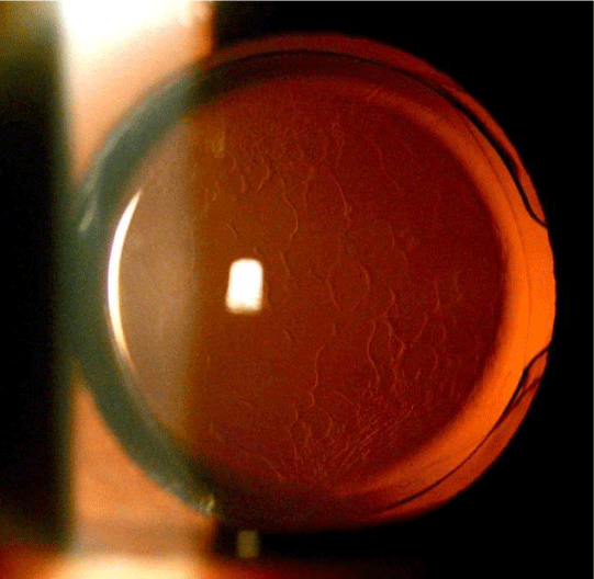
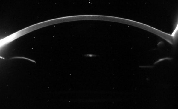
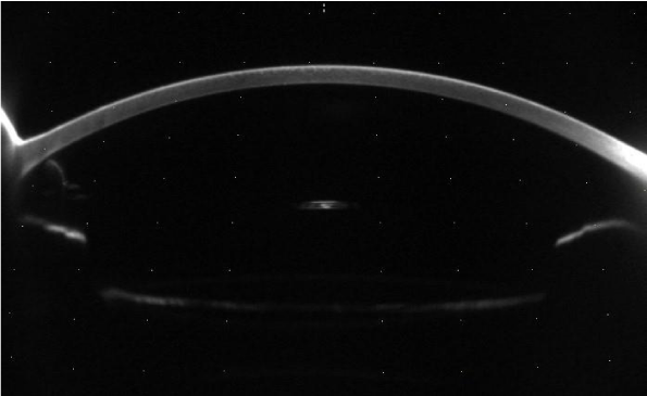
Physical examination results using Slitlamp 6 months postoperatively are presented in Figures 1,2 for OD (eye with fixOflex) and OS respectively.
We observed a very clear and stretched posterior capsule for OD eye (eye with fixOflex), whereas in contrary to the fellow eye, where characteristic wrinkles formed in the posterior capsule (OS). Moreover the transparency of the posterior capsule was higher in the right eye in comparison to that in the left eye as revealed by Scheimpflug photography (Pentacam HR, OCULUS, Germany) Figures 3,4.
Postoperative BCDVA was 1.0 for the right eye and 0.9 for the left eye. Intraocular pressure was 13mmHg for both eyes. The patient did not report subjective symptoms such as dysphotopsias or glare for either eye.
Discussion
Posterior Capsule Opacification (PCO) [3] is the most frequent, long-term complication of cataract surgery. PCO may cause light scattering, monocular diplopia, decrease of contrast sensitivity, and decrease of visual acuity [4,5]. The incidence of PCO is treaded easily and effectively in majority of the cases, with Nd:YAG laser capsulotomy. This method cleans the optical path by creating a central opening in the posterior capsule. However the method is associated with some complications like retinal detachment, IOL damage and subluxation, corneal edema, elevation of intraocular pressure and cystoid macular edema [6-9]. Some of these complications are mild and transient but other may cause permanent visual impairment. Anterior capsule opacification (ACO), also known as anterior capsule fibrosis or anterior subcapsular opacification is another possible complication of cataract surgery. ACO occurs much earlier, sometimes one month postoperatively and more often compared to PCO. Severe forms of ACO may induce contraction of the capsulorexis opening, capsule retraction and capsular phimosis. The result of this might be a decantation of the IOL and fibrotic opacification of the posterior capsule.
Attempting to eliminate the incidence of PCO and ACO researches have been focused in several strategies including surgical techniques, IOL material, IOL design, biological and pharmacological agents, and intraocular endocapsular rings [10,11]. The concept of ring insertion into the capsule was introduced by Hara et al in the context of zonular apparatus support [4]. Two years later, the first capsular tension ring (CTR) was designed for use in humans. Various similar CTRs have been designed and marketed since then [12]. The main function of these endocapsular rings is that they restore the capsule’s physical shape. CTRs have well-established use for the management of zonular weakness and dehiscence, enhancement of IOL contraption and capsule stabilisation, but also they may influence capsule opacification [13]. A new design of CTR, Peripheral Capsule Reconstructor (PCR) introduced by I.G. Pallikaris et al in a pilot study which suggested that the implantation of an endocapsular ring may contribute to the prevention of PCO and ACO after cataract surgery [1].
FixOflex intracapsular ring is a new design of the PCR and is currently under study. The complementary implantation of fixOflex aims at creating a stable environment for the lens to be placed making the positioning of the lens predictable. Besides the physiological implications of maintaining an open capsule (avoiding contact of the anterior and the posterior capsule) that avoids fibrosis and therefore loss of transparency and mechanical tensions capable of dislocating the intraocular lens, the implantation of such a ring can be seen as a method to maintain the physiological anatomical relationship between the anterior and the posterior chamber. Moreover it may serve to maintaining the anatomical spaces normally occupied by the crystalline lens and the vitreous cavity.
In the current case, we present a patient with fixOflex plus IOL in one eye and IOL alone in the other eye. Both eyes were in the average axial range as resulting from the biometry and undergone uncomplicated phacoemulsificaton procedure. At slit lamp examination an interesting finding was observed; posterior capsule in the eye with the fixOflex was stretched and clear, whereas in the fellow eye wrinkles were found at the posterior capsule. These findings are indicative of the efficacy of fixOflex implantation, as at the same patient with similar biometric measurement between eyes the implant had the desirable effect of keeping the posterior capsule stretched and prevent PCO and ACO by the peripheral footing of the implant at the capsule equator. On the other hand, the posterior capsule of the other eye found with wrinkles and formations that could indicate migration leading to incipient posterior capsular opacification. Also, evaluating another point of the fixOflex regarding the IOL placement, the position of the IOL was found effectively centered over the implant in the study eye of the patient.
The fixOflex implant is designed to function as a peripheral capsular reconstructor with complete anatomical fitting in order to prevent significantly the migration of lens epithelia cells. Furtheremore, this implant aims to maintain efficiently the IOL centration, an aspect achieved in the current patient as observed by the slit-lamp examination. A comparative study with a larger number of patients is needed to evaluate the safety and efficacy in the future.
Conclusion
Although this case report does not support an overall conclusion about the safety and efficacy of the fixoflex ring, it can serve to demonstrate its functionality and its purpose. In the case presented herein the fixoflex ring was effective in preserving the posterior capsular transparency as well as in stabilising the location of the intraocular lens. The observations from the case reported herein are inline with the other cases participating in the present clinical investigation that have not reached the 6-month postoperative interval up to date. These observations support the hypothesis that the implantation of the ring is safe end effective for the reconstructing the posterior caspule (no wringles in or around the capsule), recreating a stable barrier for the vitreous and also in preventing cell migration into the posterior capsule.
Notes on patient consent
The case reported represents the case with the longest follow-up postoperatively and is part of an ongoing clinical investigation to evaluate the safety and efficacy of the fixoflex ring. The clinical investigation pertains to a large number of cataract patients (N=120) and a 12-month follow up period. As all patients, the case reported herein has given consent to the publication of the images and other relevant clinical data.
- Pallikaris IG, Stojanovic NR, Ginis HS (2016) A new endocapsular open ring for prevention of anterior and posterior capsule opacification. Clin Ophthalmol 10: 2205-2212. Link: https://bit.ly/2GCphQg
- Hara T, Yamada Y (1991) Equator ring" for maintenance of the completely circular contour of the capsular bag equator after cataract removal. Ophthalmic Surgery 22: 358-359. Link: https://bit.ly/38gUBPT
- Apple DJ, Solomon KD, Tetz MR, Assia EI, Holland EY, et al. (1992) Posterior capsule opacification. Survey of Ophthalmology 37: 73-116. Link: https://bit.ly/3p351bT
- Schaumberg DA, Dana MR, Christen WG, Glynn RJ (1998) A systematic overview of the incidence of posterior capsule opacification. Ophthalmology 105: 1213–1221. Link: https://bit.ly/2TZwa0Z
- Pandey SK, Apple DJ, Werner L, Maloof AJ, Milverton EJ, et al. (2004) Posterior capsule opacification: a review of the aetiopathogenesis, experimental and clinical studies and factors for prevention. Indian J Ophthalmol 52: 99-112. Link: https://bit.ly/2JAwr8E
- Holweger RR, Marefat B (1997) Intraocular pressure change after neodymium: Yag capsulotomy. J Cataract Refract Surg 23: 115-121. Link: https://bit.ly/2I9PgyO
- Koch DD, Liu JF, Gill EP, Parke DW (1989) Axial myopia increases the risk of retinal complications after neodymium-yag laser posterior capsulotomy. Arch Ophthalmol 107: 986–990. Link: https://bit.ly/32kmxhV
- Aslam TM, Devlin H, Dhillon B (2003) Use of nd: Yag laser capsulotomy. Surv Ophthalmol 48: 594-612. Link: https://bit.ly/3mXEoDh
- Trinavarat A, Atchaneeyasakul L, Udompunturak S (2001) Neodymium: Yag laser damage threshold of foldable intraocular lenses. Journal of Cataract & Refractive Surgery 27: 775-780. Link: https://bit.ly/353BS8z
- Wormstone IM, Wang L, Liu CS (2009) Posterior capsule opacification. Exp Eye Res 88: 257-269. Link: https://bit.ly/2IaoJBC
- Clark DS (2000) Posterior capsule opacification. Curr Opin Ophthalmol 11: 56-64. Link: https://bit.ly/2U1RpiW
- Rupert Menapace, Oliver Findl, Michael Georgopoulos, Georg Rainer, Clemens Vass, and Karin Schmetterer. The capsular tension ring: designs, applications, and techniques. Journal of Cataract & Refractive Surgery, 26(6):898–912, 2000.
- Menapace R, Sacu S, Georgopoulos M, Findl O, Rainer G, et al. (2008) Efficacy and safety of capsular bending ring implantation to prevent posterior capsule opacification: three-year results of a randomized clinical trial. J Cataract Refract Surg 34: 1318-1328. Link: https://bit.ly/3mYe9MX

Article Alerts
Subscribe to our articles alerts and stay tuned.
 This work is licensed under a Creative Commons Attribution 4.0 International License.


 Save to Mendeley
