Journal of Clinical Research and Ophthalmology
Antenatal diagnoses of congenital cataracts and related surgical complications in a familial case of Nance-Horan Syndrome
Gayathri K Sivakumar1, Gunjan Mhapankar1, Victoria Mok Siu1,2 and Sapna Sharan1,3*
2Medical Genetics Program of Southwestern Ontario, London Health Sciences Centre, London, Ontario, Canada
3Department of Ophthalmology, Western University, London, Ontario, Canada; Ivey Eye Institute, St. Joseph’s Health Care London, London, Ontario, Canada
Cite this as
Sivakumar GK, Mhapankar G, Siu VM, Sharan S (2020) Antenatal diagnoses of congenital cataracts and related surgical complications in a familial case of Nance-Horan Syndrome. J Clin Res Ophthalmol 7(1): 026-030. DOI: 10.17352/2455-1414.000068We describe rare cases of Nance-Horan Syndrome (NHS) in two brothers with antenatal diagnoses of hyperechoic crystalline lenses and postnatal findings of bilateral dense nuclear cataracts, microphthalmia, and absent red reflexes. Here, we present the first case report to discuss the diagnosis and the intraoperative and post-operative sequelae of cataract extraction in pediatric patients with NHS.
Introduction
Nance-Horan Syndrome (NHS) was initially reported in 1974 by Walter E. Nance in the United States and Margaret B. Horan in Australia [1]. As a rare X-linked hereditary disorder, NHS is characterized by a constellation of ophthalmologic features, dental anomalies, facial dysmorphism, and intellectual delay [1,2]. With fewer than 70 NHS families described in the existing literature, the disease prevalence and incidence remain elusive. Mutations in the causative NHS gene, mapping to chromosome region Xp22.13, impair regulation of midbrain, retina, lens, tooth, and craniofacial development [1,4].
Ocular findings include dense congenital cataracts involving the fetal nucleus and posterior Y-suture with irregular tethering of the zonular fibres onto the posterior cortex [5]. Microcornea (<10 mm), microphthalmia, and nystagmus are variably present while strabismus and glaucoma are co-morbid conditions in 43% and 51% of cases, respectively [5-7]. Dental anomalies comprise supernumerary teeth, screwdriver-shaped incisors, “mulberry” molars, and diastema [1,9]. Dysmorphisms include a long face with a prominent nose and nasal bridge, large ears with anteverted and simplex pinnae, mandibular prognathism, and bradymetacarpalia [1,9]. Although ocular and dental aberrancies are hallmark features in NHS, one-third of affected males demonstrate developmental delay and intellectual disability [9]. Due to X-inactivation, heterozygous females manifest with less profound features in this genetic disorder [1,2].
There is little to no published ophthalmology literature, that provides expansive knowledge on clinical presentation, diagnosis, and management of surgical complications in NHS patients. Here, we present the first ophthalmological case report to discuss the breadth of ocular abnormalities and complications in two brothers with a diagnosis of Nance-Horan Syndrome.
Case presentation
The proband, a male newborn, delivered at full term to a healthy 24-year-old nulliparous woman, was urgently referred to the Pediatric Ophthalmology and Genetics service for absent red reflexes. The proband’s mother had an uneventful pregnancy with no history of diabetes mellitus, hypertension, systemic conditions, or gynecological infections. There was no known maternal exposure to TORCH infections or teratogens. At birth, the proband weighed 3.4 kg, had an uncomplicated neonatal history with no concerning findings related to genitourinary or gastrointestinal tract functions.
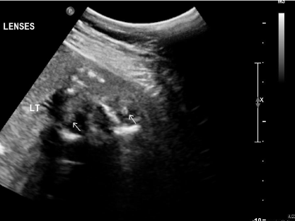
Urgent evaluation in the ophthalmology clinic revealed mild bilateral microphthalmia with a horizontal corneal diameter of 8 mm. Dense total cataracts and normal looking iris was noted bilaterally without other anterior segment anomalies. Pupils were centrally placed, symmetrical in size and reactive to light with no relative afferent pupillary defect. External ocular movements appeared to be full. Dilated fundus examination found no red reflex in either eye and leukocoria was noted bilaterally. Intra Ocular Pressures (IOP) were normal. B-scan ultrasound was unremarkable for retinal detachment, persistent fetal vasculature, hemorrhage, or malignancies in the posterior segment. Notably, history revealed that a routine antenatal 2-D fetal ultrasound at 17 weeks gestation identified bilateral hyperechogenic crystalline lenses and renal pyelectasis with an otherwise unremarkable craniofacial profile (Figure 1).
Genetic evaluation and investigation
In the initial assessment by Genetics at two weeks of age, the proband measured at the 75th percentile, 50th percentile, and 25th percentile for height, weight, and head circumference, respectively. Micrognathia was noted on physical examination with no other evidence of dysmorphic facial features. A comprehensive physical examination of all major systems was unremarkable.
A review of the family history revealed that the proband’s mother, of Scottish and Ukrainian ancestry, was diagnosed with bilateral sutural VY cataracts at six years of age. Regular follow-up by ophthalmology has not identified cataract progression or changes to visual acuity in the mother. Aside from a history of myopia, the mother had no relevant past medical history. Interestingly, on physical examination, she displayed screwdriver-shaped central incisors and small lateral incisors. The family medical history was otherwise insignificant. There were no additional cases of congenital or childhood cataracts in either parental lineage. Paternal family roots originated from Switzerland and Germany and had an unremarkable medical and ocular history. There was no consanguinity reported. There was no known family history of intellectual disability, congenital anomalies, inherited disorders, miscarriages, stillbirths, or sudden infant deaths. Family history was unremarkable for renal anomalies.
Aside from NHS, other differential diagnoses for a newborn with antenatal diagnosis of hyperechoic lenses and renal pyelectasis and postnatal findings of microphthalmia and microcornea included: (1) Lowe syndrome, characterized by mutations in OCRL1 and clinical features of aminoaciduria, failure-to-thrive, and intellectual disability; (2) Lenz syndrome, associated with alterations in NAA10 and a triad of dental, renal, and digital anomalies. Genomic analysis was performed at 18 months old to sequence genes related to congenital cataracts. Whole exome sequencing and Sanger sequencing analysis identified a pathogenic variant of the NHS gene in the proband, while the proband’s mother was heterozygous for the variant, confirming her carrier status.
Surgical intervention, Outcomes and Follow-up
At six weeks of age, the proband underwent bilateral cataract extraction with anterior vitrectomy. The right eye was operated on first, with limbal incisions at the 11 o’clock and 1 o’clock position. With the anterior chamber maintainer at 1 o’clock and 20G vitrector at 11 o’clock, anterior vitrector capsulotomy (5.0 mm), hydrodissection, and cortical matter aspiration were performed. Following a 5.0 mm posterior capsulotomy, anterior core vitrectomy was completed. Intriguingly, the iris appeared very flaccid and featureless with fewer crypts and repeatedly prolapsed at the 11 o’clock wound. Peripheral iridectomy and Miochol injection were necessitated to address this. The surgery in the left eye was performed similarly. The surgeries were otherwise uncomplicated, and the procedure was well tolerated. The fundus examination found a mild bilateral optic nerve and macular hypoplasia. Cycloplegic refraction was measured at +24.0D spherical in both eyes. Silsoft (Bausch and Lomb) aphakic contact lenses were used for visual rehabilitation. The post-operative evaluation demonstrated excellent red reflexes with clear corneas and deep/quiet anterior chambers. IOPs continued to be normal, and on the gonioscopic exam, the angles were open. An improvement in visual attention, fixation, and tracking behaviour was evident.
Two weeks post-operatively, the right eye revealed a dense Posterior Capsular Opacification (PCO), needing an urgent surgical secondary capsulectomy with anterior vitrectomy to clear the visual axis. IOPs were within the normal range. The capsular opening was enlarged to 5.0 mm. In follow up, three months later, significant PCO with capsular phimosis recurred in the right eye. The pupil was drawn towards the old incision, forming an iatrogenic corectopia and necessitating a second secondary capsulectomy with anterior vitrectomy. The posterior capsular opening was improved to 5.0 mm. Pupilloplasty was also carried out in the inferior margin of the pupil, to pull down the pupil to the visual axis. The post-operative ophthalmologic evaluation demonstrated a persistent right peaked pupil with an excellent red reflex. Peaking was attributed to synechiae, and vitreous was not noted at the site of peaking. Visual behaviour was found to be improving with aphakic contact lenses.
Three months after the initial bilateral cataract surgery, the proband had worsening peaking of the pupil in the left eye with a dull red reflex. Similar to the right eye, a dense posterior capsular opacity with iatrogenic corectopia was noted. Left secondary capsulectomy (5.0 mm opening) with anterior vitrectomy and pupilloplasty was performed to establish a clear visual axis. Healthy red reflexes were observed in both eyes post-operatively. After these multiple surgeries, the eyes were stable; visual behaviours and function continued to improve with aphakic contact lenses. IOPs were within the normal range.
Unexpectedly, ten months after the initial cataract surgery, a dull red reflex in the right eye cued a shallow retinal detachment. An urgent pars planar vitrectomy with an air-fluid exchange, endolaser retinal reattachment, and C3F8 gas exchange were performed. Despite a successful anatomical retinal attachment, the proband developed deep amblyopia. Patching for amblyopia therapy was challenging secondary to deep amblyopia, and ultimately, the family transitioned to aphakic glasses. The proband’s final visual acuity, at two years of age, was 20/1800 and 20/100 in the right and left eye, respectively.
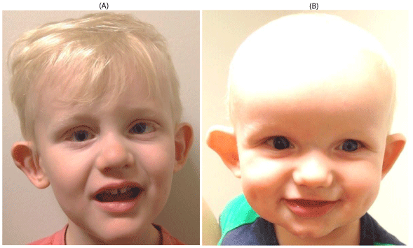
Interestingly, the mother became pregnant with a second male child whose antenatal ultrasounds demonstrated similar bilateral lenticular opacities. He was assessed by the Pediatric Ophthalmology service at two weeks of age. He was found to have bilateral microcornea, microphthalmia, and dense nuclear lenticular opacities. Normal extraocular movements were found. IOP measurements and gonioscopic examination were within normal limits. At six weeks age, he underwent bilateral cataract extraction with anterior 20 G vitrectomy. Posterior capsulectomies were 5 mm in size. Remarkably, as witnessed in his older brother, the patient’s cataract extraction became challenged by very floppy iris prolapse bilaterally, necessitating peripheral iridectomies. The limbal incisions were very tight even though made with 20 G vitrector and V lance blade. The balanced salt solution infusion flow was kept very low at 20 ml/min. Similarly, the suction rate was also kept lower at 100-150 mmHg. Limbal entry incisions were made just a little anterior when compared to the proband. The wounds were covered with conjunctiva at the end of the surgery. Fundus examination identified bilateral mild optic nerve and macular hypoplasia. Post-operatively, in both eyes, refraction was found to be +24D spherical, and the horizontal corneal diameter measured 8.0 - 8.5mm. The patient was visually rehabilitated with aphakic contact lenses. At five weeks post-cataract surgery, the right pupil was pear-shaped and displaced superiorly to the 11 o’clock position. The pupil was almost displaced to the limbus, and there was little to no visual axis. Pupilloplasty with secondary anterior capsulectomy and 20G vitrectomy was warranted (posterior capsular opening of 5 mm). The small, slit-like pupil was enlarged with the vitrector. At six months post-operatively, the eyes were stable. The right pupil has remained large and irregularly shaped post pupilloplasty; however, the visual axis has maintained clarity. The left eye did not need more surgical intervention. IOPs were within the normal range, and the filtration angles consistently remained open post-operatively. At 11 months of age, visual acuity with contact lenses (Silsoft, Bausch and Lomb) measured 20/960 in each eye, with the patient displaying sustained fixation with age-matched smooth pursuit. The post-operative pictures of these two brothers with NHS is presented in Figure 2.
In both patients, entry wounds into the anterior chamber were tight with 20G cutter during all procedures. Anterior chamber maintainer was used continuously throughout the surgery, and care was always taken to maintain adequate depth in the anterior chamber. Viscoelastic was used sparingly. Post-operative care in both patients included tobramycin eye drops for a week, and prednisolone eye drops for six weeks. Atropine eye drops were used for 4 to 5 days after surgery.
Discussion and conclusion
Congenital cataract is among the primary drivers of preventable childhood blindness and vision impairment worldwide [11]. More than 50% of all cataract presentations at birth or during early childhood have been attributed to a heterogeneous array of genetic conditions [12]. While autosomal dominant transmission of congenital cataracts is the most common form of inheritance, all patterns of Mendelian inheritance have been described [13]. The study presented here depicts the genetic, clinical, and surgical and post-surgical details of a family diagnosed with Nance-Horan Syndrome (NHS), an X-linked inherited disorder.
Albeit genetic mapping is not routinely performed in cases of congenital cataracts, the display of dental anomalies and diagnosis of childhood cataracts in the proband’s mother was suggestive of a genetic disorder in the context of the proband’s presentation. As syndromic features were absent in the proband at that point, and he was too young to manifest potential features such as dental anomalies, whole-exome sequencing followed by Sanger sequencing of variants was performed at 18 months of age. Initially, the proband’s parents were not interested in pursuing genetic testing as they felt it would not change his clinical management or their family planning, particularly as the mother did not have a history of progressive cataract complications or require surgical interventions. However, as the proband experienced multiple capsulectomies with anterior vitrectomy and a retinal detachment by 15 months of age, the potential benefits of a genetic diagnosis were discussed to guide monitoring and management of possible future comorbidities. By the time genetic testing was agreed upon, the mother was pregnant with the next child. In our study, the genetic analysis identified the proband to be hemizygous for a pathogenic variant of the NHS gene, while his mother was a heterozygous carrier for the same mutation. When genetic testing confirmed NHS, the family was provided with full genetic counselling, which included an explanation regarding the milder symptoms in the mother as compared to the severe presentation in the children, as well as the 25% recurrence risk in future pregnancies. Additionally, the family was made aware of available prenatal diagnoses for future pregnancies.
Despite cataract surgery in infancy, affected males in Nance-Horan Syndrome have pronounced reduced visual acuity, likely secondary to aberrant development of ocular structures and related predispositions for surgical complications [5,8]. We propose that pervasive ocular inflammatory and sclerotic aberrancies in NHS and a form of “contractile tissue” in the vitreous and surrounding tissues may contribute to various surgical complications, as well as the pathogenesis of the retinal detachment, noted in the proband’s case. In this case report, a very floppy iris prolapsing was encountered, necessitating iridectomy. Post-operatively, corectopia and capsular phimosis were noted as complications. Interestingly, histopathological assessment of an enucleated NHS eye has previously identified attenuated iris with neovascularization and degenerated ciliary bodies, raising the possibility of increased anterior segment inflammation in NHS eyes that contributes to iris prolapse and capsular phimosis [3]. Our patients displayed a unique susceptibility for recurrent dense PCO, which demanded multiple capsulectomies. Prompt surgical intervention should be planned to clear the visual axis. The rate of posterior capsular opacity in pediatric cataract surgery have ranged from 43.7 to 100% in the current literature [14]. At this time, there is a paucity of data on the prevalence and incidence rates of PCO in syndromic children, specifically among pediatric patients with NHS.
NHS patients may be inherently predisposed to retinal detachment. Retinal architecture has shown to be perturbed in the pathological tissue of NHS eyes, whereby the retina lacked photoreceptors, ganglion cells, and retinal pigment epithelium with notable peripheral cystoid degeneration across retinal layers. The choroid had inflammatory infiltrates and lacked choriocapillaris [3]. Mathys, et al. [10], reported analogous retino-choroidal abnormalities as well as diminished retinal function on electroretinogram with a pronounced deficit of rods. These pathological and inflammatory processes may predispose NHS patients to impaired visual function and retinal detachments. The cumulative 20-year risk of retinal detachment after pediatric cataract surgery is 7% [15]. Among pediatric patients with ocular or systemic diseases, in addition to cataracts, the overall 20-year risk of retinal detachment increases to 16%, compared to 3% in patients with isolated cataracts. Moreover, the risk of retinal detachment post-cataract surgery significantly increases to 23% in patients with intellectual disabilities. Haargaard, et al. [15], also reported a statistically significant risk of retinal detachment associated with a prior history of pars plana surgery in the pediatric population. Understandably, the increased probability of retinal detachment among those with a previous history of multiple intraocular surgeries cannot be excluded. However, with limited availability of published data surrounding complications in syndromic patients, it is challenging to concretely dissociate whether the prior history of numerous intra-ocular surgeries or the genetic disorder is the causative factor for the proband’s retinal detachment.
In the presented report, our NHS patients neither demonstrated high intraocular pressures or poorly developed anterior chamber angles with gonioscopy during pre- and post-operative assessments. Nevertheless, approximately half of the males diagnosed with NHS are expected to develop severe and unrelenting glaucoma [6]. While secondary glaucoma has been described as an innate consequence of cataract extraction in infants, glaucoma development in NHS patients may also be interceded by aberrancies in the trabecular meshwork anatomy and aqueous humour drainage system [3]. As such, clinicians must present patients with routine follow-up appointments for judicious IOP monitoring.
To our knowledge, there is little to no published literature to guide eye surgeons in the management of pediatric patients with NHS. Intra- and post-operative challenges in NHS reviewed in this report can help surgeons with anticipation and management of surgical complications. Additionally, expansive knowledge of potential complications may prime the expectations of the surgical team and parents for the likelihood of multiple intraocular surgeries and suboptimal visual outcomes in NHS patients.
- Walpole IR, Hockey A, Nicoll A (1990) The Nance-Horan Syndrome. J Med Genet 27: 632-634. Link: https://bit.ly/3fXcFjg
- Toutain A (2003) Nance-Horan syndrome. NORD Guide to Rare Disorders. Philadelphia (PA): Lippincott Williams & Wilkins; 654-655. Link: https://bit.ly/2ybxMO1
- Ding X, Patel M, Herzlich AA, Sieving PC, Chan C (2009) Ophthalmic Pathology of Nance-Horan Syndrome: Case Report and Review of the Literature. Ophthalmic Genet 30: 27-35. Link: https://bit.ly/3fYdh8y
- Gjørup H, Haubek D, Jacobsen P, Ostergaard JR (2017) Nance-Horan Syndrome-The oral perspective on a rare disease. Am J Med Genet A 173: 88-98. Link: https://bit.ly/3dYeOd4
- Sharma S, Datta P, Sabharwal JR, Datta S (2017) Nance-Horan Syndrome: A Rare Case Report. Contemporary Clinical Dentistry 8: 469-472. Link: https://bit.ly/2X4oqfn
- Lewis RA (1989) Mapping the gene for X-linked cataracts and microcornea with facial, dental, and skeletal features to Xp22: An appraisal of the Nance-Horan syndrome. Trans Am Ophthalmol Soc 87: 658-728. Link: https://bit.ly/3bI1sQi
- Nance WE, Warburg M, Bixler D, Helveston EM (1974) Congenital X-lined cataract, dental anomalies and brachymetacarpalia. Birth Defects Orig Art Ser 10: 285-291. Link: https://bit.ly/2TffNh4
- Accogli A, Traverso M, Madia F, Bellini T, Vari MS, et al. (2017) A novel Xp22.13 microdeletion in Nance-Horan Syndrome. Birth Defects Res 109: 866-868. Link: https://bit.ly/3bE0YuM
- Burdon KP, McKay JD, Sale MM, Russell-Eggitt IM, Mackey DA, et al. (2003) Mutations in a novel gene, NHS, cause the pleiotropic effects of Nance-Horan Syndrome, including severe congenital Cataract, dental anomalies, and mental retardation. Am J Hum Genet 73: 1120-1130. Link: https://bit.ly/2zPg5nD
- Mathys R, Deconinck H, Keymolen K, Jansen A, Van Esch H (2007) Severe visual impairment and retinal changes in a boy with a deletion of the gene for Nance-Horan Syndrome. Bull Soc Belge Ophthalmol 305: 49-53. Link: https://bit.ly/3bGcsxF
- Wu X, Long E, Lin H. Liu Y (2016) Prevalence and epidemiological characteristics of congenital cataract: a systematic review and meta-analysis. Sci Rep 6: 28564. Link: https://bit.ly/2TcHA1C
- Pichi F, Lembo A, Serafino M, Nucci P (2016) Genetics of Congenital Cataract. Dev Ophthalmol 57: 1-14. Link: https://bit.ly/2z9KQUz
- Khan L, Shaheen N, Hanif Q, Fahad S, Usman M (2018) Genetics of congenital cataract, its diagnosis and therapeutics. Egyptian Journal of Basic and Applied Sciences 5: 252-257. Link: https://bit.ly/2yaAy62
- Awasthi N, Guo S, Wagner BJ (2009) Posterior capsular opacification: A problem reduced but not yet eradicated. Arch Ophthalmol 127: 555-562. Link: https://bit.ly/2yVDDrb
- Haargaard B, Andersen EW, Oudin A, Poulsen G, et al. (2014) Risk of retinal detachment after pediatric cataract surgery. Invest Ophthalmol Vis Sci 55: 2947-2951. Link: https://bit.ly/2X4p6Br

Article Alerts
Subscribe to our articles alerts and stay tuned.
 This work is licensed under a Creative Commons Attribution 4.0 International License.


 Save to Mendeley
