Journal of Cardiovascular Medicine and Cardiology
Immediate effect of thoracic manual therapy on respiratory parameters, chest expansion and rom of thorax and cervical spine in mild to moderate COPD-A randomized clinical trial
Ganesh BR1 and Chandra Bahadur Khatri2*
2Post Graduate Student, CVTS Physiotherapy, KAHER’s Institute of Physiotherapy, Belgaum, Karnataka, India
Cite this as
Ganesh BR, Khatri CB (2020) Immediate effect of thoracic manual therapy on respiratory parameters, chest expansion and rom of thorax and cervical spine in mild to moderate COPD-A randomized clinical trial. J Cardiovasc Med Cardiol 7(1): 028-032. DOI: 10.17352/2455-2976.000107Background: COPD is one of the non-communicable diseases related to lung parenchyma and airways which are one of the leading causes of death and disability in India. COPD is not only considered as lung disease but also it has systematic effects that lead to co-morbidities contributing to poor performance in functional level and decreased quality of life. COPD has considerable effects in musculoskeletal system. It ranges from muscle dysfunction of respiratory system to skeletal muscle system to reduced bone density. It is also associated with decreased range of motion in cervical and thoracic region. Physiotherapy plays vital role in preventing and managing the complications in COPD patients. It has been long since exercises that consists of breathing techniques, thoracic expansion exercise and techniques to clear the secretion has been used for management of COPD. One of the emerging techniques for management of musculoskeletal dysfunctions is manual therapy. Hence current study is intended to compare the acute effects of breathing exercises and thoracic expansion exercises with high velocity low amplitude thrust.
Objectives: To evaluate and compare the immediate effect of High velocity low amplitude thrust with breathing exercises and thoracic expansion exercises on respiratory parameters, chest mobility and range of motion in cervical and thoracic spine.
Methods: Total 26 participants were divided randomly into HVLAT group (N=13) and exercise group (N=13). All the participants were assessed for baseline outcome measures and received respective treatment. Following treatment, the pulmonary function test, thoracolumbar flexibility, cervical range of motion and chest expansion were recorded and analysed.
Results: Mean age of participants in HVLAT group was 60.08±5.31 years and exercise group was 60.23±4.60 years and both groups were homogeneous in nature. Significant differences were observed only in thoracolumbar flexibility within both groups (p=0.001), increased cervical flexion (p=0.026) and chest expansion at axillary level (p=0.026) in exercise group and increased lateral flexion in HVLAT group (p=0.044) but there were no any differences in other parameters within and between two groups.
Conclusion: This study concluded that either of the intervention was insufficient to produce significant effects on respiratory parameters, range of motion and chest expansion except for thoracolumbar flexibility, cervical flexion and lateral flexion. Neither of treatment is superior to one another in terms of their acute effects.
Introduction
According to Global Initiative for Obstructive Lung Disease guidelines 2018 (GOLD 2018), Chronic Obstructive Pulmonary Disease (COPD) is ‘a common, preventable and treatable disease, characterized by persistent airflow limitation that is usually progressive and associated with an enhanced chronic inflammatory response in the airways and lungs to noxious particles or gases. Exacerbations and co morbidities contribute to the overall severity in individual patients’. COPD is the third leading cause of deaths around the globe and India stands as the capital of COPD patients in the world [1,2]. Burden of Obstructive lung disease(BOLD) along with Disability Adjusted Life Years (DALY) is increasing day by day leading to severe impairment in quality of life [3]. In India, COPD is the second leading cause of deaths which has prevalence rate range between 2 to 22% among men and 1.2 to 19% among women and more prominent in Northern states (Uttarkhand, Uttar Pradesh, Rajasthan) compared to Southern states. It commonly affects age group of above 40 [2].
Patients with COPD present not only with dyspnoea and chronic cough but also decreased exercise capacity. Lung hyperinflation, hypoxemia, hypercapnia, inflammation, malnutrition, long-term use of corticosteroids, physical inactivity, and changes in fibre-type distribution in the respiratory muscles are the main factors that contribute to respiratory muscle dysfunction in COPD. Lung hyperinflation brings mechanical disadvantage in respiratory muscles during functioning for which chest wall tries to compensate through reconfiguration and thus decrease in ability of diaphragm muscle to contract effectively. This leads to activation of accessory muscles for breathing thus increasing work of breathing [4-6]. Researches also suggest that COPD is also linked with other co morbidities such as reduced bone mineral density (50%-70%) and skeletal muscle dysfunction (32%). Skeletal muscle dysfunction can include reduced muscle strength, endurance and decreased muscle girth. COPD is also associated with decreased cervical and thoracic range of motion particularly affecting lateral bending in cervical region and rotation in both cervical and thoracic region along with cervicothoracic pain [7].
Manual therapy is one of the approaches that can be applied to correct functional or structural abnormality mainly in orthopaedic conditions [8]. Manual therapies includes various techniques that ranges from corrective exercise, chiropractic, osteopathy, physiotherapy, massage therapy, and muscle training [9]. One of the technique used in manual therapy for thoracic spine is High Velocity Low Amplitude Thrust (HVLAT). Shekelle [10]. states that there are four main hypotheses behind HVLAT manipulation: release of entrapped synovial folds or plica, relaxation of hypertonic muscle by sudden stretching, disruption of articular or periarticular adhesions, and unbuckling of motion segments that have undergone disproportionate displacements [8]. HVLAT is a safe manoeuvre that can be performed in COPD patients with very few reported cases of adverse effects and application of this technique requires a sound knowledge of anatomy and biomechanics for its effectiveness [11,12].
The immediate effects of thoracic manual therapy performed in healthy subjects by Doo Chul Shin, et al., showed a promising result [13]. Nicola R. Heneghan has performed a systematic review which concluded that there was no evidence to accept or refute the beneficial effect of manual therapy in COPD [14]. However, a systematic review done by Jaxson Wearing shows the beneficial effects of manual therapy in pulmonary functions [15]. This study is conducted to evaluate and compare the immediate effect of High velocity low amplitude thrust with breathing exercises and thoracic expansion exercises on respiratory parameters, chest mobility and range of motion in cervical and thoracic spine.
Methodology
Study design and settings
This was a randomized clinical trial study performed in tertiary care hospital on subjects with COPD from November 2018 to March 2019.
Ethical consideration
Ethical clearance was obtained from institutional ethical committee. Participants of this research were provided with informed consent sheet and their voluntary participation, safety and privacy was assured.
Participants and randomization
All the subjects in the study were scanned for inclusion and exclusion criteria and recruited accordingly. Subjects of either gender, aged 40-65years who were willing to participate in study were included along with history of mild to moderate COPD on the basis of GOLD classification. Severe COPD, presence of scoliosis or any mechanical deformity in thoracic spine, severe osteoporosis, joint hypermobilty, and pregnant women were excluded.
Subjects were evaluated for their baseline data of pulmonary functions, range of motion of cervical and thoracic region and chest expansion. Subjects were informed about procedure of the study. Randomization was done using envelope method and participants were divided into HVLAT and exercise group.
Outcome measures
Pulmonary function testing (FEV1, FVC): Vitalograph COPD-6 device was used while subject is in sitting position.
Range of motion of thorax and cervical region: Range of motion can be defined as degree of freedom available in the joint. In cervical region flexion, extension, side bending was recorded using universal goniometer. Thoracic range of motion is measured along with lumbar with the help of finger to floor distance using tape.
Chest expansion (tape method): Thoracic respiratory excursion was used for the measurement of chest expansion. Chest expansion was measured in two different levels: Upper chest and lower chest. Anatomic markers for measures of upper thoracic excursion are third intercostal space at the mid clavicular line, and fifth thoracic spinous process. Anatomic markers for measures of lower thoracic region are tip of the xiphoid process and 10th thoracic spinous process. Measurements were taken in normal breathing initially and subject is asked to take deep breath and hold. Initial and final values were noted.
Intervention
High velocity low amplitude group (HVLAT group): The subjects lying in supine/sitting position comfortably with arms crossed and therapist on the contra lateral side. To manipulate one side of the upper thorax, mid thorax and lower thorax, hand was placed on the spinous process of thoracic vertebrae on third vertebrae, seventh vertebrae and tenth vertebrae respectively. The position of hand was in pistol grip and subject was asked to take a deep breath and relax and thenar eminence was used to generate force on vertebrae in the cephalic direction.
Breathing exercises and thoracic expansion exercise group (exercise group): The subjects were asked to be seated comfortably and relaxed with hands placed on abdomen and asked to perform pursed lip breathing and thoracic expansion exercise for upper, middle and lower thoracic region. Pursed lip breathing and thoracic expansion exercise was asked to perform with 10 repetitions and 3 sets.
Statistical analysis
Statistical analysis was performed by using SPSS software version 18.0. Data were entered in excel sheet, tabulated and subjected to statistical analysis. Normality of the data was assessed using Shapiro Wilk test. To assess changes within group, paired t-test was used. Independent t-test was performed for normally distributed data to compare differences between groups and homogeneity. Chi square test was used to analyse categorical dataLevel of significance was set as 0.05 (5%).
Results
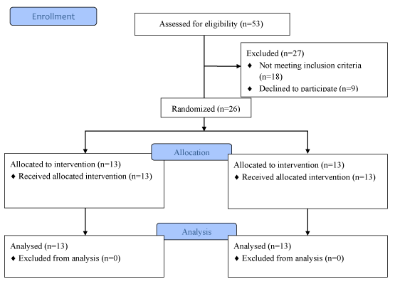
Figure 1 shows the enrolment of participants. Table 1 shows the demographic characteristics of the study population. There were 10 men and 3 women in HVLAT group and 9 men and 4 women in exercise group. Mean age in HVLAT group was 60.07±5.31 and exercise group was 60.23±4.6 years. Mean height in HVLAT group was 162±6.68 and exercise group was 163.38±7.13. Subjects in HVLAT weighted 57.84±4.09 and exercise group 54.68±5.99kg. There was no any difference between demographic characteristics which implies for the maintained homogeneity. Baseline values of pulmonary function testing range of motion of cervical region and thoracolumbar region and chest expansion in axillary and xipsternum level was also included in same table. There was no any differences in baseline demographics in HVLAT and exercise group.
Table 2 shows the mean changes in baseline characteristics within group and between groups. Significant differences within HVLAT group can be observed in side flexion to left side (p-0.44) and finger to floor distance (p=0.001). There was significant differences in flexion range of motion (p=0.026), finger to floor distance (p=0.001) and chest expansion (p=0.026) within exercise group. There were no any differences in outcomes after intervention of each technique.
Discussion
Present study was done to compare the immediate effects of HVLAT and exercises on respiratory parameters, range of motion of cervical and thoracic spine and chest expansion. It revealed that both the techniques are not superior to each other in terms of changes in baseline characteristic of outcome measures, however some significant changes can be observed within groups.
It has been believed that high velocity low amplitude thrust enhances function of sympathetic and parasympathetic system and produces relaxation of skeletal muscles along with improved blood circulation which in turn improves pulmonary funtion [13]. In our study, HVLAT have shown no any difference post intervention in PFT values. This result is consistent with previous study done by Bradley, et al., [16]. Changes in PFT values not only depend on structural compliance but also the impairment and adaptation in respiratory muscles over a period of time after COPD [5]. Though lateral flexion on left side showed increased range with statistical significance but couldn’t establish any clinical significance with improved range. But there was significant improvement in finger to floor distance. Finger to floor test was significantly improved following administration of HVLAT. This is supported by previous study done regarding immediate effect of HVLAT. It is proposed that HVLAT produces structural, neurophysiological and autonomic changes that results in increased flexibility in thoracolumbar flexibility [17]. Although this study was done in normal individuals, effects can be generalized in subjects with COPD as well without any known clinical implications. There was no any improvement seen in chest expansion at two levels after HVLAT intervention which contradicts the study done by Maji, et al., HVLAT is not sufficient to produce therapeutic effect in COPD as compared to healthy individuals in previous study [18].
Breathing exercises has been practiced in management of COPD since decades. Pursed lip breathing has shown beneficial effects in management of COPD symptoms [19]. Thoracic expansion exercises is a part active cycle of breathing technique which mainly focuses on inspiration and aids in loosening the secretion [20,21]. However, minimum dosage required to achieve clinically meaningful effects have not been known till now. In our study, there was no any change in PFT scoring following intervention of breathing exercises. Significant changes can be observed in flexion of cervical region, finger to floor distance and chest expansion at axillary level but it doesn’t add any clinical significance in improvement of signs and symptoms immediately after intervention of breathing exercises.
HVLAT and breathing exercises, when analysed individually, were not able to produce any significant changes in pulmonary function but has minimal effect on range of motion of cervical and thoracic range of motion as well as chest expansion. There was no any significance when compared with each other.
There were certain limitations of this study. Functional outcome measures would have been added in this study. Spotlight should have been thrown more in assessment of dyspnoea as it is major reason for hospital admission. Similarly, small sample size was also a limitation. Future studies can focus in addressing these limitations.
Conclusion
The present study concluded that HVLAT and breathing exercises are unable to produce significant acute effects in COPD.
Future scope
Future studies can focus more in determining minimum dosage required to produce clinically meaningful effects through application of HVLAT and breathing exercises along with addition of functional outcome measures in COPD patients.
- (2018) Global Initiative for Chronic Obstructive Lung disease (GOLD). Link: http://bit.ly/39evcn3
- Hossain MM, Sultana A, Purohit N (2018) Burden of chronic obstructive pulmonary disease in India: status, practices and prevention. Int J Pulm Respir Sci 2: 001-004. Link: http://bit.ly/39dHts3
- Chronic Respiratory diseases. World Health Organization. Link: http://bit.ly/2HcfLA1
- Charususin N, Dacha S, Gosselink R, Decramer M, Von Leupoldt A, et al. (2018) Respiratory muscle function and exercise limitation in patients with chronic obstructive pulmonary disease: a review. Expert Rev Respir Med 12: 67-79. Link: http://bit.ly/3bjO0mV
- Orozco-Levi M (2003) Structure and function of the respiratory muscles in patients with COPD: impairment or adaptation? Eur Respir J 22: 41s-51s. Link: http://bit.ly/2UuUIR3
- Heneghan N, Adab P, Jackman S, Balanos G (2015) Musculoskeletal dysfunction in chronic obstructive pulmonary disease (COPD): An observational study. Int J Ther Rehabil 22: 119-128. Link: http://bit.ly/2Uztsku
- Heneghan NR (2014) Chronic obstructive pulmonary disease and cervico-thoracic musculoskeletal dysfunction. (Doctoral dissertation, University of Birmingham) Link: http://bit.ly/37jtYG4
- Evans DW (2002) Mechanisms and effects of spinal high-velocity, low-amplitude thrust manipulation: previous theories. J Manipulative Physiol Ther 25: 251-262. Link: http://bit.ly/2ODPChI
- Grace S, Orrock P, Vaughan B, Blaich R, Coutts R (2016) Understanding clinical reasoning in osteopathy: a qualitative research approach. Chiropr Man Therap 24: 6. Link: http://bit.ly/38iEJtz
- Shekelle PG (1994) Spine update spinal manipulation. Spine 19: 858-861. Link: http://bit.ly/2UAiFpV
- Puentedura EJ, O'Grady WH (2015) Safety of thrust joint manipulation in the thoracic spine: a systematic review. J Man Manip Ther 23: 154-161. Link: http://bit.ly/2H2f3Fl
- Edmondston SJ, Singer KP (1997) Thoracic spine: anatomical and biomechanical considerations for manual therapy. Man Ther 2: 132-143. Link: http://bit.ly/2S7G9Bg
- Shin DC, Lee YW (2016) The immediate effects of spinal thoracic manipulation on respiratory functions. J Phys Ther Sci 28: 2547-2549. Link: http://bit.ly/2H5kkvK
- Heneghan NR, Adab P, Balanos GM, Jordan RE (2012) Manual therapy for chronic obstructive airways disease: a systematic review of current evidence. Man Ther 17: 507-518. Link: http://bit.ly/376oCO3
- Wearing J, Beaumont S, Forbes D, Brown B, Engel R (2016) The use of spinal manipulative therapy in the management of chronic obstructive pulmonary disease: a systematic review. J Altern Complement Med 22: 108-114. Link: http://bit.ly/2tIgCW4
- Wall BA, Peiffer JJ, Losco B, Hebert JJ (2016) The effect of manual therapy on pulmonary function in healthy adults. Sci Rep 6: 33244. Link: http://bit.ly/3bnEcIx
- Griffiths FS, McSweeney T, Edwards DJ (2019) Immediate effects and associations between interoceptive accuracy and range of motion after a HVLA thrust on the thoracolumbar junction: A randomised controlled trial. J Bodyw Mov Ther 23: 818-824. Link: http://bit.ly/2uwW9DX
- Maji B, Goyal M, Kumar SP (2018) Immediate Effects of Thoracic Spine Thrust Manipulation on Chest Expansion and Lung Function in Healthy Subjects- a pre- test and Post- Test Experimental Design. Int J Current Adv Res 9: 15616-15621. Link: http://bit.ly/38ftSAi
- Gosselink R (2004) Breathing techniques in patients with chronic obstructive pulmonary disease (COPD). Chron Respir Dis 1: 163-172. Link: http://bit.ly/38p6tg9
- Bronchiectasis Toolbox: The Active Cycle of Breathing. Link: http://bit.ly/39xAx9x
- Oxford University Hospitals. The Active Cycle of Breathing Techniques. Link: http://bit.ly/2uoUKiV

Article Alerts
Subscribe to our articles alerts and stay tuned.
 This work is licensed under a Creative Commons Attribution 4.0 International License.

 Save to Mendeley
