Journal of Clinical Microbiology and Biochemical Technology
What psoriasis can hide
Salmón-González Z1*, R Herreras-Martínez1, M Latorre-Asensio1, S Nieto1, C Garilleti2 and MS Rodríguez Duque3
2Departament of Cardiology of H.U. Marqués de Valdecilla, Santander, Spain
3Department of Pathological Anatomy of H.U. Marqués de Valdecilla, Santander, Spain
Cite this as
Salmón-González Z, Herreras-Martínez R, Latorre-Asensio M, Nieto S, Garilleti C, et al. (2023) What psoriasis can hide. J Clin Microbiol Biochem Technol 9(1): 001-002. DOI: 10.17352/jcmbt.000052Copyright
© 2023 Salmón-González Z, et al. This is an open-access article distributed under the terms of the Creative Commons Attribution License, which permits unrestricted use, distribution, and reproduction in any medium, provided the original author and source are credited.Introduction
Alpha-1 antitrypsin deficiency (DAAT) is a genetic disorder that manifests as pulmonary emphysema, liver cirrhosis, and, rarely, as the skin disease panniculitis [1], but other rare skin manifestations had been described previously in literature [2]. Despite being one of the most frequent genetic diseases, professionals need high clinical suspicion for its diagnosis.
In this article, we report a case of psoriatic erythroderma associated with DAAT. With this case, we want to underline the importance of skin involvement as part of the clinical spectrum of this disease.
Case report
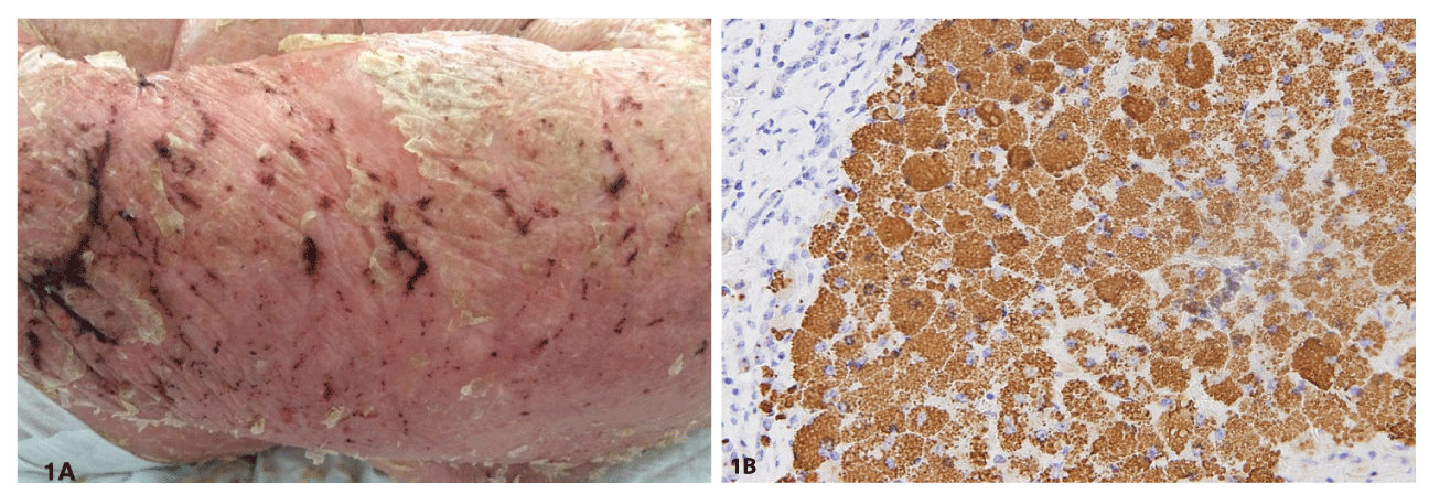
A-76 -year-old-woman with a history of psoriasis was admitted to our hospital due to a 5-day history of erythematous-scaly skin lesions on the face, trunk, and limbs findings consistent with a severe outbreak of psoriatic erythroderma (Figure 1A). Her medical history included morbid obesity, no toxic habit, hypertension, and skin psoriasis with limited involvement of the retroauricular area and the back of the hands and feet. On examination, the cardiovascular, pulmonary, and neurological systems showed no distress.
Pertinent laboratory values included creatinine 1.53 mg/dl, Urea 188 mg/dl, impaired liver function tests [Aspartate Aminotransferase (AST) 150U/l; Alanine Aminotransferase (ALT) 97U/l, alkaline phosphatase 253 U/l, total bilirubin 5.2 mg/dl] together with elevated Lactate Dehydrogenase (LDH) 450 U/l, Albumin 2.2 g/dl, activity prothrombin 52% and polyclonal hypergammaglobulinemia and alpha 1 antitrypsin value below the limit of normality; the rest of chronic liver disease study is normal. Imaging tests confirm the presence of chronic liver disease, unknown until this admission, with moderate ascites, a portal with liver discharge and splenomegaly.
The patient suffered multiple complications secondary to the decompensation of her liver diseases (spontaneous bacterial peritonitis and upper digestive hemorrhage due to esophageal varices). Despite efforts, the patient succumbed to death. Finally, the autopsy confirmed these findings and revealed ZZ genotype DAAT as the origin of the cirrhosis (Figure 1B).
Discussion
DAAT is one of the most common genetic diseases [1] that causes an increased risk of lung diseases, liver diseases, and more rarely panniculitis. It is caused by a mutation in the SERPINE 1 gene which has two alleles transmitted by codominant autosomal inheritance and located on chromosome 14q32.1; Alleles are identified with the letters according to their electrophoretic mobility: F (Fast), M (Medium), S (Slow), Z (Very Slow) or null (null). Normal alleles are called M and are present in 85% - 90% of individuals forming the MM genotype, which expresses in 100% of AAT. The most frequent deficient alleles are S and Z; Although more than 50 deficient alleles are known in clinical practice, 96% of individuals with diseases related to DAAT have ZZ genotype [1,3].
AAT is produced and secreted predominantly by hepatocytes (80%) and its main function is the inhibition of proteases and the excess elastase of neutrophils that occurs during inflammation.
The clinical manifestations are due to the absence of AAT in the tissues, as occurs in pulmonary involvement (panacinar emphysema) and in necrotizing panniculitis. On the contrary, liver dysfunction is due to the abnormal accumulation of the polymer in the endoplasmic reticulum of the hepatocyte [1,3].
Other skin diseases have been described in patients with DAAT, including pemphigus herpetiformis, Muir-Torre syndrome, Antineutrophil Cytoplasm Antibodies (ANCA) positive vasculitis, and psoriasis. As early as 1979 Beckman, et al. established a relationship between DAAT and the risk of suffering from psoriasis [4]. Later in 1984, Heng MCY, et al. [5]. Suggested that the deficiency of protease inhibitors had a fundamental role in modifying the course of psoriasis, relating the alleles that confer a lower amount of AAT (MS, MZ, ZZ) with a more severe course of psoriasis as is the case of our patient.
In conclusion, we want to highlight the importance of the diagnosis of DAAT from skin manifestations starting with panniculitis because it is the most frequent manifestation and follows the most serious cases of psoriasis, given its prevalence. Enzyme deficiency is not likely to be the direct cause of the skin lesions, but it does predispose to the development of more severe manifestations even more when other contributing factors such as toxic habits, infections, or other inflammatory processes concur. We propose including skin manifestations within the triad of classic findings that should alert the clinician to check the patient’s serum AAT levels (pulmonary emphysema, liver cirrhosis, and skin disease). This consideration may avoid delays in the diagnosis of AAT deficiency.
- Strnad P, McElvaney NG, Lomas DA. Alpha 1 -Antitrypsin Deficiency. N Engl J Med. 2020; 382:1443-1455.
- Bazex J, Bayle P, Albes B. Le déficit en alpha-1-antitrypsine. Place au sein des états pathologiques cutanés [Alpha-1-antitrypsin deficiency. Role in skin disorders]. Bull Acad Natl Med. 2002;186(8):1479-86; discussion 1486-7. French. PMID: 12669363.
- Greene CM, Miller SD, Carroll T, McLean C, O'Mahony M, Lawless MW, O'Neill SJ, Taggart CC, McElvaney NG. Alpha-1 antitrypsin deficiency: a conformational disease associated with lung and liver manifestations. J Inherit Metab Dis. 2008 Feb;31(1):21-34. doi: 10.1007/s10545-007-0748-y. Epub 2008 Jan 16. PMID: 18193338.
- Bazex J, Bayle P, Albes B. Le déficit en alpha-1-antitrypsine. Place au sein des états pathologiques cutanés [Alpha-1-antitrypsin deficiency. Role in skin disorders]. Bull Acad Natl Med. 2002;186(8):1479-86; discussion 1486-7. French. PMID: 12669363.
- Heng MC, Kloss SG. Cell interactions in psoriasis. Arch Dermatol. 1985 Jul;121(7):881-7. PMID: 2409930.

Article Alerts
Subscribe to our articles alerts and stay tuned.
 This work is licensed under a Creative Commons Attribution 4.0 International License.


 Save to Mendeley
