International Journal of Vascular Surgery and Medicine
Acute appendicitis revealed by an abscess of the anterior abdominal wall: A case report
Bannar B1*, Mahad R1, Boutakioute B1, Ouali Idrissi M1, Cherif Idrissi Ganouni N1, Maaroufi Fathiallah EL2, EL Boukhfaoui M2, Rabbani K2, Louzi A2 and Finech B2
2General and Visceral Surgery Department, AR-RAZI Hospital, Mohamed VI teaching hospital, Cadi Ayyad University, Marrakesh, Morocco
Cite this as
Bannar B, Mahad R, Boutakioute B, Ouali Idrissi M, Cherif Idrissi Ganouni N, et al. (2020) Acute appendicitis revealed by an abscess of the anterior abdominal wall: A case report. Int J Vasc Surg Med 6(1): 011-013. DOI: 10.17352/2455-5452.000038Introduction: Appendicitis is a common cause of acute abdomen; however the classic clinical signs are not often present, which can be manifested by numerous complications, but the revelation by a parietal collection remains unusual, its diagnosis can be challenging.
Presentation of case: We describe the case of a 31 years old man, who complains for pain of the right flank. The clinical examination objective an erythematous and painful swelling of the right iliac fossa, the abdominal ultrasound has objectified a collection of the right flank and right iliac fossa, The CT Scan of the Abdomen/Pelvis found a voluminous collection of soft tissues of the right anterolateral abdominal and pelvic wall, hypo dense and heterogeneous, containing air, The surgical exploration objectified a huge purulent collection with subcutaneously ceacum and appendage suppurated and perforated.
Conclusion: The onset of an abdominal wall abscess without a known cause needs to be thoroughly investigated, with consideration of a subjacent abdominal cause and appendicitis.
Introduction
Appendicitis is a prevalent cause of abdominal pain, it is a typical disease of young men, with a high incidence in those aged 10–19 years and a low frequency (5–10%) in elderly individuals aged more than 50–60 years [1]. Diagnosing acute appendicitis can be challenging, as the typical clinical history and signs are not present in 30–40% of cases [2,3].
Additionally, making a diagnosis can be more challenging in some groups of patients, including elderly individuals, which contributes to a delayed diagnosis and complicated onset [2,4]. Many clinical evaluations, such as the Alvarado score [4] and further imaging examinations, such as ultrasonography, Computed Tomography (CT), are used for the diagnosis.
Presentation of case
A-31-years old man, who complains for pain of the right flank evolving for 8 days, with fever, the clinical examination found an erythematous and painful swelling of the right iliac fossa, and an elevation of the infectious balance, The first management option was to stabilize the patient with intravenous antibiotics and , electrolyte correction before imagining.
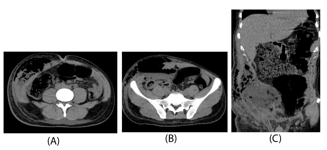
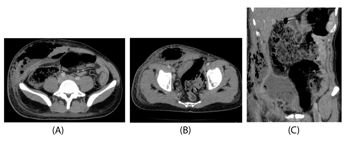
The abdominal ultrasound objectified a collection of the right flank and right iliac fossa, hypo echoic heterogeneous, poorly limited, not illuminating with color doppler, the CT scan abdomen and pelvic showed a voluminous collection of soft tissues of the right anterolateral abdominal-pelvic wall measuring: 17x10 cm and 25 cm high, quite limited, hypodense and heterogeneous contrast spontaneous, containing air (Figures 1a-c) it could be secondary to a perforation of the appendage discreetly enhanced in the periphery after contrast injection (Figures 2a-c), the caecum was subcutaneous and the appendix was not visible.
The surgical exploration objectified patient under general anesthesia, incision next to the collection with aspiration of pus and discovery of an appendix with an intact base in the center of this gangrenous collection and an appendectomy was performed with washing and drainage by two blades, one lower and the other subcutaneously.
Post-operative consequences are marked by wound suppuration, the patient is declared discharged on the 7th day of his hospitalization.
The anatomy pathology examination did not find an underlying tumor and the follow –up post operative was simple without complications.
Discussion
The onset of an abdominal wall abscess without a known cause needs to be thoroughly investigated, with consideration of a subjacent abdominal cause and appendicitis necessitates [1].
The pathophysiology of our case remains poorly explained in the literature, we have not been able to explain the exact theory of this atypical manifestation of acute appendicitis.
In some publications in the literature we found the notion of appendiculo-cutaneous fistula: rare complication of acute appendicitis this fistula is an abnormal communication between the digestive tract and the skin through the appendix [5].
The imaging examination, plays a very important role in the description and confirmation of the abscess of the abdominal wall, and to search its origin, and used to increase the diagnostic accuracy; however, in some patients, the diagnosis can only be made per operatively.
Ultrasound can objectify a collection of the anterior or subcutaneous abdominal wall ill-defined heterogeneous hypoechoic not lighting up with color doppler, sometimes seat of air bubbles. With an important infiltration of mesenteric fat and subcutaneous fat.
The CT better specifies the extent of the collection which is usually spontaneously hypodense increasing in periphery after injection of the product of contrast, the defect of the muscles of the abdominal wall, as well as the underwater situation of the cecum and the appendix which is sometimes perforated and will therefore be invisible.
Conventional treatment of appendicitis is surgical excision of the inflamed appendix as soon as the diagnosis is suspected to prevent perforation. Treatment may vary when the patient presents with complications. All patients with unexplained groing and thigh symptoms require a thorough evaluation to exclude intraabdominal disease despite minimal abdominal signs [1]. In the present patient, the complication was the formation of an abscess of the appendices with drainage to the abdominal wall. This condition was named appendicitis necessitatis by Gockel, et al. [6]. The onset of an abdominal wall abscess without a known cause requires thorough investigation to exclude abdominal diseases, and appendicitis should be considered [7].
Conclusion
The numerous anatomical variations of the appendix partially explain the pitfalls of clinical diagnosis.
In addition to the clinicobiological couple, ultrasound is often sufficient for positive diagnosis of acute appendicitis, especially in young subjects. CT is necessary and indispensable for complicated forms (especially appendicular perforations), and for most ectopic forms. We remind finally that acute appendicitis can be manifested also by a collection of the anterior abdominal wall in the right iliac fossa, even if it remains rare in the literature.
- De Souza IMAG, Nunes DADA, Massuqueto CMG, De Mendonça Veiga MA, Tamada H (2017) Complicated acute appendicitis presenting as an abscess in the abdominal wall in an elderly patient: A case report. Int J Surg Case Rep 41: 5-8. Link: https://bit.ly/3mPGA0z
- Zbierska K, Kenig J, Lasek A, Rubinkiewicz M, Wałega P (2016) Differences in the clinical course of acute appendicitis in the elderly in comparison to younger population. Pol Przegl Chir 88: 142–146. Link: https://bit.ly/3cyCsxe
- Barlas SU, Günerhan Y, Palanci Y, Işler B, Çağlayan K (2010) Epidemiological and demographic features of appendicitis and influences of several environmental factors. Turk J Trauma Emerg Surg 16: 38-42. Link: https://bit.ly/33RMxBu
- Alvarado A (2016) How to improve the clinical diagnosis of acute appendicitis in resource limited settings. World J Emerg Surg 11: 1-4. Link: https://bit.ly/3iTNNds
- El Haoudi K, Majdoub KI (2014) Fistule appendiculo-cutanée: complication rare de l’appendicite aiguë. Pan Afr Med J 19: 194. Link: https://bit.ly/2G3WLX0
- Gockel I, Jäger F, Shah S, Steinmetz C, Junginger T (2007) Appendicitis necessitatis. Der Chirurg 78: 840-842. Link: https://bit.ly/33RRt9r
- Singal R, Gupta S, Mittal A, Gupta S, Singh M, et al. (2012) Appendico-cutaneous fistula presenting as a large wound: a rare phenomenon-brief review; Acta Med. Indones 44: 53-56. Link: https://bit.ly/2G4cIww
Article Alerts
Subscribe to our articles alerts and stay tuned.
 This work is licensed under a Creative Commons Attribution 4.0 International License.


 Save to Mendeley
