International Journal of Vascular Surgery and Medicine
Diagnosis and indications for revascularization in Takayasu’s Arteritis: Report of two cases and literature review
Magaye Gaye*, Adama Sawadogo, Papa Adama Dieng, Ndèye Fatou Sow, Souleymane Diatta, Momar Sokhna Diop, Papa Salmane Ba, Assane Ndiaye, Amadou Gabriel Ciss and Mouhamadou Ndiaye
Cite this as
Gaye M, Sawadogo A, Dieng PA, Sow NF, Diatta S, et al. (2017) Diagnosis and indications for revascularization in Takayasu’s Arteritis: Report of two cases and literature review. Int J Vasc Surg Med 3(3): 036-039. DOI: 10.17352/2455-5452.000026Takayasu’s arteritis (TA) is an inflammatory disease of large vessels that predominantly affects the aorta and its main branches such as supra-aortic trunks, renal and digestive arteries. The diagnosis is based on criteria proposed by the American College of Rheumatology and modified by Sharma. These vascular lesions present a problem of surgical indications because of their pathogenic particularity. In this work, we report our experience on the diagnosis and management of two cases of TA. The case 1 was a 62-year-old female patient diagnosed with stenosis of the common carotid artery and the right subclavian artery. A bypass between the carotid artery and the subclavian artery was indicated but not performed. The second patient was a 23-year-old female patient diagnosed with renovascular hypertension. Investigations showed a significant stenosis of the left renal artery. She underwent angioplasty-stenting of the left renal artery and the result was good. Her echocardiography showed left ventricular and atrial hypertrophy and both. The two patients had no indirect signs of myocardial ischemia and arterial pulmonary injuries.
Introduction
Takayasu’s arteritis (TA) is a chronic inflammatory disease of the vessels. The aorta and its main branches such as the supra-aortic trunks, the renal and digestive arteries are predominantly affected. This inflammation leads to thickening of the vascular wall due to fibrosis that is the most characteristic early sign of Takayasu’s disease [1,2]. This thickening leads to stenosis and formation of thrombus which are responsible for various complications such as ischemia [3]. The diagnosis of this disease is based on a set of radio-clinic arguments including the clinical condition, the topography of involved artery, its appearance (stenosis or ectasia) and the association with cutaneous and visceral lesions [4]. The criteria proposed by the American College of Rheumatology [3] as well as the modified criteria of Sharma [5] can be used in case vasculitis to make the diagnosis. There are 3 major criteria including stenosis or occlusion of the middle segment of the left or right subclavian artery in angiography and characteristic symptoms lasting at least one month (claudication, pulseless or asymmetric pulsation, fever, cervicalgia, amaurosis, visual disturbances, syncope, dyspnea and palpitations). There are 10 minor criteria that include sensitivity of the carotid artery to palpation, brachial arterial pressure > 140/90 mmHg, stenosis or occlusion of the middle portion of the left carotid artery or distal third of the brachiocephalic trunk in the angiography and many others. Therefore, a rating based on clinical, radiological and histological criteria appears to be more useful in practice [4]. On the other hand, coronary and arterial pulmonary lesions can be observed during this disease. Indeed, the presence of 2 major criteria or 1 major criterion + 2 minor or 4 minor criteria suggests a high probability of Takayasu’s disease [5]. Above all, the treatment of TA is medical and consists of giving corticosteroid and even immunosuppressive treatment sometimes. Revascularization is considered in case of non-response to medical treatment or in case of complications such as visceral ischemia, aneurysm or thrombosis. Regarding the revascularization, open surgery and endovascular treatment are currently discussed [6]. We report two cases of Takayasu arteritis with arterial complications that were managed in our department. Then we will discuss the diagnosis and indications of revascularization while we will be reviewing the current data of the literature.
Clinical Cases
Case 1
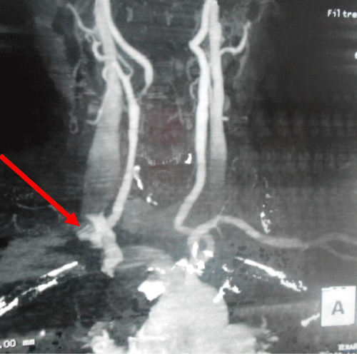
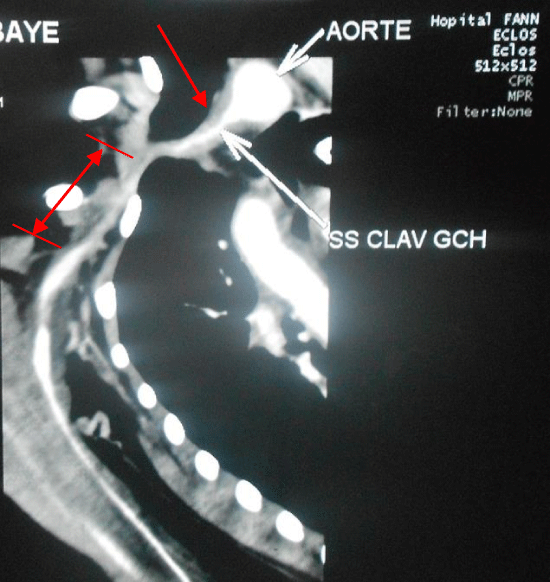
Mrs. DB was 62 years old. She consulted for bilateral impotence of the upper limbs associated with paresthesia and a feeling of coldness of the hands. She was diabetic type 1 and was being treated with insulin. Also, she was treated for hypertension. The clinical examination did not found trophic changes or upper limb deficit. At the right arm, the pulses were abolished on humeral, radial and ulnar arteries. Clinically, she had no cardiac and pulmonary signs. In addition, a right carotid bruit was auscultated. The arterial ultrasound showed many lesions including a severe occlusion of the right subclavian artery ostium (Figure 1), a stenosis (greater than 60%) of the middle segment of the right common carotid artery, an occlusive stenosis of the right internal carotid artery and a stenosis (50% of the lumen) of the left common carotid artery. The angio CT of the supra-aortic trunks also showed more lesions that consisted of a severe stenosis of the proximal right subclavian artery, an occlusion of the vertebral arteries on the V0 and V1 segments and a stenosis of the right internal carotid artery (Figures 1,2). There was not calcification. The echocardiography showed a conserved ejection fraction of left ventricle and a lack of myocardial ischemia. CRP and platelet counts were normal. Taking into account the clinical arguments and complementary investigations, the diagnosis of Takayasu’s disease was made. Immunosuppressive therapy was started in addition to naftidrofuryl and acetylsalicylic acid. Six months later, the symptomatology was persisting. Then an arteriography was prescribed. It showed a 5 cm-long occlusion at the origin of the right subclavian artery. Also, it revealed an occlusion of the right renal artery. It was impossible to catheterize the right subclavian artery and as such it was impossible to perform endovascular procedure for revascularization. Subclavian artery bypass was indicated but was not performed because of refusal of the patient. She discharged from hospital but later, she was missing during the follow up.
Case 2
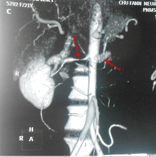
RB was a 23-year-old female patient who was suffering from hypertension for 3 years. Despite her triple anti-hypertensive therapy (bisoprolol, amlodipine and indapamide), her blood pressure remained high. On examination, RB presented good general condition. In the upper left limb there was an abolition of radial and humeral pulse without signs of ischemia. The left carotid pulse was absent. Clinically, she had no pulmonary and cardiac signs. The Doppler ultrasound of the supra-aortic trunks revealed diffuse wall thickening in the brachiocephalic artery and common carotid with significant reduction of their lumen responsible of damping of internal carotid flow. The flow of the right vertebral artery is damped on its segment 2. The angio CT showed a long and severe stenosis of the axillary artery (6 cm) and of the left axillary artery on 13 mm in a context of diffuse parietal arterial thickening (Figure 3). In addition, there was bilateral stenosis of the renal arteries just after theirs origins. The length was about 13 mm on the right and 10 mm on the left (Figure 4). The echocardiogram noted a left atrial and ventricular hypertrophy, a conserved ejection of the left ventricle and a lack of indirect signs of myocardial ischemia. The biology revealed an increased erythrocyte sedimentation rate (42 mm Hour 1), the CRP was also increased to 96 mg/L with leucocytosis at 16,700 / mm3. The Waaler Rose test was negative. The final diagnosis was vascular lesions in a context of Takayasu’s disease. Endovascular revascularization of the renal arteries was decided. The perioperative arteriography confirmed a stenosis of 90% of the left renal artery and 35% of the right. Angioplasty-stenting was performed on the left. The postoperative course was uneventful and the patient was discharged from the hospital on day 3. The second postoperative control on day 45 was good; in particular the blood pressure was normalized (120/70 mm Hg). However a follow up of the other vascular lesions is still ongoing.
Discussion
Takayasu’s arteritis (TA) was described for the first time in 1908 by Mikito Takayasu, a Japanese ophthalmologist [7]. Like in our two cases, it is a pathology that mainly affects women in about 80 to 100% of the cases [8]. It occurs in the young patients during theirs 20’s or 30’s [1]. Indeed in the series of Kerr et al. [9], only 2 of 10 patients were aged over 40 years old at the period when diagnosis was made. In fact, the delay between the first symptoms and the diagnosis is very variable and the disease can be discovered late like in our case 1 (62 years old) because the signs are not very specific during the pre-occlusive phase of this disease. In addition, the absence of cephalic, ophthalmic and articular signs made us eliminate the Horton’s disease. On the other hand, a biopsy of the temporal artery would be more formal. The diversity and disparity of clinical signs led the American College of Rheumatology to suggest diagnostic criteria in 1990 [3] which were modified by Sharma in 1996 [7]. This intends to facilitate the diagnosis. Thus, the case 1 presented 2 major criteria and the case 2 had 1 major criterion and 3 minor criteria. Taking into account the Sharma score, both our patients were diagnosed with TA with a sensitivity of 92.5% and a specificity of 95% [5]. No biopsy or biomolecular tests were prescribed because the pathogenesis of TA remains elusive and there is no biological specific test [10,11]. New imaging tools such as computerized tomography or magnetic resonance angiography, fludeoxyglucose positron emission tomography-computerized tomography and recently contrast-enhanced ultrasonography are frequently used in the diagnosis and to assess vascular inflammation. Accumulating evidence shows that biological agents such as anti-tumor necrosis factor agents, tocilizumab and rituximab could be used effectively in refractory cases [12]. According to literature data, the corticosteroid therapy is the first-line treatment and in the event of failure the addition of methotrexate may stabilize the disease [13]. The medical treatment was different in our 2 patients. In the case 1, it consisted of an arterial vasodilator, a platelet aggregation inhibitor and an immunosuppressive drug in the place of corticosteroid due to diabetes whereas the case 2 benefited from antihypertensive and corticosteroid therapy. This is mainly related to the difference in the clinical features of the patients. In the literature, no therapeutic protocol is unanimously recognized [8] although corticosteroid is recommended during the acute resurgences. The efficacy of anticoagulants and antiplatelet agents has not been demonstrated because hypercoagulability and thrombosis are not common complications of TA [4].
The Takayasu arteritis surgical treatment is indicated in case of failure of medical treatment. Revascularization can be done by endovascular or by bypass. It was necessary in our patients who were already in the occlusive stage of the disease. For the case 1, the failure to catheterize the right subclavian artery indicated subclavian artery bypass. Moreover, the indication for revascularization of the right common carotid artery was relevant due to the significant stenosis of this artery. Open surgery is an effective method according to Kim et al. [6] as they used 15 surgical bypasses on 25 patients (60%) with lesions of the supra-aortic trunks. This would avoid major complications such as stroke. The case 2 underwent endovascular treatment: angioplasty - stenting of the renal arteries with a good result. This indication was justified by the Chaudhry’s study which suggests renal revascularization in all cases of renal artery stenosis ≥ 70% with or without reno-vascular hypertension [11]. It is a less invasive and less morbid method compared with surgical bypass and which shortens the hospitalization delay [11]. To refresh, the case 2 stayed only 3 days in the hospital. Angioplasty only is associated with a high rate of re-stenosis [6], hence the interest of stenting. The long-term prognosis depends on major complications such as aortic insufficiency, retinopathy, arterial aneurysms and increasing erythrocyte sedimentation rate [11]. According to current data regarding TA revascularization, conventional surgery is indicated in large vessel lesions [6,14], whereas endovascular techniques remain more effective in stenosis of the renal artery [11]. This attitude is explained by the pathogenesis of the vascular lesions of this disease. Indeed, the latter is characterized by a pan-arteritis associating adventitial thickening, focal infiltration of the tunica media and intimal hyperplasia. The association of these micro-lesions results in stenosis or occlusion [15]. Thus, in the case of angioplasty-only or associated with stenting there is a continuous intimal cell proliferation that will create a secondary stenosis [6]. Other therapeutic modalities are being explored for the treatment of TA such as TNF inhibitors and active angioplasty balloons but they have not proved their worth yet [15].
Conclusion
The surgical management of vascular lesions of Takayasu’s disease remains accessible in the developing countries because conventional open surgery is much more appropriate than endovascular surgery. The main challenge is the early diagnosis of these vascular lesions.
- Quéméneur T, Hachulla EH, Lambert M, Perez-Cousin M, Queyrel V, et al. (2006) Maladie de Takayasu. Press Med 35: 847-856. Link: https://goo.gl/49jht4
- De Souza AW, de Carvalho JF (2014) Diagnosis and classification criteria of Takayasu arteritis. J Autoimmun 48: 79-83. Link: https://goo.gl/mzp6Jb
- Arend WP, Michel BA, Bloch DA, Hunder GG, Calabrese LH, et al. (1990) The American College of Rheumatology criteria for the classification of Takayasu arteritis. Arthritis Rheum 33: 1129-1134. Link: https://goo.gl/69jP9m
- Jacq F, Emmerich J (1998) Maladie des vaisseaux. Ed Doin Paris. pp 187-93.
- Sharma BK, Jain S, Suri S, Numano F (1996) Diagnostic criteria for Takayasu arteritis. Int J Cardiol 54: 141-147. Link: https://goo.gl/M9qwD6
- Kim YW, Kim DI, Park YJ, Yang SS, Lee GY, et al. (2012) Surgical bypass vs endovascular treatment for patients with supra-aortic arterial occlusive disease due to Takayasu arteritis. J Vasc Surg 55: 693-700. Link: https://goo.gl/jWL4Xj
- Takayasu MA (1908) A case with unusual changes of the central vessels in the retina. Acta Soc Ophtalmol Jpn ; 12: 554-5.
- Lambert M, Hachulla E, Harton PY, Perez-Cousin M, Beregi JP, et al. (1998) Atérite de Takayasu :Explorations vasculaires et prise en charge thérapeutique thérapeutique. Expérience à propos de 16 cas. Rev Méd Interne 19: 878-884. Link: https://goo.gl/9FbDqx
- Kerr GS, Hallahan CW, Gordano J, Leavitt RY, Fauci AS, et.al. (1994) Takayasu arteritis. Ann Intern Med 120: 919-929. Link: https://goo.gl/i8rUU1
- Monach PA (2014) Biomarkers in vasculitis. Curr Opin Rheumatol 26: 24-30. Link: https://goo.gl/EdK95f
- Chaudhry MA, Latif F (2013) Takayasu's arteritis and its role in causing renal artery stenosis.Am. J Med Sci 346: 314-318. Link: https://goo.gl/qQxBSv
- Seyahi E (2017) Takayasu arteritis. An update Curr Opin Rheumatol 29: 51-56. Link: https://goo.gl/TtoUUX
- Abdelmajid Bouzerda, Ali khatouri (2016) manifestations cardiaques de la maladie de Takayasu: A propos d'une observation et revue de la littérature. Pan Afr Med J 24: 82. Link: https://goo.gl/YCkR8w
- Keser G, Direskeneli H, Aksu K (2014) Management of Takayasu arteritis a systematic review. Rheumatology 53: 793-801. Link: https://goo.gl/h2T1Xk
- Andrews J, Mason JC (2007) Takayasu's arteritis-recent advances in imaging offer promise. Rheumatology 46: 6-15. Link: https://goo.gl/WGXRLU
Article Alerts
Subscribe to our articles alerts and stay tuned.
 This work is licensed under a Creative Commons Attribution 4.0 International License.


 Save to Mendeley
