International Journal of Spine Research
New therapy option: Maisonneuve fracture without transsyndesmotic fixation
Dachang Feng, Zhaofa Liu* and Haitao Chen
Cite this as
Feng D, Liu Z, Chen H (2022) New therapy option: Maisonneuve fracture without transsyndesmotic fixation. Int J Spine Res 4(1): 009-012. DOI: 10.17352/ijsr.000022Copyright License
© 2022 Feng D, et al. This is an open-access article distributed under the terms of the Creative Commons Attribution License, which permits unrestricted use, distribution, and reproduction in any medium, provided the original author and source are credited.Ankle fracture is one of the common injuries in the orthopedic department, the Maisonneuve fracture is a specific type of ankle injury. This fracture is usually caused by rotational force. According to the Lauge -Hansen classification, it is a pronation and external rotation type injury, often resulting in inferior tibiofibular injury. Because it is extremely unstable, it is usually treated surgically.
Operative treatment includes medial malleolus fixation, reduction of the inferior tibiofibular joint and screw fixation. When the fibula fractured is without shortening or dislocation, it is still controversial if the inferior tibiofibular joint needs fixation. This study aims to introduce a new method-Maisonneuve without transsyndesmotic fixation and analysis the follow-up result.
Introduction
Ankle fracture is one of the common injuries in the orthopedic department. The Maisonneuve fracture was first reported by the French doctor Maisonneuve in 1840 [1-3]. It’s a specific type of ankle injury. It includes injury of the medial structure (medial malleolus fracture or deltoid ligament tear), inferior tibiofibular syndesmosis injury, and proximal 1/3 fracture of the fibula. Associated with the posterior tibiofibular ligament or fractures of the posterior malleolus injures sometimes.
This fracture is usually caused by rotational force. According to the Lauge - Hansen classification, it is a pronation and external rotation type injury, often resulting in inferior tibiofibular injury. Because it is extremely unstable, it is usually treated surgically.
Operative treatment includes medial malleolus fixation, reduction of the inferior tibiofibular joint, and screw fixation [4-6]. When the fibula fractured is without shortening or dislocation, it is still controversial if the inferior tibiofibular joint needs fixation. The aim of this study is to introduce a new method-Maisonneuve without transsyndesmotic fixation and analysis the follow-up result.
Case description
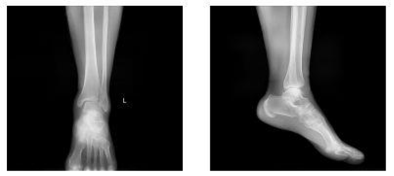
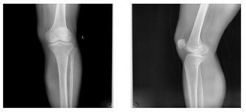
A 56-year-old woman was sent to the emergency department by her family. She sprained her left ankle when she went down the stairs. She had difficulty walking because of a painful left ankle. Specialized physical examination found tenderness in the anterolateral distal tibia and fibular neck. X-ray showed fractures in the tibia lateral border and fibular neck, with no evidence of inferior tibiofibular joint dislocation (Figures 1,2). A computed tomography scan confirmed the presence of Tillaux-Chaput and Volkmann fractures (Figures 3-5).
Operative process
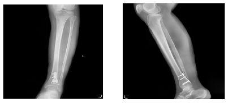
The anesthetic for the operation was lumbar anesthesia. A posterolateral approach was used to expose the Volkmann fracture. An anterior tibial approach was used to expose the Tillaux-Chaput fracture. The inferior tibiofibular joint was checked carefully. Fractures were reduced and then fixed. Volkmann fracture was fixed by buttress plate, and Tillaux-Chaput fracture was performed by hollow screw. Hook test intraoperative was negative. Anatomical reduction and perfect stability were confirmed by X and CT postoperatively (Figures 6.7). CT scan in the transverse section confirmed that the anterior and posterior edge of the distal fibula is in an arc with the fractures of the tibia. It meant there was no dislocation of the distal fibula. The anterior and posterior ligaments of the inferior tibiofibular ligament are in good condition.
Three weeks after surgery, the patient is required to touch down weight-bearing in a plaster splint. Patients are then allowed partial weight-bearing after six weeks and progress to weight-bearing. Ankle joint function exercises can be done three weeks after surgery. X noted that the fracture healed without shortening or rotation of the fibula when twelve weeks after surgery (Figure 8). The AOFAS score is 92 at twelve weeks follow-up [7,8].
Discussion
Fracture of the fibula in approximately one-third is an important feature of Maisonneuve fracture [2]. The damage mechanism of Maisonneuve fracture is the 3 or 4 stages of PER(pronation–external rotation) injury following Lauge–Hansen classification [9-11]. The first is medial malleolus fracture or deltoid ligament rupture, even the inside of the structure is complete [12]. The second is the rupture of the AITFL(anterior inferior tibiofibular ligament) or avulsion fracture of the AITFL attachment point, the rupture of the IOL(interosseous ligament)and IOM(interosseous membrane), and the third is a fracture of the proximal fibula sometimes even without proximal fibula fractures [13]. If the rotational force doesn’t end, it’s the fourth stage. It is the rupture of the PITFL(posterior inferior tibiofibular ligament) or the avulsion fracture of the PITFL.
It is well known that the mainstream treatment option is the fixation of medial malleolus fracture, reduction of the inferior tibiofibular joint, and screw fixation [14,15]. Some scholars have also reported the efficacy of suture-button devices in the treatment of Maisonneuve fracture [16,17]. There are some defects in screw fixation or suture-button devices. The inferior tibiofibular screw was surgically removed. Treatment costs are more. How to control the pressure and reduction on the tibiofibular joint. Thordarson et.al confirmed contact pressures on inferior tibiofibular malreduction resulting in poorer joint function [18]. In addition, there is a risk of screw breakage in screw fixation [19]. The efficacy of suture-button devices is proven [17]. But previous research by Coetzee et.al suggested that suture-button devices do not guarantee axial stability of the fibula, they recommended plate and screw fixation for Maisonneuve fracture [20]. Klitzman, et al. summarized that the mobility of the suture button may promote physiological healing of the inferior tibiofibular ligament [21]. Fraissler L, et al. summarized a study that ankle arthroscopy can guide the reduction of the tibiofibular joint and check the location and direction of screws to reduce errors, patients can get a better prognosis [22].
When the fibular fracture is in normal length/rotation, inferior tibiofibular joint fixation or suture-button devices is still controversial.
Four factors of ankle stability are medial malleolus, deltoid ligament(mostly deep layer ), lateral malleolus, and inferior tibiofibular ligament. We image the theory-the stability of the ankle joint as a ring. When three-quarters of the ring is stable structurally, the ankle is stable. So it is with Maisonneuve’s injury as well. In this type, no fracture with medial malleolus, the deltoid ligament is damaged, the fibularis in normal length, but exists rotation due to Tillaux-Chaputand Volkmann fracture. According to the ring theory, Volkmann fracture fixation was performed by buttress plate, and Tillaux-Chaput was by hollow screw. Hook test intraoperative was negative. A short leg cast protects the ankle for 6 weeks. According to follow-up, the treatment was effective.
According to the theory of PER injury, the fourth stage is the rupture of the PITFL(posterior inferior tibiofibular ligament) or the avulsion fracture of the PITFL. Bone and ligament strength varies in different populations. Avulsion fractures of the anterior and posterior inferior tibiofibular ligaments are more common because the epiphyseal is not closed and the ligaments are strong in children. The rupture of the anterior and posterior inferior tibiofibular ligament is more common and avulsion fractures at the ligament attachment point are rare because the bone is stronger than the ligament in young and middle-aged people. Avulsion fractures at the ligamentous attachment point are more common because of osteoporosis in the elderly. Volkmann fracture occurs instead of the rupture of the posterior inferior tibiofibular ligament in this type. We consider the mechanism is pronation–external rotation with ankle plantar flexion. Ankle plantarflexion causes Volkmann fracture, not avulsion fracture. This is similar to an ankle fracture in a child, the posterior inferior tibiofibular ligament (PITFL) is well, but a posterior ankle epiphyseal fracture occurs. Similar Maisonneuve fractures may be more pronounced in children because ligaments are stronger than epiphyses.
When Maisonneuve fracture with these features-avulsion fractures of the attachment point on the inferior tibiofibular ligament, the inferior tibiofibular joint may not be fixed. When the fibula is not shortening and the avulsion fracture is firmly fixed, the inferior tibiofibular joint will be stable.
Conclusion
No transsyndesmotic fixation for Maisonneuve is not the mainstream treatment but is a new option for Maisonneuve fractures in some cases(children and elderly ). Postoperative follow-up confirmed that patients with similar Maisonneuve with the new technique can achieve good function and avoid the need for tibiofibular screw removal, thereby reducing surgical costs. There are still a few defects -only 14 patients.
- Duchesneau S, Fallat LM. The Maisonneuve fracture. J Foot Ankle Surg. 1995 Sep-Oct;34(5):422-8. doi: 10.1016/S1067-2516(09)80016-1. PMID: 8590875.
- Maisonneuve JG. Recherches sur la fracture du perone. Arch Gen Med. 1840; 7:165-187.
- Merrill KD. The Maisonneuve fracture of the fibula. Clin Orthop Relat Res. 1993 Feb;(287):218-23. PMID: 8448946.
- Babis GC, Papagelopoulos PJ, Tsarouchas J, Zoubos AB, Korres DS, Nikiforidis P. Operative treatment for Maisonneuve fracture of the proximal fibula. Orthopedics. 2000 Jul;23(7):687-90. doi: 10.3928/0147-7447-20000701-15. PMID: 10917243.
- Miller AN, Carroll EA, Parker RJ, Helfet DL, Lorich DG. Posterior malleolar stabilization of syndesmotic injuries is equivalent to screw fixation. Clin Orthop Relat Res. 2010 Apr;468(4):1129-35. doi: 10.1007/s11999-009-1111-4. Epub 2009 Oct 2. PMID: 19798540; PMCID: PMC2835618.
- Stufkens SA, van den Bekerom MP, Doornberg JN, van Dijk CN, Kloen P. Evidence-based treatment of maisonneuve fractures. J Foot Ankle Surg. 2011 Jan-Feb;50(1):62-7. doi: 10.1053/j.jfas.2010.08.017. PMID: 21172642.
- Ibrahim T, Beiri A, Azzabi M, Best AJ, Taylor GJ, Menon DK. Reliability and validity of the subjective component of the American Orthopaedic Foot and Ankle Society clinical rating scales. J Foot Ankle Surg. 2007 Mar-Apr;46(2):65-74. doi: 10.1053/j.jfas.2006.12.002. PMID: 17331864.
- Kitaoka HB, Alexander IJ, Adelaar RS, Nunley JA, Myerson MS, Sanders M. Clinical rating systems for the ankle-hindfoot, midfoot, hallux, and lesser toes. Foot Ankle Int. 1994 Jul;15(7):349-53. doi: 10.1177/107110079401500701. PMID: 7951968.
- Bissuel T, Gaillard F, Dagneaux L, Canovas F. Maisonneuve Equivalent Injury With Proximal Tibiofibular Joint Dislocation: Case Report and Literature Review. J Foot Ankle Surg. 2017 Mar-Apr;56(2):404-407. doi: 10.1053/j.jfas.2016.10.003. Epub 2017 Jan 20. PMID: 28117256.
- LAUGE-HANSEN N. Fractures of the ankle. II. Combined experimental-surgical and experimental-roentgenologic investigations. Arch Surg. 1950 May;60(5):957-85. PMID: 15411319.
- Porter DA, Jaggers RR, Barnes AF, et al. Optimal management of ankle syndesmosis injuries. Open Access J Sports Med. 2014; 5:173–182.
- He JQ, Ma XL, Xin JY, Cao HB, Li N, Sun ZH, Wang GX, Fu X, Zhao B, Hu FK. Pathoanatomy and Injury Mechanism of Typical Maisonneuve Fracture. Orthop Surg. 2020 Dec;12(6):1644-1651. doi: 10.1111/os.12733. Epub 2020 Sep 7. PMID: 32896104; PMCID: PMC7767678.
- Liu GP, Li JG, Gong X, Li JM. Maisonneuve injury with no fibula fracture: A case report. World J Clin Cases. 2021 May 26;9(15):3733-3740. doi: 10.12998/wjcc.v9.i15.3733. PMID: 34046477; PMCID: PMC8130071.
- van den Bekerom MP, Raven EE. Current concepts review: operative techniques for stabilizing the distal tibiofibular syndesmosis. Foot Ankle Int. 2007 Dec;28(12):1302-8. doi: 10.3113/FAI.2007.1302. PMID: 18173998.
- van den Bekerom MP, Hogervorst M, Bolhuis HW, van Dijk CN. Operative aspects of the syndesmotic screw: review of current concepts. Injury. 2008 Apr;39(4):491-8. doi: 10.1016/j.injury.2007.11.425. Epub 2008 Mar 7. PMID: 18316086.
- Curtis MJ, Michelson JD, Urquhart MW, Byank RP, Jinnah RH. Tibiotalar contact and fibular malunion in ankle fractures. A cadaver study. Acta Orthop Scand. 1992 Jun;63(3):326-9. doi: 10.3109/17453679209154793. PMID: 1609601.
- Westermann RW, Rungprai C, Goetz JE, Femino J, Amendola A, Phisitkul P. The effect of suture-button fixation on simulated syndesmotic malreduction: a cadaveric study. J Bone Joint Surg Am. 2014 Oct 15;96(20):1732-8. doi: 10.2106/JBJS.N.00198. PMID: 25320200.
- Thordarson DB, Motamed S, Hedman T, Ebramzadeh E, Bakshian S. The effect of fibular malreduction on contact pressures in an ankle fracture malunion model. J Bone Joint Surg Am. 1997 Dec;79(12):1809-15. doi: 10.2106/00004623-199712000-00006. PMID: 9409794.
- Lambers KT, van den Bekerom MP, Doornberg JN, Stufkens SA, van Dijk CN, Kloen P. Long-term outcome of pronation-external rotation ankle fractures treated with syndesmotic screws only. J Bone Joint Surg Am. 2013 Sep 4;95(17):e1221-7. doi: 10.2106/JBJS.L.00426. PMID: 24005206.
- Coetzee JC, Ebeling P. Treatment of syndesmosis disruptions with tightrope fixation. Tech Foot Ankle Surg. 2008; 7(3):196-202.
- Klitzman R, Zhao H, Zhang LQ, Strohmeyer G, Vora A. Suture-button versus screw fixation of the syndesmosis: a biomechanical analysis. Foot Ankle Int. 2010 Jan;31(1):69-75. doi: 10.3113/FAI.2010.0069. PMID: 20067726.
- Fraissler L, Mattiassich G, Brunnader L, Holzer LA. Arthroscopic findings and treatment of maisonneuve fracture complex. BMC Musculoskelet Disord. 2021 Sep 24;22(1):821. doi: 10.1186/s12891-021-04713-8. PMID: 34560870; PMCID: PMC8461909.
Article Alerts
Subscribe to our articles alerts and stay tuned.
 This work is licensed under a Creative Commons Attribution 4.0 International License.


 Save to Mendeley
