International Journal of Spine Research
Lumbar lordosis in patients undergoing non-instrumented spinal stenosis
Anna Isart*, Gemma Vila-Canet, David Cancer, Augusto Covaro, Enric Caceres, Ana Garcia De Frutos and Maite Ubierna
Cite this as
Isart A, Vila-Canet G, Cancer D, Covaro A, Caceres E, et al. (2021) Lumbar lordosis in patients undergoing non-instrumented spinal stenosis. Int J Spine Res 3(1): 011-015. DOI: 10.17352/ijsr.000020strong>Copyright © 2021 Isart A, et al. This is an open-access article distributed under the terms of the Creative Commons Attribution License, which permits unrestricted use, distribution, and reproduction in any medium, provided the original author and source are credited.
Objective: To study the behaviour of Lumbar Lordosis (LL) after non-instrumented decompression surgery in patients diagnosed with Lumbar Spinal Stenosis (LSS).
Methods and materials: Retrospective analysis of patients undergoing non-lumbar instrumented decompression surgery for lumbar spine stenosis, operated on between January 2011 and December 2017. The variables collected were age, sex, affected segment, and presence or not of degenerative spondylolisthesis (ELS). The Lumbar Lordosis (LL) parameter was analysed using conventional radiology in standing position pre and postoperatively.
Results: 64 patients were selected, 17 women and 47 men, with an average age of 68 (35-83). 65% stenosis was located in a single level, and 39.1% had degenerative ELS grade I. The average follow-up was 26 months (6m-104m). A preoperative LL angle of 43.2º (9.8º-70.8º) and 47º (8º-76º) were found at the postoperative follow-up, with an average difference of 3.8º (-15.7º-20.2º). 9.4% (6 patients) of degenerative ELS evolved to grade II, and 8 patients needed reoperation for different reasons.
In patients with ELS, we found a greater increase in postoperative LL (5.59º) than in patients without ELS (2.61º) (p = 0.08).
No statistically significant relationship was found between the behaviour of the LL with the number of decompressed levels (p = 0.43) and the need for reoperation (p = 0.26).
Conclusions: According to our study, the technique of posterior decompression without instrumentation of the lumbar spine stenosis is not associated with a decrease of lumbar lordosis parameter. Conversely, there is a slight tendency for LL to increase in cases where a degenerative ELS is present.
Abbreviations
LL: Lumbar Lordosis, LSS: Lumbar Spinal Stenosis, ELS: Spondylolisthesis, MRI: Magnetic Resonance Imaging, VAS: Visual Analog Scale
Introduction
Lumbar Spinal Stenosis (LSS) is a narrowing of the spinal canal, lateral recesses and vertebral foramina by surrounding bone and soft tissues that compromise neural structures resulting in degenerative changes [1,2].
When conservative treatment (combination of anti-inflammatory drugs, physical therapy and conditioning, and epidural injections) fails, patients are usually referred for surgical treatment. The aim of surgery is to decompress the spinal canal and dural sac from degenerative bone and ligamentous overgrowth with laminectomy. This is the surgical technique recommended when there isn’t significant lumbar pain and there isn’t spinal instability [1-4].
The alignment of the lumbar spine has pathological changes with regard to LSS. It is recognized that the incidence of LSS in adults correlates closely with the loss of Lumbar Lordosis (LL). Patients with LSS tend to lean forward to relieve their symptoms, and preoperative LL could be improved when their symptoms reduce after decompression surgery without additional corrective procedures [5-12]. In a review by Ogura, et al. [13], in 2019, it is remarked that LL was increased after decompression, with significant improvement observed in 5 of the 6 studies. Roussouly, et al. [14], reported the standard sagittal parameters in a normal population is in average value for LL was 61.4º with a range from 41.2º to 81.9º.
The aim of the present study is to investigate the behavior of Lumbar Lordosis (LL) before and after non-instrumented decompression surgery in patients diagnosed with lumbar spinal stenosis. The hypothesis is that patients with LSS no decrease the level of LL after non-instrumented decompression surgery.
Materials and methods
Retrospective study of patients with degenerative pathology of the lumbar spine, undergoing non-lumbar instrumented decompression surgery for lumbar spine stenosis. All patients have been operated on by the same team of surgeons during the period of time from January 2011 to December 2017.
The inclusion criteria included patients diagnosed with lumbar spinal stenosis and patients with persistent neurological symptomatology after conservative treatment for 3-6 months (all of them were previously treated with medication and pain clinic with combination of anti-inflammatory drugs, physical therapy and conditioning, and epidural injections). Patients without significant lower back pain and radiological functional studies without mechanical instability was observed in. All the patients had a Magnetic Resonance Imaging (MRI) image corresponding to the symptomatology. In the MRI, it showed spinal stenosis divided into two categories: central or lateral recess involvement. In addition, some patients had lumbar juxtafacet cysts. For each patient, the levels to be decompressed were determined based on neurological examination and preoperative MRI. They also had standing x-rays (posteroanterior and lateral) of the lumbar pre and postoperative. As a result, they all had an operation of decompression and/or posterolateral non-instrumented fusion.
The exclusion criteria included patients diagnosed with other lumbar pathology, patients operated on with lumbar instrumentation, and patients without pre or postoperatively x-rays.
Epidemiological data was collected including age and gender, diagnosis of lumbar spinal stenosis, presence or not of degenerative spondylolisthesis (ELS) and we classify it according to the Meyerding classification [15], and the Lumbar Lordosis (LL) parameter. The LL parameter was analyzed using conventional radiology in standing position pre and postoperatively at the final follow-up (minimum 6 months postoperatively) using an image analysis software on a computer screen. The LL was measured according to Cobb’s method from the upper endplates of the L1 and the S1 vertebrae [16]. And by telephone interview, we asked about postoperative satisfaction (yes/no) and postoperative Visual Analog Scale (VAS) at the end of follow-up.
The surgical technique of decompression was unilateral laminectomy for lateral recess stenosis with partial facetectomy and midline decompression for central LSS with partial facetectomy if needed. The facetectomy was limited to less than 50% bilaterally.
Statistical analysis
A descriptive analysis of the sample was carried out. Qualitative variables are described with frequencies and percentages, and continuous numerical variables with mean and standard deviation (or median and 25th and 75th percentiles). To check the degree of relationship between the angle difference and the variables of age, VAS and time (months), the Pearson correlation test was used, and to check if there were angle differences between the different groups of dichotomous variables, the Student T test for independent data was used. P values lower than 0.05 were considered statistically significant.
Results
During the aforementioned period of time, 64 patients were selected who met the study inclusion criteria, with a gender distribution of 17 women (26.56%) and 47 men (73.43%), with an average age of 68 years (range 35-83). The average follow-up time was 26 months (range 6-104).
In 42 patients (65.6%), stenosis was located in a single level, in 19 patients (29.7%) were located in two levels, and in three patients (4.7%) were located in three levels. The most frequent vertebral levels were L4-L5 (60.94%), L3-L4 (28.13%) and L5-S1 (10.93%). 35 patients (54.7%) had a central lumbar spinal stenosis (in this patients, central decompression was performed) and 29 patients (45.3%) had a lateral recess stenosis (in all this cases, unilateral laminectomy was performed). Seven patients showed stenosis because of a juxtafacet cyst. In 25 patients (39.1%), there was degenerative ELS grade I; in this study all the ELS were degeneratives (Table 1).
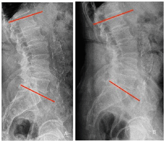
The average preoperative Lumbar Lordosis (LL) was 43.2 degrees (9.8º-70.8º) and 47 degrees (8º-76º) at the postoperative follow-up (minimum 6 months postoperatively), with an average difference of increase of 3.8º (-15.7º-20.2º) (Figure 1A,B).
Eight patients (12.5%) needed reoperation. Six patients suffered early complications within 3 months of surgery: two wound infections, 2 hematomas and 2 insufficient decompressions. Other two patients were operated for recurrent stenosis by disc bulging, one at 13 months of surgery and other 3 years after first surgery). In this series, there aren’t patients that needed a new surgery with instrumentation.
We found a mean postoperative VAS at the end of follow-up of 2.48 (0-8) with no relationship with postoperative LL variation (p=0.197). When we asked about postoperative satisfaction we found 58 satisfied patients (90.6%) and 6 dissatisfied patients (9.4%), with no relation to the evolution of LL, finding a mean LL in satisfied patients of 3.79º and 3.6º in dissatisfied patients (p=0.947).
In patients with ELS, we found a greater increase in postoperative lumbar lordosis (46.41º preoperative LL and 52º postoperative LL, increasing 5.59º) than in patients without ELS (41.28º preoperative LL and 45.22º postoperative LL, increasing to 2.61º) with a significant margin (p = 0.08).
In six patients (9.4%), degenerative ELS grade I evolves to grade II. The patients without progression of ELS had a greater increase of postoperative lumbar lordosis (6.98º) than patients with evolution of ELS (2.62º) (p=0.017).
In general, we observed a greater increase in postoperative lumbar lordosis in patients with juxtafacet cyst diagnosis (p=0.031), but there weren’t any differences between central or lateral recess lumbar spinal stenosis and the lumbar lordosis evolution (p=0.258).
No statistically significant relationship was found between the behavior of the LL with the gender distribution (p=0.355), the number of decompressed levels (p = 0.436), the number of months of x-rays postoperatively (p=0.439), and the need for reoperation (p = 0.26).
Discussion
This study data shows that the mean preoperative LL was 43.2 degrees and increased to 47 degrees at the postoperative follow-up after decompressive laminectomy without instrumentation, with a slight average improvement of 3.8º. Patients with LSS tend to walk with flexion posture in order to decrease the epidural pressure. After surgery for posterior decompression, they return to an upright posture so increasing the lumbar lordosis [17-19].
Our hypothesis confirmed that patients with LSS don’t decrease LL after non-instrumented decompression surgery, and we observed an increase of LL of 3.8º comparable to the mean increased reported in the literature, with a values between (1.7 and 6º) [13,17, 20-23]. The range of lumbar lordosis in the current series did not differ much from that of the general elderly population. This series, therefore, represents the typical patients with lumbar canal stenosis without overt deformity [2,24].
According to Madkouri [7], the loss of lordosis in LSS patients can be explained by two mechanisms, one which is adaptive and pain-relieving, and which can be improved by isolated decompression surgery. The other which is related to the aging of the spine (degenerative disc disease and muscle atrophy) and which cannot be improved by decompression surgery alone.
Jeon, et al. [17], studied the shift of LL in the first and second year postoperative, they observed that the LL increased at the 1-year follow-up, and was maintained at this level at the 2-year follow-up. However, we studied the postoperative x-rays with a minimum of six months and a maximum of 8.6 years, and we didn’t find any differences in the valuation time (p=0.439).
We didn’t observe differences between the LL and the number of decompressed levels (p=0.43). Instead, Fujiis, et al. [20], observed that the increase in LL was significantly lower in the group of 2, 3, 4 or 5 levels decompressed as compared with those in group 1 level (p= 0.02), and most of the patients are included in the 2 or 3 level decompression group, in contrast, in our study 65% of the patients are in the single level decompression group.
The patients with ELS have a higher LL angle (46.41º) than other patients with LSS (41.28º) (P=0.08) in the preoperative condition. The same situation was found by Yoshida [25] in his study where the patients of ELS are significantly higher (43.1º) than in the patients without ELS (35.5º) (p<0.005). The subgroup of patients of our study with ELS that didn’t evolve during the postoperative period have a higher LL angle than the group as a whole (p=0.017).
In a second study by Minamide and Yoshida [26], they only analyzed patients with ELS, and they divided those patients into two groups, with and without instability. They observed that the progression of ELS increased by 7% in patients with instability and increased by 8.2% in patients without instability. In our study, we observed that ELS evolved in 9.4% of patients, similar to published values.
Ogura [27] and Sigmundsson [28] studied the satisfaction after decompression surgery without fusion for lumbar spinal stenosis. Ogura observed a 75% of satisfaction with a postoperative VAS of 1.6. Sigmundsson found 64,1% of satisfaction in patients without instrumentation, with a postoperative VAS of 1. In our study, we detected a 90.6% of satisfaction but with a postoperative VAS of 2.48.
There are some limitations associated with this study. Firstly, it is a retrospective study. Secondly, we only studied the LL and we didn’t study more parameters, and we also didn’t use the total x-ray of the spine. Thirdly, we didn’t apply scores to assess the improvement of function and pain.
Conclusion
According to our study, the technique of posterior decompression without instrumentation of the lumbar spine stenosis is not associated with a decrease of lumbar lordosis parameter. Conversely, there is a slight tendency for LL to increase in cases where a degenerative ELS is present.
- Ulrich NH, Burgstaller JM, Pichierri G, Wertli MM, Farshad M, et al. (2017) Decompression Surgery Alone Versus Decompression Plus Fusion in Symptomatic Lumbar Spinal Stenosis: A Swiss Prospective Multicenter Cohort Study With 3 Years of Follow-up. Spine (Phila Pa 1976) 42: E1077-E1086. Link: https://bit.ly/3zVpnYH
- Chang HS (2018) Influence of Lumbar Lordosis on the Outcome of Decompression Surgery for Lumbar Canal Stenosis. World Neurosurg 109: e684-e690. Link: https://bit.ly/3CVs12A
- Ha DH, Kim TK, Oh SK, Cho HG, Kim KR, et al. (2020) Results of Decompression Alone in Patients with Lumbar Spinal Stenosis and Degenerative Spondylolisthesis: A Minimum 5-Year Follow-up. Clin Orthop Surg 12: 187-193. Link: https://bit.ly/3kW6eBy
- Covaro A, Vilà-Canet G, García de Frutos A, Ubierna M, Ciccolo F, et al (2017) Management of degenerative lumbar spinal stenosis: an evidence-based review. EFORT Open Rev 1: 267-274. Link: https://bit.ly/3op98RB
- Farrokhi MR, Haghnegahdar A, Rezaee H, Rad MR (2016) Spinal sagittal balance and spinopelvic parameters in patients with degenerative lumbar spinal stenosis; a comparative study. Clin Neurol Neurosurg 151: 136-141. Link: https://bit.ly/39QroLh
- Abbas J, Hamoud K, May H, Hay O, Medlej B, et al. (2010) Degenerative lumbar spinal stenosis and lumbar spine configuration. Eur Spine J 19: 1865-1873. Link: https://bit.ly/3D1X64z
- Madkouri R, Brauge D, Vidon-Buthion A, Fahed E, Mourier KL, et al. (2018) Improvement in Sagittal Balance After Decompression Surgery without Fusion in Patients with Degenerative Lumbar Stenosis: Clinical and Radiographic Results at 1 Year. World Neurosurg 114: e417-e424. Link: https://bit.ly/3CTlm8Y
- Bredow J, Oppermann J, Scheyerer MJ, Gundlfinger K, Neiss WF, et al. (2015) Lumbar lordosis and sacral slope in lumbar spinal stenosis: standard values and measurement accuracy. Arch Orthop Trauma Surg 135: 607-612. Link: https://bit.ly/3F5AVfF
- Ogura Y, Shinozaki Y, Kobayashi Y, Kitagawa T, Yonezawa Y, et al. (2019) Impact of sagittal spinopelvic alignment on clinical outcomes and health-related quality of life after decompression surgery without fusion for lumbar spinal stenosis. J Neurosurg Spine 25: 1-6. Link: https://bit.ly/3CYG6fQ
- Shin EK, Kim CH, Chung CK, Choi Y, Yim D, et al. (2017) Sagittal imbalance in patients with lumbar spinal stenosis and outcomes after simple decompression surgery. Spine J 17: 175-182. Link: https://bit.ly/2Y2kBv8
- Zárate-Kalfópulos B, Reyes-Tarrago F, Navarro-Aceves LA, García-Ramos CL, Reyes-Sánchez AA, et al. (2019) Characteristics of Spinopelvic Sagittal Alignment in Lumbar Degenerative Disease. World Neurosurg 126: e417-e421. Link: https://bit.ly/39NlZol
- Radovanovic I, Urquhart JC, Ganapathy V, Siddiqi F, Gurr KR, et al. (2017) Influence of postoperative sagittal balance and spinopelvic parameters on the outcome of patients surgically treated for degenerative lumbar spondylolisthesis. J Neurosurg Spine 26: 448-453. Link: https://bit.ly/3EYYS8F
- Ogura Y, Kobayashi Y, Shinozaki Y, Ogawa J (2020) Spontaneous correction of sagittal spinopelvic malalignment after decompression surgery without corrective fusion procedure for lumbar spinal stenosis and its impact on clinical outcomes: A systematic review. J Orthop Sci 25: 379-383. Link: https://bit.ly/3F7JTcn
- Roussouly P, Gollogly S, Berthonnaud E, Dimnet J (2005) Classification of the normal variation in the sagittal alignment of the human lumbar spine and pelvis in the standing position. Spine 30: 346–353. Link: https://bit.ly/39Vrl0w
- Meyerding HW (1932) Spondylolisthesis. Surg Gynecol Obstet 54: 371-377. Link: https://bit.ly/39VrqkQ
- Cobb JR (1948) Outline for the study of scoliosis. Instructional Course Lect 5: 261-275. Link: https://bit.ly/3m9lwSZ
- Jeon CH, Lee HD, Lee YS, Seo HS, Chung NS (2015) Change in sagittal profiles after decompressive laminectomy in patients with lumbar spinal canal stenosis: a 2-year preliminary report. Spine (Phila Pa 1976) 40: E279- E285. Link: https://bit.ly/3maYMCj
- Takahashi K, Miyazaki T, Takino T, Matsui T, Tomita K (1995) Epidural pressure measurements. Relationship between epidural pressure and posture in patients with lumbar spinal stenosis. Spine 20: 650-653. Link: https://bit.ly/3ijBBVk
- Lee JH, Kim KT, Suk KS, Lee SH, Jeong BO, et al. (2010) Analysis of spinopelvic parameters in lumbar degenerative kyphosis: correlation with spinal stenosis and spondylolisthesis. Spine (Phila Pa 1976) 35: E1386- E1391. Link: https://bit.ly/39NdxFA
- Fujii K, Kawamura N, Ikegami M, Niitsuma G, Kunogi J (2015) Radiological improvements in global sagittal alignment after lumbar decompression without fusion. Spine (Phila Pa 1976) 40: 703-709. Link: https://bit.ly/3kR80UO
- Dohzono S, Toyoda H, Takahashi S, Matsumoto T, Suzuki A, et al. (2016) Factors associated with improvement in sagittal spinal alignment after microendoscopic laminotomy in patients with lumbar spinal canal stenosis. J Neurosurg Spine 25: 39-45. Link: https://bit.ly/3uxOGiE
- Chang HS (2018) Effect of Sagittal Spinal Balance on the Outcome of Decompression Surgery for Lumbar Canal Stenosis. World Neurosurg 119: e200-e208. Link: https://bit.ly/2Y2ld3U
- Ogura Y, Shinozaki Y, Kobayashi Y, Kitagawa T, Yonezawa Y, et al. (2019) Impact of decompression surgery without fusion for lumbar spinal stenosis on sagittal spinopelvic alignment: minimum 2-year follow-up. J Neurosurg Spine 1-7. Link: https://bit.ly/3zVJhmg
- Yoshida G, Yasuda T, Togawa D, Hasegawa T, Yamato Y, et al. (2014) Craniopelvic alignment in elderly asymptomatic individuals: analysis of 671 cranial centers of gravity. Spine 39: 1121-1127. Link: https://bit.ly/3olvdAE
- Yoshida T, Kawakami M, Teraguchi M, Kagotani R, Minetama M, et al. (2019) A comparative study of gait characteristics associated with and without degenerative lumbar spondylolisthesis in patients with lumbar spinal stenosis. J Orthop Sci 24: 590-595. Link: https://bit.ly/2WqCiUR
- Minamide A, Yoshida M, Simpson AK, Nakagawa Y, Iwasaki H, et al. (2018) Minimally invasive spinal decompression for degenerative lumbar spondylolisthesis and stenosis maintains stability and may avoid the need for fusion. Bone Joint J 100-B: 499-506. Link: https://bit.ly/3maGhxB
- Ogura Y, Kobayashi Y, Shinozaki Y, Kitagawa T, Yonezawa Y, et al. (2020) Factors Influencing Patient Satisfaction After Decompression Surgery Without Fusion for Lumbar Spinal Stenosis. Global Spine J 10: 627-632. Link: https://bit.ly/2YaPu13
- Sigmundsson FG, Jönsson B, Strömqvist B (2017) Determinants of patient satisfaction after surgery for central spinal stenosis without concomitant spondylolisthesis: a register study of 5100 patients. Eur Spine J 26: 473-480. Link: https://bit.ly/3mgcOTf
Article Alerts
Subscribe to our articles alerts and stay tuned.
 This work is licensed under a Creative Commons Attribution 4.0 International License.


 Save to Mendeley
